Rare and Interesting Case of Eye Choriodal Melanoma Presenting as a Congestive Glaucoma in a 55 Years Male Patient-Juniper
Publishers
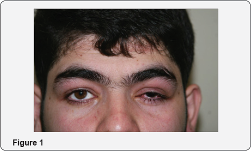
55 years male patient presented as a case of a
glaucoma in 2011 at ER ophthalmological dept of medical hospital
Srinagar Kashmir, he presented with severe pain marked redness of left
eye and marked loss of vision on exam he had sever ciliary congestin
corneal odema very shallow a chamber and dilated pupil no clear view of
fundus and IOP of more than 550mm Hg. He was put on emergency medication
of a c glaucoma in form of topical LUPITISS eye drops one drop l eye od
2 britiblu eye drops i drop L eye bd 3 osmotic diuretic 20 percent
mannitol 250cc IV 4 500mf of injection diamox I we even with this
medication pt dod not respond even after more than 12 hours of this
medication he was advised admission which he refused however no a aor
B-scan ultrasound was done pt after couple of days sought second
ophthalmic consultation this time the con ophthalmologist performed
B-scan ultrasound and picked up a solid retinal detachment of l eye to
confirm melanoma.ch he was referred for MRI scan of orbits and brain
unfortunately for patient and treating ophthalmologist the report of
scan was very unsatisfactory the radiologist gave a very unsatisfactory
report that pt has pan ophthalmitis l eye however ophthalmologist
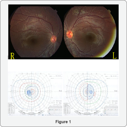
requested him that this is a case of CD melonoma [1],
and it has to be confirmed by MRI in this process p both pt and got
confused and patient left the treatment he was unfortunately going from
one oph to other as a painful blind eye for two years key words
melanoma.ch is the commonest tumor it can be benign or malignant
LUPITISS t is prostaglandin analogue and t stands for temilola beta
blocker beta blocker 3 britiblu is brimolol 3 mannitol is osmotic
duretic 4 diamox is carbonic anhydrase inhibitor od means one dop once a
day 5bd is one drop twice a day glaucoma is a v=condition where i o
gets raised in eye pathogenesis of IOP aqueous is produced by ciliary
process of ciary epethelium im post chamber where [2],
the aqueous nourishes lens then through pupil it comes in ant chamber
and aqua nourishes cornea and then it is dained though angle of
filtration through canal of schlem and finally aqueous comes in aqua
veins so any condition which causes obstruction in drainage of aqueous
will result in rise of IOP which is called glaucoma classification of
glaucoma cong glaucoma occurs during intra uterine life 2 infantile
glaucoma if from birth to 3 years 3 juvenile off 3 years to 17 years 2
primary glaucoma open and closed angle 3 sec glaucoma which occurs sec
to any intraocular disease 4 ocular hypertension sec to systematic
tension dx of cong glaucoma also called buphthalmos big eye ball a large
cornea we do under anesthesia in these neonates and measure [3], IOP and measure corneal diameter treatment modalities which r surgical have been
I. Goniotomy
II. Goniopexy
III.Trabeculotomy
IV.Sinostomy
In open angle glaucoma there is no pain moderate rise of IOP causes headache there are 3 features
a. Fundus changes
b. Changes in visual fields
Fundus causing glaucomatous cupping and even optic
atrophy most the times pts of open angle glaucoma are diagnosed late due
to lack of pain and pt may come when pt already [4],
had glaucomatous cupping and optic atrophy that's why we are doing
glaucoma screening after 40 years of age where we do routine measuring
of IOP refraction and fundus examination in this screening we are able t
pick up cases of open angle glaucoma treatment is medical and surgical
first [5],
we put it on medical therapy and if it doesn't not respond to it or it
not coming for regular ophthalmic evaluation or pt belongs to fat of
place lack of ophthalmic advice one should do surgery the surgical
modalities are 1 trabeculectomy advantages of this procedure
1. It is cosmetically better
2. It can be repeated
3. It is a physiologically better operation
4. It communicates the ant c with canal of schlem by splitting the trabecular mesh work
Nowadays, we have better options of lased and
operation of laser choice is an argon laser trabeculoplasty closed angle
glaucoma comes very early due [6],
to severe pain redness and marked loss of vision in most of the these
case they respond to anti glaucoma medication after the eye settled down
we do laser iridotomy even in normal eye of these pts we do polylectic
laser iridotomy then we have ocular hypertension due to systemic
hypertension key is control of b p ten we have a miscellaneous group of
glaucoma 1 neo vascularisation glaucoma due to c are c occlusion it is
said to subside in 3 months last l by least we have absolute glaucoma pt
has painful blind eye treatment modalities.
a) Cyclodiathermy
b) Cyclo croprexy
Sometimes due to severe pain we may inject
retrobulbar injection of xylocaine or alcohal to revealve pain sometimes
one may have to enuck leation if all measures fail introduction ch
melanoma is the most common primary malignan intraocular tumor and the
second most common type of primary malignant melanoma in the body it
most often whites of northern European descent essential update early
treatment may prevent metastatic deaths in patients with melanoma.ch in a
prospective cohort study of 3072 patients with melanoma. ch researchers
we found for some pts early treatment rather than watching and waiting
might better prevent metastatic deaths signs and symptoms melanoma.ch
remain asymptomatic for prolonged period of time may be found
incidentally during ophthalmoscope in general the more the their origin
the longer the delay of any symptoms melanoma.ch present with following
symptoms:
a. Blured vision
b. Paracentral scotoma
c. Progressive and painless visual loss
d. Floaters
e. Sever ocular pain
f. Weight loss marked fatigue cough or change in bowel or bladder suggests primary non ocular malignancy with melanoma.
ophthalmological examination may reveal following
A. Small melanoma.ch typically takes the form of a
nodular dome shaped and well circumscribed mass under the retinal
pigment epethelium
B. Melanoma.ch grow they adopt more irregular configurations
C. Diffuse melanoma.ch characterized by growth
throughout the choroid with minimal elevation are more difficult to
diagnose and often cause significant exudative redetachment
D. Melanoma.ch may have variable coloration some are more pigmented and others are less
E. If tumor is light colored its abnormal vascularisation usually can be seen ohthalmoscopicaly
F. Overlying melanoma. ch
There are usualy tet pig epethelial changes eg drusen
dx 1 liver enzyme are indicated in any pt with uveal melanoma is the
most common site of melanoma.ch metastases so one has to do
a. Alkaline phosphates
b. Glutamic oxaloacetic trans aminase
c. Lactase dehyrogenase
Gamma glutamyl transpeptidase a scan and B-scan
ultrasound MRI scan ct scan f f angoigraphy can help point toits dx 2
small ch melanomas may show floresene angigrahkic changes similar to
choriodal nevi 3 large melanomas may show patchy pattern of early
hypoflourence and hyper flourescence followed by late intense staining
simultaneous flourescence of retinal [7-8], and ch circulation within is fiarly distintive of melanomas.ch management
I. Observation may be acceptable for post uveal tumors not well established in particular tumors of less than
II. To 2.5mm and 10mm in diameter can be observer until growth is documented
III. Plaque brachy therapy is a widely accepted alternative to enucleation medium sised post uveal melanomas less than 10mm
IV. External beam irradiation with protons and helium ions
V. Pp vasectomy
VI. Block excision
VII. Laser photocoagulation
VIII. Orbital exenteration in extreme cases.
The above mentioned pt b was seen by me in 2013 his
all investigations l f t c bc k f t lipid profile f b s were within
normal limits ultrasound abdomen normal no evidence of any metastases i
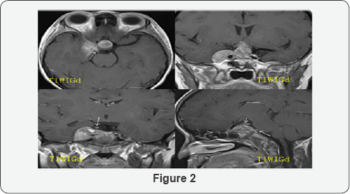
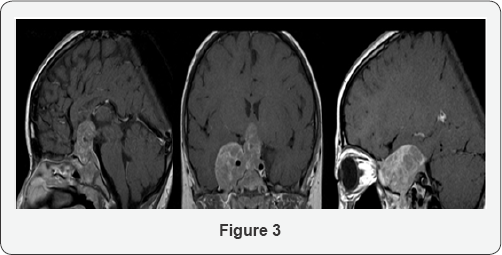
requested for MRI scan of orbits and brain it was very satisfactory
showing that optic nerve optic tract Chiasm a basal ganglia pit gland
thalmoid brain ventricles and cerebral hemispheres all were normal
discussion we have aired mentioned that scleral cavity can be
1. Antto lens so it will cause visual impairment
2. Involve vitrous causind floaters
3. Involve angle of filtration and present and a c glaucoma as was seen in our case.
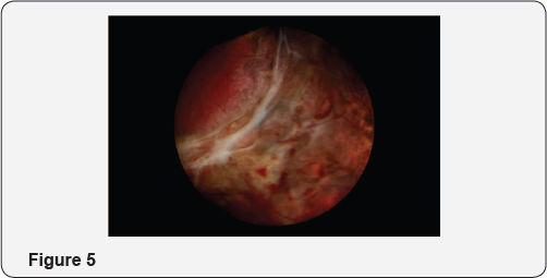
So after all above mentioned investigations under gowhar ahamed I removed whole cornea lens vitrous choriod and retina [9-11],
so we were left with intact intra scleral cavity optic nerve and all
ocular muscles i stitched ant and pst lips of sclera with 6 zero vicryl
kept a drain in intra scleral cavity which was removed after 24 hours
later i put a confirmer to give shape to orbit for 15 days after removal
of confirmer i fitted a well matching and fitting prosthesis the
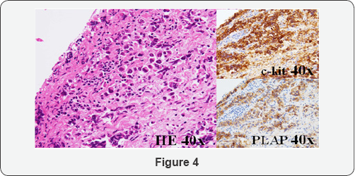
removed tissues were sent for histopathological examination did not show
any evidence of malignancy 3 years [12-15], have passed all repeat tests od blood ultra sonography b abdomen are normal.
So any pt f ac c glaucoma if does not respond to
treatment please do a or b scan ultrasound to pick up solid ret det and
rule out m melanoma.ch by MRI review of literature 1 vertex varies can
present as Uveal melanoma however it is a benign condition and temporary
2 in dept of ophthalmology manchester a pt came with a c glaucoma
vision in normal eye unaided was 6,18 have did not respond to treatment
MRI showed melanoma.ch so Enucleation was done 2 in Saudi medical lornal
and cairio institute of ophthalmology and nophthalmology one pt came
with BSEC glaucoma with imp of 56mmhg pt did not respond to treatment
MRI was done it revealed melanoma.ch so Enucleation was done.
For more Open Access
Journals in Juniper Publishers please
click on: https://juniperpublishers.com
For more articles
in JOJ Ophthalmology (JOJO) please click
on: https://juniperpublishers.com/jojo/index.php
For more about juniper publishers please click on:
https://www.juniperpublishersgroup.com/