Thursday, 2 February 2023

Attitudes and Beliefs of Undergraduate Students to Spectacle Wear - Juniper Publishers

 JOJ Ophthalmology- Juniper Publishers
 

Abstract

Purpose: The aim of the study was to determine the attitudes and beliefs of KNUST undergraduates to spectacle wear.

Method: A descriptive cross-sectional study was done. Close ended questionnaires were distributed to 500 KNUST undergraduates to collect data on their attitudes and beliefs to spectacle wear.

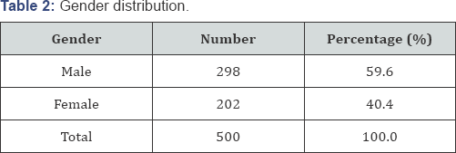
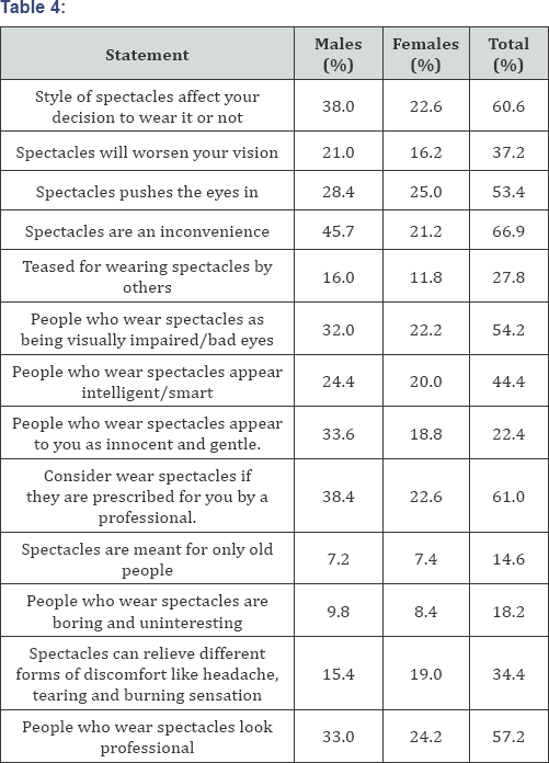
Results: A total of 500 undergraduates from the Kwame Nkrumah University of Science and Technology (KNUST) were included in this study. 298 were males (59.6%) and 202 (40.4%) were females with age ranging from 17-32 years (mean 21.51±1.93SD). 76% of the study population had heard of refractive error (near-sightedness, far sightedness or astigmatism) before. 57.4% felt that spectacles could be used to correct refractive error. 61% agreed they would wear spectacles if they were prescribed by an eye doctor. 53.4% believed eyeglasses cause the eyes to be pushed in and 66.9% believed eyeglasses are an inconvenience. Two-thirds (66%) did not know that eyeglasses could be used to relieve other forms of ocular discomfort like headache and tearing. 54.2% of respondents saw people who wore eyeglasses as visually handicapped, while 14.6% believed that eyeglasses were meant for old people. 27.8% of the respondents believed that they would be teased if they wore glasses. 57.2% said people who wear spectacles look professional.

Conclusion: Spectacles are still the most commonly preferred modality for the correction of refractive errors in the world today. Acceptance of glasses for the correction of refractive errors among KNUST undergraduates is not encouraging. There still lingers in the minds of the educated population, certain misconceptions regarding refractive errors and the various methods of correction which needs to be addressed. Counseling, mass media, support groups and the provision of information about refractive errors in schools and college textbooks, will help in dispelling the misconceptions and the distorted facts about these.

Keywords: Refractive error; Near-sightedness; Far sightedness; Astigmatism glasses; Spectacle; Students; Blindness; Perception; Psychosocial

Background

Uncorrected refractive is a major leading cause of preventable visual impairment. Global estimates indicate that more than 2.3 billion people in the world suffer from poor vision due to refractive error; of which 670 million people are considered visually impaired because they do not have access to corrective treatment. Current data suggests that more than 90% of people with uncorrected refractive error, worldwide, reside in rural and low-income countries [1].

Refractive errors, if uncorrected, results in an impaired quality of life of people. There is difficulty performing daily life tasks, such as learning, working and driving thus depriving them of the advantages in living a complete life. The commonest and cheapest treatment modality for correcting refractive errors is spectacles or eye glasses. The zeal with which patients comply with wearing of spectacle will be triggered by the level of their understanding of refractive errors and their attitudes towards eye glasses [2].

In Ghana and Africa at large, it is widely believed that children or young people should not have eye problems and as such should have no need to wear glasses. This therefore imposes some kind of stigma on anyone who wears glasses [3]. Others believe that glasses damage and weaken the eyes and they only wear them when it is absolutely necessary or on special occasions [4-6], have concluded from their studies that only a small percentage of young people who are living with marked uncorrected refractive errors actually have their spectacle prescription on. These studies have all focused on children, parents' attitude and teachers' involvement. The present study focused on undergraduates; because they are more mature and better informed to make educated decisions for themselves. With this background, this study seeks to find out the attitude and beliefs of KNUST undergraduates to spectacle wear. This is in order to improve acceptance of glasses when prescribed thus reducing blindness and visual impairment due to uncorrected refractive errors.

Methods

It was a descriptive cross-sectional study done among undergraduate students in Kwame Nkrumah University of Science and Technology (KNUST). KNUST, located in Kumasi in the Ashanti Region, is one of the largest public universities in Ghana. A convenience sampling technique was used to select the participants.

The sample size was calculated using the formula: n= Z2p(1-p)/ d2

Where:

‘p’ is the assumed population proportion = 0.75 'd' is the margin of error= 0.04

With a (CI) confidence interval of 95% 'Z'= 1.96

n = 1.962 x 0.75(1-0.75)/ 0.042 = 451

10/100X451=46

Minimum sample size = 497 participants. A sample size of 500 students was used in the study.

Informed consent was obtained from all participants before the study and aim of the study was described to them. Participants were also told they could withdraw from the study at any time. Confidentiality and anonymity was ensured with records and the information collected from participants and they were used solely for the purpose of the study. The study was approved by the Ethics and Research Committee of the Kwame Nkrumah University of Science and Technology, and was carried out in accordance with the tenets of the declaration of Helsinki.

The data was collected and gathered using structured questionnaires. The questionnaires were given to the students and they were instructed to make sure their attitudes and beliefs toward spectacle wear were best presented by ticking or circling the right answers among the alternatives. The other components of the questionnaire were the demography of the participant, psychosocial aspects, the perceptions about refractive errors and the various treatment options. The questionnaires were taken back the same day they were given as soon as the participants answered them. This was done in a period of one week. Data analysis was performed using SPSS version 23.0 (IBM Corporation, Armonk, NY, USA) and the results were calculated in percentages and presented in tables.

Results
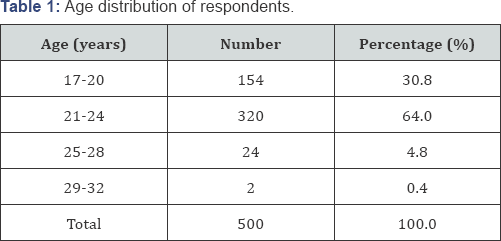
Shown in Table 1 & 2 are age and gender distribution of the participants respectively. 500 students took part in this study and there were 298 males (59.6%) and 202 (40.4%) females with age ranging from 17-32 years (mean age: 21.51±1.93). Table 3 shows correspondents' knowledge of refractive error correction methods. 57.4% of the study subjects responded that spectacles are one of the treatment plan to correct refractive errors, 38.6% felt contact lenses could be used to correct refractive errors, 21.8% felt drugs could be used to correct refractive errors, 23.0% considered surgery to be appropriate to correct refractive errors.
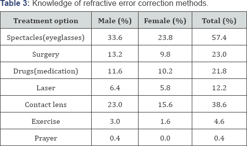
76.0% among the 500 students knew what refractive error (near-sightedness, long sightedness or astigmatism) is. 65.0% of the total population knew of at least one of the various methods of correction of refractive errors.

Table 4 comprises the psychosocial questions and their answers given adjacent to the questions. 60.6% of the total study population said the attractive style of spectacles will affect their decision to wear them. 61.0% of the total population agreed they would put on their eye glasses if they are a prescription from an eye care professional. 37.2% and 53.4% think spectacles will worsen their vision and cause the eyes to be sunken in respectively. 66.9% of the total population felt that spectacles are an inconvenience. 27.8% of the respondents felt that they would be teased for wearing spectacles by others. 54.2% also see people who wear spectacles as being visually impaired. 14.6% think spectacles are only meant for old people while 34.4% think spectacles can relieve different forms of discomfort like headache, tearing and burning sensation. 57.2% of the study participants also think people who wear spectacles look professional.
Discussion

Majority of the total population in this study (76.0%) know what refractive error is or had heard of refractive error before. This compares favorably to the study by [7] where a proportion of only 32.0% of the 500 study participants knew of refractive error in the University of Benin, Nigeria or gone to examine their eyes before despite the fact that they had had an eye problem before. The number (32.0%) is quite low compared to the figure obtained in this study and may be due to time gap. Approximately 65.0% of the respondents in this study knew of the various refractive error correction methods which is relatively high. As part of the treatment modalities which were reported, eye glasses were higher, contact lenses were next, with surgery and drugs following. This is in agreement with statistics from a previous study where it was reported that 50.0% of the study population agreed that eye glasses are a treatment option for correcting refractive error. Almost two- thirds of the participants (61%) in this study would not mind wearing spectacles if they are prescribed by an eye professional for them. This is in agreement with previous studies by [8,9], where 38.4% and 45.0% respectively also confirmed they would wear prescribed eye glasses [7]. Also reported that 49.8% of their participants would not mind wearing spectacles that will correct their refractive error.

Approximately 57.4% had in mind that spectacles could correct refractive errors yet very few of the respondents (34.4%) knew eye glasses could also be used to prevent headaches, glare from sunlight, burning sensation and other discomforts associated with the eye. 77.0% of the correspondents in this study did not know they could do refractive surgery in order to improve their eyesight and decrease or eliminate their dependency on spectacles. This agrees with the result obtained by where it was estimated that 83.0% of the interviewees were not aware of refractive surgery as a method of correcting refractive error. This is not a problem of much concern here in Ghana since modern machinery and equipment to carry out such an operation on the eyes are not available and considering the fact that Ghana is a developing country. 4.60% and 0.42% of the correspondents in this study thought prayers and exercises could be used to correct refractive errors respectively [4]. Confirmed that 31.0% of the subjects in their study felt that diet, yoga and traditional medicine could cure refractive errors.

The difference in the findings above could be due to the fact that, their study was conducted in a rural setting whereas this study was conducted in an urban centre where the level of education is higher. However, a misconception like this could result in them refraining from seeking appropriate treatment. 53.4% of participants in this study feared to use glasses with the belief that eye glasses cause sunken eyes. This is in line with two previous studies where people despised the use of eye glasses even when given to them for by an eye professional [10,11]. The fear of spectacles damaging the eyes was also a significant hindrance to spectacle use in a Nigerian study [1]. In studies on Chinese children, a common reason for not wearing spectacles was the belief that spectacles weakened the eyes [12]. In a report among secondary school students in Tanzania, this 'fear' was referred to as 'parental concerns about the safety of spectacle use' and it was listed among the barriers against the use of spectacles by students [5].

It has been speculated that spectacle wear could interfere with normal emmetropization (which depends on the growth of the eye, the refractive state, and the visual stimulation) in infants. Yet, the long term effect of spectacles on normal changes in the refractive error of the human eye is negligible [13]. The fear of spectacles damaging the eyes should be directly addressed and the concerns should be alleviated during consultations at eye care clinics. A greater proportion of the study population (66.90%) also said eye glasses are an inconvenience and will not wear it even if prescribed by an eye doctor. This indicates the need to change their perspective towards the use of spectacles through the dissemination of knowledge and information.

More males (16.0%) than females (11.8%) believed that they will be laughed at for wearing glasses as they regarded spectacles as one that makes beauty fade. 27.8% of the total population in this study said they will be mocked if they wear spectacles. This is similar to a previous study by [4] where it was reported that a large percentage of respondents (35.0%) were teased for wearing eyeglasses. It was generally the peers who had negative attitude to glasses wear and victimized other children with refractive errors who wear glasses. According to [4] 31.0% respondents were ready to hide their glasses to avoid rejection before marriage. An attitude like this could result in serious problems like psychosocial maladjustment, anxiety, depression loneliness, lowered self-esteem and behavioral problems.

37.2% of the respondents in this study believed that the continuous use of glasses would progressively cause their vision to worsen. This agrees with the result obtained by [4] in Dakshina where 30% of the participants thought that glasses would worsen their vision if used continually. In a study which was conducted in Pakistan by [14,15], 69.0% of the people thought that using spectacles would cause their vision to deteriorate and therefore tried to avoid it. The reason for the difference in numbers between the above two studies could be the difference in the study setting and in the educational level between the two populations, ours being an urban setting with a higher educational level of the respondents, our findings were contrary to those of the Pakistan study [7], confirmed that, 57.0% of the participants considered other people who wore glasses as visually impaired. In this study, 54.20% of the respondents regarded people who wore glasses as visually handicapped. The above two findings are similar. Children are discouraged with this inaccurate view by their teachers and parents and may decide not to wear them when they grow [16,17].

Conclusion

From this study, eyeglasses are still atop of all the various methods used to correct refractive error. Knowledge of refractive errors among KNUST undergraduates is not encouraging. There still lingers in the minds of the educated populace, certain misconceptions regarding refractive errors and the various methods used in correcting them [18-25]. All patients visiting the eye clinics should be made to be aware of the good outcomes eye glasses offer to the eyes more especially when their present eye condition calls for a spectacle prescription. This would help to erase any false information and deep rooted taboos regarding spectacle usage. Students should also be educated on the disadvantages associated with not wearing a prescribed eye glass and the basics of refractive errors as well as its other treatment modalities. There is also the need for people to have their eyes checked at the eye clinics and at outreach programs organised by recognised eye care units. This would help in the early diagnosis of refractive error if there exist any [26-31].


For more Open Access Journals in Juniper Publishers please click on: https://juniperpublishers.com/index.php

For more articles in JOJ Ophthalmology (JOJO) please click on: https://juniperpublishers.com/jojo/index.php


Thursday, 24 March 2022

Juniper Publishers- JOJ Ophthalmology


Visual Rehabilitation and Tolerability Using Hybrid Contact Lenses of Patients with Moderate to Severe Keratoconus-Juniper Publishers

Introduction

Keratoconus is non inflamatuary and progressive disease of the younger age group in which corneal thinning occurs and cornea assumes a conical shape associated with abnormal curvature. This changes often results in irregular astigmatism and myopia and leads to mild to marked visual impairment [1]. Corneal topography is a non-invasive technique to detect and monitor the progression of keratoconus. Contact lenses were used to improve visual conditions in keratoconus. Rigid gas permeable (RGP) lenses are most used type of contact lenses but in patient with severe disease RGP lens is insufficient for visual impairment and comfort [2]. Irregular astigmatism which developed after ectasia is not corrected with glasses and soft contact lenses. Mini-scleral, semi- scleral and scleral contact lenses are safe options in the management of irregular corneas. Because of heavy costs of scleral lenses, hybrid lenses with the comfort of the soft lenses and the optical quality of the hard lenses are developed [2]. This article reports the efficiency and accuracy of hybrid contact lens known as Clear Kone (Synerg Eyes Inc., Carlsbad, CA) in patients with moderate to severe keratoconus.

Methods

This study was performed in Medical Park Hospital, Antalya, Turkey. The patients were requested to sign informed consent forms. Patients had been previously diagnosed with keratoconus by corneal topography (Pentacam HR, Oculus, Wetzlar, Germany) and bio microscopic findings of Fleisher ring and Vogt lines. Inclusion criteria were keratoconus patients with severe visual problems and indication of intra corneal ring or transplantation surgery by another eye center. Cross linking was performed for all patients before the study. All patients were not appropriate for rigid gas permeable (RGP) lenses. All eyes were fitted with Clear Kone hybrid keratoconus lenses. The fitting is based on the concept of sagittal depth called as vault in relation to the cornea. Skirt curvature was determined as steep, median or flat according to limbus. Proper fitting was observed with using sodium fluorescein. After fitting, control of lens movement, vision and corneal epitelium were performed, 3 hours later. Corneal topography findings, pachymetry and refraction and vision were determined every 6 months.

Results

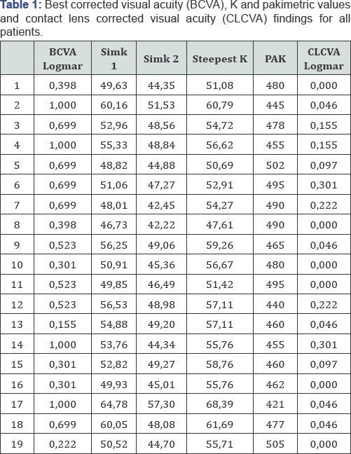
In this study, 19 eyes of 11 patients (6 men and 5 women) with a mean age of 26, 4 (16-43) were evaluated. Keratometry findings are between55-75 (Kmax). Before using contact lens, uncorrected and best corrected visual acuity with glasses were 0, 74±0, 3 LogMAR and 0, 58±0, 22 Log MAR respectively. Visual acuity with hybrid contact lenses was 0, 09±0, 05 LogMAR (Table 1). Mean follow-up was 7, 2 months (4-12 months). One patient didn't tolerate because of corneal edema.

Discussion

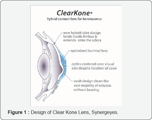
The first treatment choice for a patient with keratoconus is using a RGP lens [3]. Therefore, most of these patients, visions not corrected by glasses or soft contact lenses, have already tried the RGP lenses. The potential challenges associated with fitting rigid lenses are suboptimal initial comfort on non adapted eyes, the potential for lens decentration and the risk of lens ejection. Soft contact lenses have more comfort but less visual correction especially in advanced disease. Aim of hybrid lens is to combine the preferred properties of rigid and soft contact lenses [4]. Hybrid lenses are combines of a center-zone rigid lens and a peripheral zone soft skirt. Clear Kone lens which is used this study requires the determination of two fitting parameters, vault for rigid component and skirt curvature for soft component [5]. Due to design of the hybrid lens, most of the refraction power is provided by the tear layer, which increases the optical quality and oxygen supply of the cornea. Additionally, a little space between cornea and hard part of the lens prevents mechanical abrasion of the cornea [4] (Figure 1).

In our study, all patients have moderate to severe keratoconus and discomfort while using RGP lenses. Because of this condition, by other eye centers, corneal ring or keratoplasty were advised. Before the surgical treatment, we wanted to try another non- invasive management, hybrid lens fitting. Disadvantage of using hybrid lens, Clear Kone Synerg Eyes, is that this process is time consuming and requires more patience. Because of this, for appropriate final lens and shortening of process, we used the parameters of RGP lenses. This approach improved patients' compliances. Clear Kone lenses with hard central part and tears between lens and cornea improve vision. In our study, all patients had a good visual outcome. Soft peripheral part provides stability and comfort. Except one patient, 10 of 11 patients had a good comfort.

According to findings of this study, using hybrid lens on the keratoconus patients can be good choise for the patients with moderate to severe disease with discomfort of soft or hard lenses before thinking surgical management.

For more Open Access Journals in Juniper Publishers please click on: https://juniperpublishers.com

For more articles in  JOJ Ophthalmology (JOJO) please click on: https://juniperpublishers.com/jojo/index.php

For more about juniper publishers  please click on: https://www.juniperpublishersgroup.com/


Saturday, 25 December 2021

Juniper Publishers- JOJ Ophthalmology


Giant Solitary Trichilemmal Cyst of the Upper Eye Lid: Masquerading Lacrimal Gland Tumor: A Clinico Radio Pathological Case Report-Juniper Publishers


Introduction

Trichilemmal cyst, also known as a pilar cyst, forms from the outer root sheath of hair follicle. They are common benign tumors most often found on the scalp [1]. The involvement of the eyelids is quite rare. It presents as a smooth, firm, mobile, and round nodule without a visible punctum. There is often an autosomal dominant inheritance pattern as the lesion is frequently familial [2]. Till now, there are only few case reports of trichilemmal cyst of the eyelid [3-6].

Case Report

A 58 year old mahemedian male from Bulendsar, UP, India was referred to Sharda Hospital because of a persistent static small swelling upper lid for last 10 years which is progressively increased over last three months. It was horizontally disposed [7-8], approximately 12mm/8mm in horizontal and vertical dimension. According to the patient this tumour first appeared as upper lid nodule. But for the last three months, it was growing rapidly and he was unable to open the eyelid. Patient developed ptosis and difficulties in vision due to large tumour and weight of the mass. There is drooping of upper eye lid since then. He is a non vegeterian and none of his family members had similar problem. An ophthalmic examination revealed a visual acuity of 20/40 in each eye due to presence of cataract which was not improved with pin hole or refraction [9-10]. The right upper lid has covered total cornea and almost 3/4th of palpebral apperature. A closer examination of anterior segment of both eyes were apparently normal expect early cataractous changes. Both the fundii were normal.

Examination of upper lid showed a large swelling arising from the upper lid, involving the 1/3rd of the right upper lid. Surface was smooth, overlying skin stretched. The mass was not with any surface blood vessels, with no colour or temperature changes, no expansible impulse on coughing and on auscultation no bruit was heard. Texture of the tumour mass was firm and solid. Intra Ocular Pressure by Applanation Ton+ometry was 16mm hg in both the eyes. Schirmer value was 18mm and that of BUT 14 in both the eyes. Plain X Ray of right orbit showed a large, well-defined, solid solitary mass at lateral aspect of upper lid just adjacent to lacrimal gland. measuring 3.1x2.1cm, over Routine Blood Investigations were within normal limit A clinical diagnosis of epithelial cyst was made. The patient under wentan excisional biopsy of the right upper lid. A skin incision was made along the lid crease [11-12], the skin was undermined over the mass which was unexpectedly easy in view of its adherence to mass on preoperative examination and the mass was easily separated from the underlying orbicularis. The mass was removed in to and primary skin closure was done. The excised mass was white in color. Post operatively, the lid contour was normal and there was no ptosis. No recurrence or evolution of new lesion was noted on follow-up. The patient is highly satisfied for cosmetic and free of growth. Histopathological examination of the specimen revealed the mass was lined by stratified squamous epithelium that lacks a granular cell layer and was filled with compact «wet» keratin.

(A) Clinical photograph of the right eye showing a growth over lateral aspect of upper lid with ptosis

(B) Photomicrograph from the removed growth showing (haematoxylin and eosin, original magnification x250, inset x325).

Comment

Trichilemmal cysts of the body may run in families and they may or may not be inflamed. They are solitary in 30% of cases and multiple in 70% of cases. These cysts are derived from the outer root sheath of the hair follicle. Their origin is unknown, but it has been suggested that they are produced by budding from the external root sheath as a genetically determined structural aberration. They arise preferentially in areas of high hair follicle concentration, therefore, 90% of cases occur on the scalp. Rarely, these cysts may grow more extensively and form rapidly multiplying trichilemmal tumors, also called proliferating trichilemmal cysts, which are benign but may grow aggressively at the cyst site. Very rarely, trichilemmal cysts can become malignant. They are lined by stratified squamous epithelium that lacks a granular cell layer and are filled with compact “wet”keratin. Areas consistent with proliferation can be found in some cysts. In rare cases, this leads to formation of a tumor, known as a proliferating trichilemmal cyst. The tumor is clinically benign, although it may display nuclear atypia, dyskeratosis, and mitotic figures which often can be mis leaded as the diagnosis of squamous cell carcinoma. Surgical excision is the only mode of management.

For more Open Access Journals in Juniper Publishers please click on: https://juniperpublishers.com

For more articles in  JOJ Ophthalmology (JOJO) please click on: https://juniperpublishers.com/jojo/index.php

For more about juniper publishers  please click on: https://www.juniperpublishersgroup.com/


Tuesday, 14 December 2021

Juniper Publishers- JOJ Ophthalmology


An Interesting Case of Golden Hars Syndrome in 3 Years Male Child-Juniper Publishers


Introduction

Now we have defined d h syndrome as a complex of cong limmal dermoid with cong pre auricular skin tag aor appendage limbal dermoids sometimes are bilateral rarely. Usually they are unilateral involving whole of cornea or may be cofined to conjunctiva only incidence of l dermoids are 1 in 10000 or 500 in 2700 they are graded according to involvement of cornea grade 1. when only corneal epithelium is involved 2 grade 2 des membrane 3 grade 3 whole of ant segment of eye is involved inferno temporal involvement of limbal dermoid is the commonest about 70 percent.

Case Report

6 months back a 3 years male child was seen by me in my office with parents having noticed a small palish. White infero temporal limbal region left eye with cong presence of pre auricular skin appendage ft delivered child after lsc s no other cong anamolies seen vision mydriatic refraction ant segment and fundi were normal child had grade 1 limbal dermoid with the commonest site being infer temporal.

Results and Discussion

Golden hars is not a vision threatening disease most of these pts have normal intelligence and vision only in cases where visual axis is involved the we think of surgery conclusion my case was a simple case of gowhar ahmad grade 1 limbal dermoid so it only need observation in cases where visual axis is involved and vision is threatened then the surgical modalities are

    1. Visual

    2. Cosmotic

We do

    1. Lamellar keratoplaty

    2. Amionotic membrane graft

    3. Stem cell graft

Their families of golden hars syndrome in Greece 2 in middle east in gulf war children born in different military hospital had golden hars syndrome 3 sometimes optic nerve drusen is associated with g h syndrome.

For more Open Access Journals in Juniper Publishers please click on: https://juniperpublishers.com

For more articles in  JOJ Ophthalmology (JOJO) please click on: https://juniperpublishers.com/jojo/index.php

For more about juniper publishers  please click on: https://www.juniperpublishersgroup.com/


Wednesday, 8 December 2021

Juniper Publishers- JOJ Ophthalmology

 Visual Rehabilitation and Tolerability Using Hybrid Contact Lenses of Patients with Moderate to Severe Keratoconus-Juniper Publishers


Introduction

Keratoconus is non inflamatuary and progressive disease of the younger age group in which corneal thinning occurs and cornea assumes a conical shape associated with abnormal curvature. This changes often results in irregular astigmatism and myopia and leads to mild to marked visual impairment [1]. Corneal topography is a non-invasive technique to detect and monitor the progression of keratoconus. Contact lenses were used to improve visual conditions in keratoconus. Rigid gas permeable (RGP) lenses are most used type of contact lenses but in patient with severe disease RGP lens is insufficient for visual impairment and comfort [2]. Irregular astigmatism which developed after ectasia is not corrected with glasses and soft contact lenses. Mini-scleral, semi- scleral and scleral contact lenses are safe options in the management of irregular corneas. Because of heavy costs of scleral lenses, hybrid lenses with the comfort of the soft lenses and the optical quality of the hard lenses are developed [2]. This article reports the efficiency and accuracy of hybrid contact lens known as Clear Kone (Synerg Eyes Inc., Carlsbad, CA) in patients with moderate to severe keratoconus.

Methods

This study was performed in Medical Park Hospital, Antalya, Turkey. The patients were requested to sign informed consent forms. Patients had been previously diagnosed with keratoconus by corneal topography (Pentacam HR, Oculus, Wetzlar, Germany) and bio microscopic findings of Fleisher ring and Vogt lines. Inclusion criteria were keratoconus patients with severe visual problems and indication of intra corneal ring or transplantation surgery by another eye center. Cross linking was performed for all patients before the study. All patients were not appropriate for rigid gas permeable (RGP) lenses. All eyes were fitted with Clear Kone hybrid keratoconus lenses. The fitting is based on the concept of sagittal depth called as vault in relation to the cornea. Skirt curvature was determined as steep, median or flat according to limbus. Proper fitting was observed with using sodium fluorescein. After fitting, control of lens movement, vision and corneal epitelium were performed, 3 hours later. Corneal topography findings, pachymetry and refraction and vision were determined every 6 months.

Results

In this study, 19 eyes of 11 patients (6 men and 5 women) with a mean age of 26, 4 (16-43) were evaluated. Keratometry findings are between55-75 (Kmax). Before using contact lens, uncorrected and best corrected visual acuity with glasses were 0, 74±0, 3 LogMAR and 0, 58±0, 22 Log MAR respectively. Visual acuity with hybrid contact lenses was 0, 09±0, 05 LogMAR (Table 1). Mean follow-up was 7, 2 months (4-12 months). One patient didn't tolerate because of corneal edema.

Discussion

The first treatment choice for a patient with keratoconus is using a RGP lens [3]. Therefore, most of these patients, visions not corrected by glasses or soft contact lenses, have already tried the RGP lenses. The potential challenges associated with fitting rigid lenses are suboptimal initial comfort on non adapted eyes, the potential for lens decentration and the risk of lens ejection. Soft contact lenses have more comfort but less visual correction especially in advanced disease. Aim of hybrid lens is to combine the preferred properties of rigid and soft contact lenses [4]. Hybrid lenses are combines of a center-zone rigid lens and a peripheral zone soft skirt. Clear Kone lens which is used this study requires the determination of two fitting parameters, vault for rigid component and skirt curvature for soft component [5]. Due to design of the hybrid lens, most of the refraction power is provided by the tear layer, which increases the optical quality and oxygen supply of the cornea. Additionally, a little space between cornea and hard part of the lens prevents mechanical abrasion of the cornea [4] (Figure 1).

In our study, all patients have moderate to severe keratoconus and discomfort while using RGP lenses. Because of this condition, by other eye centers, corneal ring or keratoplasty were advised. Before the surgical treatment, we wanted to try another non- invasive management, hybrid lens fitting. Disadvantage of using hybrid lens, Clear Kone Synerg Eyes, is that this process is time consuming and requires more patience. Because of this, for appropriate final lens and shortening of process, we used the parameters of RGP lenses. This approach improved patients' compliances. Clear Kone lenses with hard central part and tears between lens and cornea improve vision. In our study, all patients had a good visual outcome. Soft peripheral part provides stability and comfort. Except one patient, 10 of 11 patients had a good comfort.

According to findings of this study, using hybrid lens on the keratoconus patients can be good choise for the patients with moderate to severe disease with discomfort of soft or hard lenses before thinking surgical management.

For more Open Access Journals in Juniper Publishers please click on: https://juniperpublishers.com

For more articles in  JOJ Ophthalmology (JOJO) please click on: https://juniperpublishers.com/jojo/index.php
For more about juniper publishers  please click on: https://www.juniperpublishersgroup.com/


Friday, 3 December 2021

Juniper Publishers- JOJ Ophthalmology

Why Should SLT be the First-Choice Treatment for Glaucoma? -Juniper Publishers


Introduction

Glaucoma is a chronic, degenerative optic neuropathy which is characterized by changes in the optic disc and visual field loss. Intraocular pressure is considered the major risk factor for the progression of glaucomatous optic neuropathy involving the death of retinal ganglion cells and their axons. Selective Laser Trabeculoplasty (SLT) uses a 532nm frequency-doubled, q-switched neodymium, yttrium-aluminum-garnet laser that delivers a low-energy, large spot, it was designed to selectively target pigmented trabecular meshwork (TM) cells while sparing adjacent cells and tissues from thermal damage.

Discussion

Glaucoma is the second leading cause of blindness worldwide. Glaucoma affects more than 70 million people worldwide with approximately 10% being bilaterally blind, making it the leading cause of irreversible blindness in the world [1]. Meta-analysis of many studies showed that there was no statistically significant difference in IOP-reduction or treatment success between SLT and medical treatment [2].

As a first-line therapy for glaucoma, medications have many disadvantages and drawbacks. Patients have to accept their adverse effects, repeated application of medications and continuous medical costs. The potency of treatments may be undercut when people are non-compliant. The introduction to SLT, which uses a less severe energy to generate IOP-lowering effect without dangerous adverse reactions, has again brought up the question: can laser device treatments defeat eye drops as the main therapy for Glaucoma especially OAG? In the initial released research, SLT was used as the adjunct treatment to medication. Later several researches had recommended that SLT might be the main treatment for POAG or OHT [3]. Melamed et al. [4] found 40 in 45 eyes (89%) which experienced SLT as the main treatment had an IOP loss of 5mmHg or more. Mean IOP- reduction was 7.7±3.5mmHg (30%) at 18 months post-SLT. A potential, multicenter study by McIlraith et al. [5] involved 100 eyes (61 patients) with recently clinically diagnosed POAG or OHT. IOP reduction was 8.3mmHg (31.0%) in the SLT team (74 eyes) in contrast to 7.7mmHg (30.6%) in the latanoprost team (26 eyes) (P=0.208 and P=0.879). The amount of IOP reduction was mathematically considerably less in the latanoprost team than in the SLT team [5].

Selective laser trabeculoplasty (SLT) has been proven secure, well recognized, low-cost and very efficient at reducing intraocular pressure (IOP) as primary treatment in several types of glaucomas. The maintenance of trabecular meshwork (TM) structure and the confirmed effectiveness in decreasing IOP tend to make SLT a very affordable and secure alternative to argon laser trabeculoplasty (ALT). SLT may also be effective for situations of unsuccessful ALT and is a technique that may also be repeatable, compared with ALT. SLT is also a simple procedure for an ophthalmologist to learn and proceed. SLT has been confirmed to be efficient as main therapy and can be an effective adjunct therapy with medications for early stages of glaucoma. Furthermore, SLT can be regarded as a primary treatment choice for patients who cannot accept or who are noncompliant with their glaucoma medications, without disrupting the success of the forthcoming surgery [6].

Conclusion

SLT is easy to perform and well tolerated by patients and provides the benefits of ALT while using much less energy with less obvious harm to the TM. It seems to have comparative effectiveness to eye drops and to be a safe and effective glaucoma treatment without conformity threats or wide spread adverse reactions. Therefore, SLT is now considered the first line therapy for various types of glaucoma and can be an effective adjunct in the early types of glaucoma.

For more Open Access Journals in Juniper Publishers please click on: https://juniperpublishers.com

For more articles in  JOJ Ophthalmology (JOJO) please click on: https://juniperpublishers.com/jojo/index.php
For more about juniper publishers  please click on: https://www.juniperpublishersgroup.com/