Saturday, 25 December 2021

Juniper Publishers- JOJ Ophthalmology


Giant Solitary Trichilemmal Cyst of the Upper Eye Lid: Masquerading Lacrimal Gland Tumor: A Clinico Radio Pathological Case Report-Juniper Publishers


Introduction

Trichilemmal cyst, also known as a pilar cyst, forms from the outer root sheath of hair follicle. They are common benign tumors most often found on the scalp [1]. The involvement of the eyelids is quite rare. It presents as a smooth, firm, mobile, and round nodule without a visible punctum. There is often an autosomal dominant inheritance pattern as the lesion is frequently familial [2]. Till now, there are only few case reports of trichilemmal cyst of the eyelid [3-6].

Case Report

A 58 year old mahemedian male from Bulendsar, UP, India was referred to Sharda Hospital because of a persistent static small swelling upper lid for last 10 years which is progressively increased over last three months. It was horizontally disposed [7-8], approximately 12mm/8mm in horizontal and vertical dimension. According to the patient this tumour first appeared as upper lid nodule. But for the last three months, it was growing rapidly and he was unable to open the eyelid. Patient developed ptosis and difficulties in vision due to large tumour and weight of the mass. There is drooping of upper eye lid since then. He is a non vegeterian and none of his family members had similar problem. An ophthalmic examination revealed a visual acuity of 20/40 in each eye due to presence of cataract which was not improved with pin hole or refraction [9-10]. The right upper lid has covered total cornea and almost 3/4th of palpebral apperature. A closer examination of anterior segment of both eyes were apparently normal expect early cataractous changes. Both the fundii were normal.

Examination of upper lid showed a large swelling arising from the upper lid, involving the 1/3rd of the right upper lid. Surface was smooth, overlying skin stretched. The mass was not with any surface blood vessels, with no colour or temperature changes, no expansible impulse on coughing and on auscultation no bruit was heard. Texture of the tumour mass was firm and solid. Intra Ocular Pressure by Applanation Ton+ometry was 16mm hg in both the eyes. Schirmer value was 18mm and that of BUT 14 in both the eyes. Plain X Ray of right orbit showed a large, well-defined, solid solitary mass at lateral aspect of upper lid just adjacent to lacrimal gland. measuring 3.1x2.1cm, over Routine Blood Investigations were within normal limit A clinical diagnosis of epithelial cyst was made. The patient under wentan excisional biopsy of the right upper lid. A skin incision was made along the lid crease [11-12], the skin was undermined over the mass which was unexpectedly easy in view of its adherence to mass on preoperative examination and the mass was easily separated from the underlying orbicularis. The mass was removed in to and primary skin closure was done. The excised mass was white in color. Post operatively, the lid contour was normal and there was no ptosis. No recurrence or evolution of new lesion was noted on follow-up. The patient is highly satisfied for cosmetic and free of growth. Histopathological examination of the specimen revealed the mass was lined by stratified squamous epithelium that lacks a granular cell layer and was filled with compact «wet» keratin.

(A) Clinical photograph of the right eye showing a growth over lateral aspect of upper lid with ptosis

(B) Photomicrograph from the removed growth showing (haematoxylin and eosin, original magnification x250, inset x325).

Comment

Trichilemmal cysts of the body may run in families and they may or may not be inflamed. They are solitary in 30% of cases and multiple in 70% of cases. These cysts are derived from the outer root sheath of the hair follicle. Their origin is unknown, but it has been suggested that they are produced by budding from the external root sheath as a genetically determined structural aberration. They arise preferentially in areas of high hair follicle concentration, therefore, 90% of cases occur on the scalp. Rarely, these cysts may grow more extensively and form rapidly multiplying trichilemmal tumors, also called proliferating trichilemmal cysts, which are benign but may grow aggressively at the cyst site. Very rarely, trichilemmal cysts can become malignant. They are lined by stratified squamous epithelium that lacks a granular cell layer and are filled with compact “wet”keratin. Areas consistent with proliferation can be found in some cysts. In rare cases, this leads to formation of a tumor, known as a proliferating trichilemmal cyst. The tumor is clinically benign, although it may display nuclear atypia, dyskeratosis, and mitotic figures which often can be mis leaded as the diagnosis of squamous cell carcinoma. Surgical excision is the only mode of management.

For more Open Access Journals in Juniper Publishers please click on: https://juniperpublishers.com

For more articles in  JOJ Ophthalmology (JOJO) please click on: https://juniperpublishers.com/jojo/index.php

For more about juniper publishers  please click on: https://www.juniperpublishersgroup.com/


Tuesday, 14 December 2021

Juniper Publishers- JOJ Ophthalmology


An Interesting Case of Golden Hars Syndrome in 3 Years Male Child-Juniper Publishers


Introduction

Now we have defined d h syndrome as a complex of cong limmal dermoid with cong pre auricular skin tag aor appendage limbal dermoids sometimes are bilateral rarely. Usually they are unilateral involving whole of cornea or may be cofined to conjunctiva only incidence of l dermoids are 1 in 10000 or 500 in 2700 they are graded according to involvement of cornea grade 1. when only corneal epithelium is involved 2 grade 2 des membrane 3 grade 3 whole of ant segment of eye is involved inferno temporal involvement of limbal dermoid is the commonest about 70 percent.

Case Report

6 months back a 3 years male child was seen by me in my office with parents having noticed a small palish. White infero temporal limbal region left eye with cong presence of pre auricular skin appendage ft delivered child after lsc s no other cong anamolies seen vision mydriatic refraction ant segment and fundi were normal child had grade 1 limbal dermoid with the commonest site being infer temporal.

Results and Discussion

Golden hars is not a vision threatening disease most of these pts have normal intelligence and vision only in cases where visual axis is involved the we think of surgery conclusion my case was a simple case of gowhar ahmad grade 1 limbal dermoid so it only need observation in cases where visual axis is involved and vision is threatened then the surgical modalities are

    1. Visual

    2. Cosmotic

We do

    1. Lamellar keratoplaty

    2. Amionotic membrane graft

    3. Stem cell graft

Their families of golden hars syndrome in Greece 2 in middle east in gulf war children born in different military hospital had golden hars syndrome 3 sometimes optic nerve drusen is associated with g h syndrome.

For more Open Access Journals in Juniper Publishers please click on: https://juniperpublishers.com

For more articles in  JOJ Ophthalmology (JOJO) please click on: https://juniperpublishers.com/jojo/index.php

For more about juniper publishers  please click on: https://www.juniperpublishersgroup.com/


Wednesday, 8 December 2021

Juniper Publishers- JOJ Ophthalmology

 Visual Rehabilitation and Tolerability Using Hybrid Contact Lenses of Patients with Moderate to Severe Keratoconus-Juniper Publishers


Introduction

Keratoconus is non inflamatuary and progressive disease of the younger age group in which corneal thinning occurs and cornea assumes a conical shape associated with abnormal curvature. This changes often results in irregular astigmatism and myopia and leads to mild to marked visual impairment [1]. Corneal topography is a non-invasive technique to detect and monitor the progression of keratoconus. Contact lenses were used to improve visual conditions in keratoconus. Rigid gas permeable (RGP) lenses are most used type of contact lenses but in patient with severe disease RGP lens is insufficient for visual impairment and comfort [2]. Irregular astigmatism which developed after ectasia is not corrected with glasses and soft contact lenses. Mini-scleral, semi- scleral and scleral contact lenses are safe options in the management of irregular corneas. Because of heavy costs of scleral lenses, hybrid lenses with the comfort of the soft lenses and the optical quality of the hard lenses are developed [2]. This article reports the efficiency and accuracy of hybrid contact lens known as Clear Kone (Synerg Eyes Inc., Carlsbad, CA) in patients with moderate to severe keratoconus.

Methods

This study was performed in Medical Park Hospital, Antalya, Turkey. The patients were requested to sign informed consent forms. Patients had been previously diagnosed with keratoconus by corneal topography (Pentacam HR, Oculus, Wetzlar, Germany) and bio microscopic findings of Fleisher ring and Vogt lines. Inclusion criteria were keratoconus patients with severe visual problems and indication of intra corneal ring or transplantation surgery by another eye center. Cross linking was performed for all patients before the study. All patients were not appropriate for rigid gas permeable (RGP) lenses. All eyes were fitted with Clear Kone hybrid keratoconus lenses. The fitting is based on the concept of sagittal depth called as vault in relation to the cornea. Skirt curvature was determined as steep, median or flat according to limbus. Proper fitting was observed with using sodium fluorescein. After fitting, control of lens movement, vision and corneal epitelium were performed, 3 hours later. Corneal topography findings, pachymetry and refraction and vision were determined every 6 months.

Results

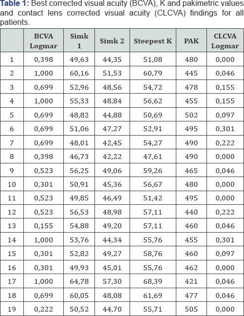
In this study, 19 eyes of 11 patients (6 men and 5 women) with a mean age of 26, 4 (16-43) were evaluated. Keratometry findings are between55-75 (Kmax). Before using contact lens, uncorrected and best corrected visual acuity with glasses were 0, 74±0, 3 LogMAR and 0, 58±0, 22 Log MAR respectively. Visual acuity with hybrid contact lenses was 0, 09±0, 05 LogMAR (Table 1). Mean follow-up was 7, 2 months (4-12 months). One patient didn't tolerate because of corneal edema.

Discussion

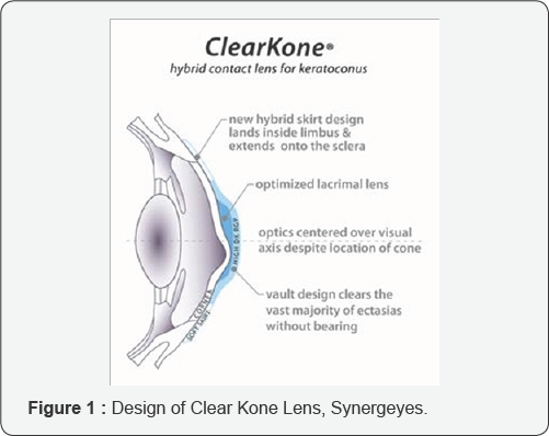
The first treatment choice for a patient with keratoconus is using a RGP lens [3]. Therefore, most of these patients, visions not corrected by glasses or soft contact lenses, have already tried the RGP lenses. The potential challenges associated with fitting rigid lenses are suboptimal initial comfort on non adapted eyes, the potential for lens decentration and the risk of lens ejection. Soft contact lenses have more comfort but less visual correction especially in advanced disease. Aim of hybrid lens is to combine the preferred properties of rigid and soft contact lenses [4]. Hybrid lenses are combines of a center-zone rigid lens and a peripheral zone soft skirt. Clear Kone lens which is used this study requires the determination of two fitting parameters, vault for rigid component and skirt curvature for soft component [5]. Due to design of the hybrid lens, most of the refraction power is provided by the tear layer, which increases the optical quality and oxygen supply of the cornea. Additionally, a little space between cornea and hard part of the lens prevents mechanical abrasion of the cornea [4] (Figure 1).

In our study, all patients have moderate to severe keratoconus and discomfort while using RGP lenses. Because of this condition, by other eye centers, corneal ring or keratoplasty were advised. Before the surgical treatment, we wanted to try another non- invasive management, hybrid lens fitting. Disadvantage of using hybrid lens, Clear Kone Synerg Eyes, is that this process is time consuming and requires more patience. Because of this, for appropriate final lens and shortening of process, we used the parameters of RGP lenses. This approach improved patients' compliances. Clear Kone lenses with hard central part and tears between lens and cornea improve vision. In our study, all patients had a good visual outcome. Soft peripheral part provides stability and comfort. Except one patient, 10 of 11 patients had a good comfort.

According to findings of this study, using hybrid lens on the keratoconus patients can be good choise for the patients with moderate to severe disease with discomfort of soft or hard lenses before thinking surgical management.

For more Open Access Journals in Juniper Publishers please click on: https://juniperpublishers.com

For more articles in  JOJ Ophthalmology (JOJO) please click on: https://juniperpublishers.com/jojo/index.php

For more about juniper publishers  please click on: https://www.juniperpublishersgroup.com/


Friday, 3 December 2021

Juniper Publishers- JOJ Ophthalmology

Why Should SLT be the First-Choice Treatment for Glaucoma? -Juniper Publishers


Introduction

Glaucoma is a chronic, degenerative optic neuropathy which is characterized by changes in the optic disc and visual field loss. Intraocular pressure is considered the major risk factor for the progression of glaucomatous optic neuropathy involving the death of retinal ganglion cells and their axons. Selective Laser Trabeculoplasty (SLT) uses a 532nm frequency-doubled, q-switched neodymium, yttrium-aluminum-garnet laser that delivers a low-energy, large spot, it was designed to selectively target pigmented trabecular meshwork (TM) cells while sparing adjacent cells and tissues from thermal damage.

Discussion

Glaucoma is the second leading cause of blindness worldwide. Glaucoma affects more than 70 million people worldwide with approximately 10% being bilaterally blind, making it the leading cause of irreversible blindness in the world [1]. Meta-analysis of many studies showed that there was no statistically significant difference in IOP-reduction or treatment success between SLT and medical treatment [2].

As a first-line therapy for glaucoma, medications have many disadvantages and drawbacks. Patients have to accept their adverse effects, repeated application of medications and continuous medical costs. The potency of treatments may be undercut when people are non-compliant. The introduction to SLT, which uses a less severe energy to generate IOP-lowering effect without dangerous adverse reactions, has again brought up the question: can laser device treatments defeat eye drops as the main therapy for Glaucoma especially OAG? In the initial released research, SLT was used as the adjunct treatment to medication. Later several researches had recommended that SLT might be the main treatment for POAG or OHT [3]. Melamed et al. [4] found 40 in 45 eyes (89%) which experienced SLT as the main treatment had an IOP loss of 5mmHg or more. Mean IOP- reduction was 7.7±3.5mmHg (30%) at 18 months post-SLT. A potential, multicenter study by McIlraith et al. [5] involved 100 eyes (61 patients) with recently clinically diagnosed POAG or OHT. IOP reduction was 8.3mmHg (31.0%) in the SLT team (74 eyes) in contrast to 7.7mmHg (30.6%) in the latanoprost team (26 eyes) (P=0.208 and P=0.879). The amount of IOP reduction was mathematically considerably less in the latanoprost team than in the SLT team [5].

Selective laser trabeculoplasty (SLT) has been proven secure, well recognized, low-cost and very efficient at reducing intraocular pressure (IOP) as primary treatment in several types of glaucomas. The maintenance of trabecular meshwork (TM) structure and the confirmed effectiveness in decreasing IOP tend to make SLT a very affordable and secure alternative to argon laser trabeculoplasty (ALT). SLT may also be effective for situations of unsuccessful ALT and is a technique that may also be repeatable, compared with ALT. SLT is also a simple procedure for an ophthalmologist to learn and proceed. SLT has been confirmed to be efficient as main therapy and can be an effective adjunct therapy with medications for early stages of glaucoma. Furthermore, SLT can be regarded as a primary treatment choice for patients who cannot accept or who are noncompliant with their glaucoma medications, without disrupting the success of the forthcoming surgery [6].

Conclusion

SLT is easy to perform and well tolerated by patients and provides the benefits of ALT while using much less energy with less obvious harm to the TM. It seems to have comparative effectiveness to eye drops and to be a safe and effective glaucoma treatment without conformity threats or wide spread adverse reactions. Therefore, SLT is now considered the first line therapy for various types of glaucoma and can be an effective adjunct in the early types of glaucoma.

For more Open Access Journals in Juniper Publishers please click on: https://juniperpublishers.com

For more articles in  JOJ Ophthalmology (JOJO) please click on: https://juniperpublishers.com/jojo/index.php

For more about juniper publishers  please click on: https://www.juniperpublishersgroup.com/

Tuesday, 30 November 2021

Juniper Publishers- JOJ Ophthalmology


Rare and Interesting Case of Eye Choriodal Melanoma Presenting as a Congestive Glaucoma in a 55 Years Male Patient-Juniper Publishers

Abstract

55 years male patient presented as a case of a glaucoma in 2011 at ER ophthalmological dept of medical hospital Srinagar Kashmir, he presented with severe pain marked redness of left eye and marked loss of vision on exam he had sever ciliary congestin corneal odema very shallow a chamber and dilated pupil no clear view of fundus and IOP of more than 550mm Hg. He was put on emergency medication of a c glaucoma in form of topical LUPITISS eye drops one drop l eye od 2 britiblu eye drops i drop L eye bd 3 osmotic diuretic 20 percent mannitol 250cc IV 4 500mf of injection diamox I we even with this medication pt dod not respond even after more than 12 hours of this medication he was advised admission which he refused however no a aor B-scan ultrasound was done pt after couple of days sought second ophthalmic consultation this time the con ophthalmologist performed B-scan ultrasound and picked up a solid retinal detachment of l eye to confirm melanoma.ch he was referred for MRI scan of orbits and brain unfortunately for patient and treating ophthalmologist the report of scan was very unsatisfactory the radiologist gave a very unsatisfactory report that pt has pan ophthalmitis l eye however ophthalmologist requested him that this is a case of CD melonoma [1], and it has to be confirmed by MRI in this process p both pt and got confused and patient left the treatment he was unfortunately going from one oph to other as a painful blind eye for two years key words melanoma.ch is the commonest tumor it can be benign or malignant LUPITISS t is prostaglandin analogue and t stands for temilola beta blocker beta blocker 3 britiblu is brimolol 3 mannitol is osmotic duretic 4 diamox is carbonic anhydrase inhibitor od means one dop once a day 5bd is one drop twice a day glaucoma is a v=condition where i o gets raised in eye pathogenesis of IOP aqueous is produced by ciliary process of ciary epethelium im post chamber where [2], the aqueous nourishes lens then through pupil it comes in ant chamber and aqua nourishes cornea and then it is dained though angle of filtration through canal of schlem and finally aqueous comes in aqua veins so any condition which causes obstruction in drainage of aqueous will result in rise of IOP which is called glaucoma classification of glaucoma cong glaucoma occurs during intra uterine life 2 infantile glaucoma if from birth to 3 years 3 juvenile off 3 years to 17 years 2 primary glaucoma open and closed angle 3 sec glaucoma which occurs sec to any intraocular disease 4 ocular hypertension sec to systematic tension dx of cong glaucoma also called buphthalmos big eye ball a large cornea we do under anesthesia in these neonates and measure [3], IOP and measure corneal diameter treatment modalities which r surgical have been

    I. Goniotomy

    II. Goniopexy

    III.Trabeculotomy

    IV.Sinostomy

Case Report

In open angle glaucoma there is no pain moderate rise of IOP causes headache there are 3 features

    a. Fundus changes

    b. Changes in visual fields

Fundus causing glaucomatous cupping and even optic atrophy most the times pts of open angle glaucoma are diagnosed late due to lack of pain and pt may come when pt already [4], had glaucomatous cupping and optic atrophy that's why we are doing glaucoma screening after 40 years of age where we do routine measuring of IOP refraction and fundus examination in this screening we are able t pick up cases of open angle glaucoma treatment is medical and surgical first [5], we put it on medical therapy and if it doesn't not respond to it or it not coming for regular ophthalmic evaluation or pt belongs to fat of place lack of ophthalmic advice one should do surgery the surgical modalities are 1 trabeculectomy advantages of this procedure

    1. It is cosmetically better

    2. It can be repeated

    3. It is a physiologically better operation

    4. It communicates the ant c with canal of schlem by splitting the trabecular mesh work

Nowadays, we have better options of lased and operation of laser choice is an argon laser trabeculoplasty closed angle glaucoma comes very early due [6], to severe pain redness and marked loss of vision in most of the these case they respond to anti glaucoma medication after the eye settled down we do laser iridotomy even in normal eye of these pts we do polylectic laser iridotomy then we have ocular hypertension due to systemic hypertension key is control of b p ten we have a miscellaneous group of glaucoma 1 neo vascularisation glaucoma due to c are c occlusion it is said to subside in 3 months last l by least we have absolute glaucoma pt has painful blind eye treatment modalities.

    a) Cyclodiathermy

    b) Cyclo croprexy

Sometimes due to severe pain we may inject retrobulbar injection of xylocaine or alcohal to revealve pain sometimes one may have to enuck leation if all measures fail introduction ch melanoma is the most common primary malignan intraocular tumor and the second most common type of primary malignant melanoma in the body it most often whites of northern European descent essential update early treatment may prevent metastatic deaths in patients with melanoma.ch in a prospective cohort study of 3072 patients with melanoma. ch researchers we found for some pts early treatment rather than watching and waiting might better prevent metastatic deaths signs and symptoms melanoma.ch remain asymptomatic for prolonged period of time may be found incidentally during ophthalmoscope in general the more the their origin the longer the delay of any symptoms melanoma.ch present with following symptoms:

    a. Blured vision

    b. Paracentral scotoma

    c. Progressive and painless visual loss

    d. Floaters

    e. Sever ocular pain

    f. Weight loss marked fatigue cough or change in bowel or bladder suggests primary non ocular malignancy with melanoma.

ophthalmological examination may reveal following

    A. Small melanoma.ch typically takes the form of a nodular dome shaped and well circumscribed mass under the retinal pigment epethelium

    B. Melanoma.ch grow they adopt more irregular configurations

    C. Diffuse melanoma.ch characterized by growth throughout the choroid with minimal elevation are more difficult to diagnose and often cause significant exudative redetachment

    D. Melanoma.ch may have variable coloration some are more pigmented and others are less

    E. If tumor is light colored its abnormal vascularisation usually can be seen ohthalmoscopicaly

    F. Overlying melanoma. ch

There are usualy tet pig epethelial changes eg drusen dx 1 liver enzyme are indicated in any pt with uveal melanoma is the most common site of melanoma.ch metastases so one has to do

    a. Alkaline phosphates

    b. Glutamic oxaloacetic trans aminase

    c. Lactase dehyrogenase

Gamma glutamyl transpeptidase a scan and B-scan ultrasound MRI scan ct scan f f angoigraphy can help point toits dx 2 small ch melanomas may show floresene angigrahkic changes similar to choriodal nevi 3 large melanomas may show patchy pattern of early hypoflourence and hyper flourescence followed by late intense staining simultaneous flourescence of retinal [7-8], and ch circulation within is fiarly distintive of melanomas.ch management

    I. Observation may be acceptable for post uveal tumors not well established in particular tumors of less than

    II. To 2.5mm and 10mm in diameter can be observer until growth is documented

    III. Plaque brachy therapy is a widely accepted alternative to enucleation medium sised post uveal melanomas less than 10mm

    IV. External beam irradiation with protons and helium ions

    V. Pp vasectomy

    VI. Block excision

    VII. Laser photocoagulation

    VIII. Orbital exenteration in extreme cases.

Introduction

The above mentioned pt b was seen by me in 2013 his all investigations l f t c bc k f t lipid profile f b s were within normal limits ultrasound abdomen normal no evidence of any metastases i requested for MRI scan of orbits and brain it was very satisfactory showing that optic nerve optic tract Chiasm a basal ganglia pit gland thalmoid brain ventricles and cerebral hemispheres all were normal discussion we have aired mentioned that scleral cavity can be

    1. Antto lens so it will cause visual impairment

    2. Involve vitrous causind floaters

    3. Involve angle of filtration and present and a c glaucoma as was seen in our case.

Discussion

So after all above mentioned investigations under gowhar ahamed I removed whole cornea lens vitrous choriod and retina [9-11], so we were left with intact intra scleral cavity optic nerve and all ocular muscles i stitched ant and pst lips of sclera with 6 zero vicryl kept a drain in intra scleral cavity which was removed after 24 hours later i put a confirmer to give shape to orbit for 15 days after removal of confirmer i fitted a well matching and fitting prosthesis the removed tissues were sent for histopathological examination did not show any evidence of malignancy 3 years [12-15], have passed all repeat tests od blood ultra sonography b abdomen are normal.

Conclusion

So any pt f ac c glaucoma if does not respond to treatment please do a or b scan ultrasound to pick up solid ret det and rule out m melanoma.ch by MRI review of literature 1 vertex varies can present as Uveal melanoma however it is a benign condition and temporary 2 in dept of ophthalmology manchester a pt came with a c glaucoma vision in normal eye unaided was 6,18 have did not respond to treatment MRI showed melanoma.ch so Enucleation was done 2 in Saudi medical lornal and cairio institute of ophthalmology and nophthalmology one pt came with BSEC glaucoma with imp of 56mmhg pt did not respond to treatment MRI was done it revealed melanoma.ch so Enucleation was done.

For more Open Access Journals in Juniper Publishers please click on: https://juniperpublishers.com

For more articles in  JOJ Ophthalmology (JOJO) please click on: https://juniperpublishers.com/jojo/index.php

For more about juniper publishers  please click on: https://www.juniperpublishersgroup.com/


Monday, 1 November 2021

Juniper Publishers- JOJ Ophthalmology


Endophthalmitis after Lower Lid Blepharoplasty; A Rare Complication-Juniper Publishers

Case Presentation

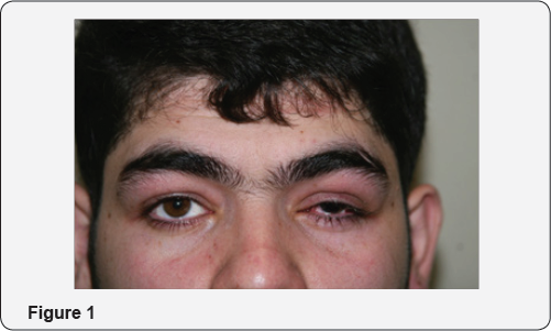
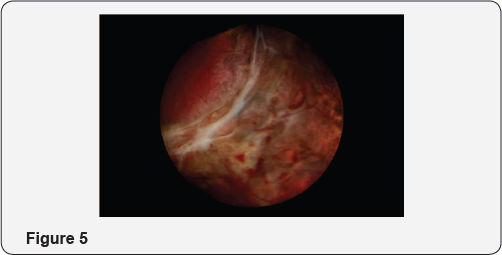
A 22-year- old man referred to the emergency ward because of vision loss in his right eye five days after bilateral lower lid blepharoplasty, his right eye showed ciliary injection, cornea showed mild edema and anterior chamber showed 4+cells and faint hypopyon in slit lamp examination, There was severe chemosis, without any site of laceration. However, fundus examination showed severe fibrinous reaction and vitritis. (Figures 1-5 are related to the post-operative status.)

Discussion

Based on the diagnosis of undetermined uveitis the patient underwent diagnostic vitrectomy, interestingly there was a site of laceration in the posterior fundus, leading to peritomy and its repair associated with severe vitreous inflammation and retinal necrosis and vasculitis. The patient underwent pars plana vitrectomy associated with intra vitreal antibiotic injection and silicone oil injection; the culture of the vitreous documented staphylococcus aurous in the vitreous sample. This rare complication has been rarely described in the literature [1,2].

Conclusion

Although rare, doing blepharoplasty a common cosmetic in inexperienced hand could lead to a severe devastating intra ocular complication.

For more Open Access Journals in Juniper Publishers please click on: https://juniperpublishers.com
For more articles in  JOJ Ophthalmology (JOJO) please click on: https://juniperpublishers.com/jojo/index.php
For more about juniper publishers  please click on: https://www.juniperpublishersgroup.com/