Thursday 28 November 2019

Allergic Bilateral Blepharoconjunctivitis and Periorbital Edema Following Rituximab, Bendamustine and Bortezomib Chemotherapy- Juniper Publishers


JUNIPER PUBLISHERS- JOJ Ophthalmology


Purpose: To report a case of bilateral allergic blepharoconjunctivitis and periorbital edema following chemotherapy with combination regimen of rituximab, bendamustine and bortezomib.
Case report: A 59-year-old man presented with bilateral periorbital swelling and blurry vision following three cycles of rituximab, bendamustine and bortezomib combination chemotherapy for mantle cell lymphoma. At presentation, visual acuity was 20/25 and 20/20 in the right and left eyes, respectively. Slit lamp examination disclosed a bilateral periorbital myxedema of both upper and lower eyelids, as well as bulbar and palpebral conjunctivitis. The patient was prescribed conservative topical medications and monitored with serial ophthalmic examinations. At 6 weeks follow up, ophthalmic symptoms improved with the resolution of blepharoconjunctivitis and periorbital edema.
Conclusion: Bilateral allergic blepharoconjunctivitis and periorbital edema were reported following rituximab, bendamustine and bortezomib chemotherapy. Ophthalmic examination and preventive measures may be warranted particularly in patients with combination chemotherapy regimen.
Keywords: Blepharoconjunctivitis; Periorbital edema; Rituximab; Bendamustine; Bortezomib; Chemotherapy


Introduction

Ocular toxicities of anti-cancer medications are often underestimated as compared to the more serious adverse effects in other organ systems [1-3]. Concurrent to the development of new chemotherapeutic agents and new combination regimens, there has been an increase in the number of reported ophthalmic adverse events following chemotherapy. Although there is a wide spectrum of ocular toxicities induced by cancer therapy [2-4] little is known about the ophthalmic toxicities of newer target-specific cellular molecules intended for specific molecular and biologic pathways. We present a subject with mantle cell lymphoma who developed bilateral allergic blepharoconjunctivitis and periorbital edema following chemotherapy with combination regimen of rituximab, bendamustine and bortezomib.


Case Report

A 59-year-old man with stage IV mantle cell lymphoma presented to the outpatient Eye Clinic at Stroger Hospital of Cook County with the complaint of bilateral periorbital swelling and pain, burning sensation, and blurry vision for 1 week. The symptoms started 1 week following the third cycle of rituximab, bendamustine and bortezomib chemotherapy, improved without medications in the time interval between the third and fourth cycles, and progressed again right after the fourth chemotherapy cycle at the day of ophthalmic examination. There was no past ophthalmic history and his past medical history included peptic ulcer disease. His medications were tylenol, acyclovir, docusatesenna and pantoprazole. At the initial ophthalmic evaluation, visual acuity (VA) was 20/25 and 20/20 in the right and left eyes, respectively. There was a bilateral periorbital swelling and redness which was not tender on palpation. The patient’s pupils were round and reactive without an afferent pupillary defect. Extraocular motility was full and visual fields by confrontation demonstrated no defect. Intraocular pressure was 12mmHg and 13mmHg in the right and left eye, respectively.
The slit-lamp biomicroscopy disclosed a bilateral periorbital edema of upper and lower eyelids, and bulbar and palpebral conjunctivitis. Anterior segment showed corneal irritation and dry eyes bilaterally, and dilated fundus examination was unremarkable. Review of systems disclosed right supraclavicular, infra auricular and bilateral axillary lymphadenopathies, and a rash in the upper chest and upper extremities. Due to temporal relation between the patient’s ophthalmic presentation and chemotherapy, his symptoms were attributed to an allergic reaction to his chemotherapy regimen. Oral diphenhydramine, topical hydrocortisone lotion, ocular cyclosporine and artificial tears were prescribed. The patient was instructed to perform warm compresses and lid scrubs daily, and to return to the clinic two weeks later for follow up or sooner if urgent symptoms including further vision loss and eye pain occurred. At 6 weeks follow up, patient showed improvement with resolution of blepharoconjunctivitis and periorbital edema in both eyes.


Discussion

Conjunctivitis concurrent with periorbital edema have been reported following chemotherapy with 5-fluorouracil and methotrexate [5]. Moreover, isofosfamate [6] Deoxycoformycin [7] doxorbucin [8] Cystosinarabinoside [9] docetaxel, [10] and cyclophosphamide [11] have also been associated with conjunctivitis. Other chemotherapeutic drugs, such as carboplatin, [12] have been linked to periorbital edema. To our knowledge there have been no reports of bilateral allergic blepharoconjunctivitis and periorbital edema following chemotherapy with combination regimen of rituximab, bendamustine and bortezomib.
Rituximab is a monoclonal antibody against the CD20 antigen of B cells and is used to treat non-Hodgkin B-cell lymphomas. The most common reported ophthalmic side effects of rituximab include transient ocular edema, conjunctivitis, burning sensation, and transient visual changes, [13] similar to the ophthalmic symptoms of the patient in the current study. Bendamustine is nitrogen mustard which is used in the treatment of chronic lymphocytic leukemia and lymphomas [14]. To our knowledge, there have been no reported ocular adverse effects of bendamustine, but other agents with similar mechanism including cyclophosphamide may cause blepharoconjunctivitis and reversible blurred vision [15]. Bortezomib is a proteasome inhibitor and is approved for treating multiple myeloma and mantle cell lymphoma [16]. There have been no reported ocular adverse effects for bortezomib.
The exact underlying mechanism of bilateral blepharoconjunctivitis and periorbital edema in the patient in the current study is not clear; however, since the ophthalmic symptoms started right after each exposure to chemotherapy medications, it is more likely that the chemotherapy medications caused the symptoms. Furthermore, both the ophthalmic symptoms and the skin reactions in the upper chest and upper extremities happened simultaneously, which is suggestive of allergic reaction to combination chemotherapy. It is not clear if either rituximab or bendamustine monotherapy or the combination chemotherapy caused the ocular symptoms in this patient; however, it is probable that the combination therapy may have increased the risk for development of ocular toxicities.


Conclusion


In summary, bilateral allergic blepharoconjunctivitis and periorbital edema were reported following rituximab, bendamustine and bortezomib chemotherapy. Ophthalmic examination and preventive measure of allergic reaction may be warranted particularly in patients with combination chemotherapy regimen.

For more Open Access Journals in Juniper Publishers please click on: https://juniperpublishers.com


Wednesday 27 November 2019

Inferior Rectus Myositis after an Uneventful Repair of Blowout Fracture- Juniper Publishers

JUNIPER PUBLISHERS- JOJ Ophthalmology

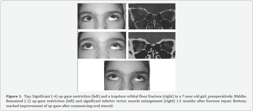
PA 7-year-old girl who had undergone a successful orbital floor blow-out fracture repair continued to have up-gaze and down gaze restriction post-operatively. She was observed for 6 weeks when an orbital imaging showed inferior rectus enlargement. Enlarged IR muscle associated with pain and restriction of up and down gaze led to a provisional diagnosed of myositis to which oral steroid was commenced and resulted in full recovery of up-gaze in 2 months.
Keywords: Blowout fracture; Inferior rectus; Myositis; Orbital fracture


Background

Early repair of a white-eyed blowout orbital floor fracture has been recommended in order to avoid permanent ischemic damage to the entrapped inferior rectus (IR) muscl0e [1]. However, even after a proper surgical repair of orbital floor fracture, up-gaze restriction may persist predominantly in children [1,2]. This may result from necrosis of muscle [2], IR muscle fibrosis [1], residual entrapped IR muscle sheath or peri- muscular tissue [3], and preoperative severe injury and swelling of the IR muscle [4].
This is, to the best of our knowledge, the first report of IR myositis after an uneventful white eyed blow out fracture repair. Iran University Eye Research Center ethic approval and patient’s parents’ consent were obtained.


Case Presentation

A 7 year-old girl was referred 2 days after a facial trauma. On examination, there was no or little eyelid swelling but marked painful restriction of up-gaze (-4) and moderate restriction of down gaze (-2) on the right eye. There was also hypoesthesia on the right cheek area. Vision (20/20 on both eyes) and ocular examinations were otherwise normal. Coronal Computed tomography (CT) scan showed a trapdoor floor fracture with inferior rectus entrapment which was extended from mid-globe to mid-orbit sections (Figure 1). Forced duction test was performed just before starting the operation 4 days after trauma which was strongly positive. Using trans-conjunctival approach, the entrapped muscle and peri-orbital tissue were released and the fracture site was covered by a properly fashioned porous polyethylene (Medpor, USA) sheet (0.85mm). The IR muscle was found to be discolored but viable. Forced duction test showed no restriction at the end of operation. Postoperatively, she was instructed to take oral systemic antibiotic (Cephalexin 250 mg, 4 times daily for 5 days), topical antibiotic and topical steroid (4 times daily for a week). A day after operation, there was less limitation of motion in down gaze (-1) and up-gaze (-2). However, she continued to have the same degrees of restriction associated with mild pain throughout post-operative follow ups at 1, 4, and 6 weeks. Since the repair was uneventful and postoperative follow up did not show improvement of muscle restriction, an orbital CT scan was requested. It showed no residual entrapment but significantly enlarged IR muscle. Increased thickness of IR muscle associated with pain on movement led to a provisional diagnosis of post-operative IR myositis and or persistent intra-sheath hematoma. Therefore, oral prednisolone (1mg/kg/day) was commenced and tapered within 6 weeks time. Up- and down restriction improved a week after its commencement. Completely normal examination was observed 1 year afterward with no recurrence of restriction and pain.


Conclusion

Possible explanations for persistent up-gaze restriction after a successful blowout fracture repair are: residual entrapment of any part of orbital soft-tissue [3] especially in the presence of posterior floor fracture [5], strangulation and necrosis of IR muscle [2], IR muscle fibrosis [1] and preoperative severe injury and swelling of the IR muscle [4]. Younger patients seem to recover longer than adults and a satisfactory force duction test at the end of the operation does not guarantee free voluntary movement of the involved eye [1,2]. In the presenting case, up- and down gaze restriction mildly improved a day after operation, but remained the same up to 1.5 months then after. In order to assess the possibility of residual IR entrapment, an orbital CT scan was requested which showed no entrapment but significantly enlarged IR muscle. In the context of up and down gaze restriction, pain, and enlarged IR muscle an orbital inflammatory myositis and or persistent IR hematoma were the provisional diagnoses. A rapid response to oral steroid was in favor of post-operative IR myositis. Post strabismus surgery extraocular myositis has been previously reported [6]. Whereas, to the best of our knowledge, there has been no any report of IR myositis after orbital fracture repair. Preoperative IR muscle swelling was reported to be a useful indicator of longer recovery after orbital floor fracture repair [4]. On reviewing the case, there was no significant IR enlargement preoperatively. Since this patient was treated with systemic steroid, it is not possible to comment on whether post-operative myositis is self limited if left untreated. The myositis did not recur one year after steroid treatment which may imply that it was due to either trauma or intra-operative manipulation. In conclusion, post-operative IR myositis should be considered in cases with residual up-gaze limitation after an uneventful orbital floor fracture repair.

For more Open Access Journals in Juniper Publishers please click on: https://juniperpublishers.com


Friday 15 November 2019

The Gas Ionization by Plasma Technology for Noninvasive Techniques in Oculoplastic-Juniper Publishers

The Gas Ionization by Plasma Technology for Noninvasive Techniques in Oculoplastic

Purpose: The objective of the research is to highlight the new non -invasive techniques there are in oculoplastic using plasma technology (4th stage of matters, gas ionization) aimed at resolving multiple eyesores problems in the ophthalmic area. We will talk about how through new techniques in oculoplastic we can solve problems like, xanathelasmas, Hemangiomas, eyelidcysts, ektropion, entropion, scars, syrignomas, papillomas, benign tumors, chalazion and benign tumors in difficult areas such as gray line.
Method: The methodology that is followed to solve the above-mentioned cases is the use of Plasma Exeresis using PLEXR machine. After first diagnosis and analysis in each region, we proceed to address and resolve the musing the Plasma Exeresis. The techniques of Plasma Exeresis are either spot by spot or spray method, depending on the case. There is no use of searing iron, the area is not cauterized but sublimated without complications, without scarring, and without affecting the optic nerve.
Results: Complete resolution of problem areas from the first session without the phenomena of recurrence. In case such as ektropion and entropion resolve can occur after 2-3 sessions.
Conclusion: In the past the solution to such problems requires surgical rooms, microscopes, stitches, and long period of recuperation. Now not only the patient can bloodlessly, more economically, without surgery and rehabilitation to get rid of these specific problems but also the doctor to perform all these methods, without complications in his clinic.


Introduction

In 2017, due to the fact that the advancements in the medical industry are changing rapidly, the need for direct and non-painful solutions to problems patients is increasing. The Exeresis Plasma method comes to cover this need because the use of it becomes necessary at the Oculoplastic and at the ophthalmology field. Now the doctor can proposing outright solutions to the patient without mentioning the word surgery thus eliminate the fear of the patient for the surgery. Our aim is to showthe new bloodless techniques in oculoplastic that their effectiveness equals that of surgery but without the complications and discomfort that may occur from a surgery [1-3].


Diagnosis

We examined and studied 347 patients who were experiencing problems as xanthelasmas (38), hemangiomas (4), eyelid cysts (20), entropion (2) ektropion (10), syrignomas (100), papillomas (100) benign nodules (45), chalazion (8) and benign tumors in difficult areas such as the gray line (20). The age range is from 25-78 years of age, smoking and non-smoking men and women. All incidents had as common fact the fear of surgery that is why the suggestive method was with Plasma Exeresis [3-6] (Figure 1).


Method

All patients accepted antisepsis in the areas that we would apply the Exeresis Plasma method. Then in all cases, except from those of ektropion, entropion, chalazion and benign tumors in the gray line, we applied numbing cream for 30 minutes. After the 30 minutes we removed the cream and started the process. The area was anesthetized and the patient did not felt anything during the treatment. In cases of ektropion, entropion, chalazion and benign tumors in the gray line, applied numbing injection with xylocaine [6-9].
Once anesthetized the area either with anesthetic cream or anesthetic injection we started implementing Plasma Exeresis. For implementing the method we used Plexr machine which has three pistols with different intensity. Those pistols do not use electric power and they are not connected with cables. Depending on the site and the problem, we use the corresponding pistol. Sublimation methods are 2. Spot by spot method or Spray Method. All cases were treated with the two methods either separately or in combination [10,11] (Figure 2).


Analysis

  1. Xanthelasmas: Spray Method
  2. Hemangiomas: Spot by spot method
  3. Cysts: depending on the shape and size of the cyst, combination of 2 methods.
  4. Ektropion-Entropion : Spot by spot with specific direction
  5. Scars: Combination of 2 techniques depending on the scar
  6. Syrrigkoma: Spot by spot Method
  7. Papillomas: depending on the size either Spray Method or Spot by Spot Method
  8. Benign Tumors: Spot by spot around the tumor to anesthetize the area and then Spray method to sublimate the tumor
  9. Chalazio: If it’s in the gray line follow the spray method, if it is in tarsus use the spray method and the spot by spot method combined
  10. Benign tumors at the gray line: Spray Method


Results

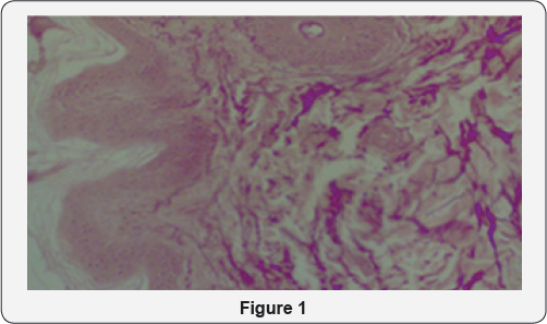
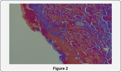
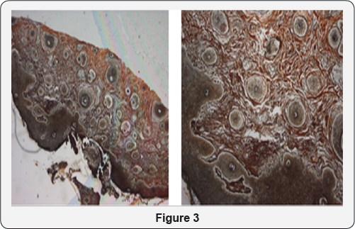
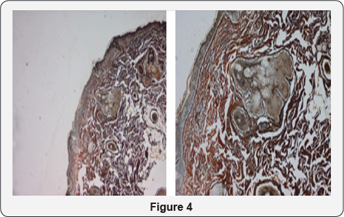
The result in all cases is the complete sublimation of the problems from the first session, without scarring, sutures, and long recovery periods. In cases of ectropion and entropion we may need a second session depending on the degree of ectropion / entropion (2nd or 3rd degree). Using Plasma Exeresis method is effective not only because the patient avoids surgery having the same effect with less cost but also for histological reasons. Histological analyzes before and after show that in regions that was applies Plasma Exeresis, the area not only reconstructed but created and type collagen III after application [12,13] (Figure 3 & 4).


Conclusion

After 347 successful cases in 1 year period, it is a fact that now every doctor can provide permanent and effective treatments to the patient using the Plasma technology (ionization of gas4th station of matter) in his clinic. The patient with low cost and without the fear of surgery solves his problem without blood, stitches and painful procedures. Finally it is worth to mention that the use of Plasma analysis is not only effective in ophthalmology and in other specialties as well.

Incidence of Myopia in Relation to Close Work at Indus Medical College Hospital, Tando Mohammad Khan-Juniper Publishers


JUNIPER PUBLISHERS- JOJ Ophthalmology


Incidence of Myopia in Relation to Close work at Indus Medical College Hospital, Tando Mohammad Khan
Purpose: To evaluate the incidence of myopia in relation with close near work particularly in those children involved in using small screen tablets and mobiles phones.
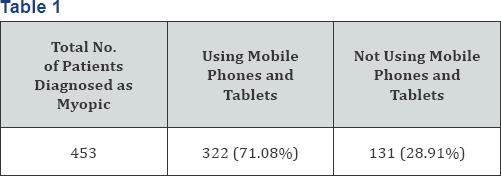
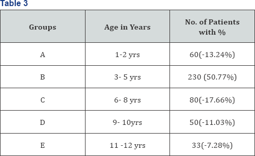
Material and methods: This study was conducted between January 2016 to June 2016 to know the incidence of myopic refractive error in children between 1 to 12 years in outpatient department of Indus medical college hospital and particularly the effect of close work like using tablets and mobile phones upon these children .We included 453 children who were diagnosed as myopic and having astigmatism , most of the children 322(71.08%) were using tablets and mobile while 131 (28.91%) were not involve in using mobile and tablets. 197 (43.48%) were boys while remaining 256(56.51%) were girls . They were divided into different age groups i.e. 60 (13.24%) children, were in age group A, majority of children 230(50.77%) belonged to group B, 80 (17.66%) were in age group C, 50 (11.03%) in group D and 33(7.28%) were in age group E. All of the patients in groups A to C and those non co-operative from group D and E were under gone cycloplegic refraction.
Results: Out of 453 children 203(44.81%) found to have myopia of up to -3.0D, 87(19.20%) have myopia of up to -2.0D, 67(14.79%) have myopia of up to -4.0D and 96(21.19%) having myopia of -1.0D. majority of patients i.e. 301(66.44%) having astigmatism of up to -1D, 83(18.32%) have astigmatism of up to -2.0D, 52(11.47%) were having astigmatism of up to - 3.0D and 17(3.75%) having astigmatism of up to -4.0D. Majority of patients (though female ratio higher than male ratio) were having myopic astigmatism of up to -1.0D , in the age group B i.e. between ages of 3 to 5 years and were involved in using mobiles and tablets.
Conclusion: Myopia is the more prevalent refractive error all over the world .in our study it is more prevalent in children between the ages 2 to 5 years of age particularly involved in close near work like apart from studying they were using mobiles and tablets particularly small screen for longer time. So better to use large screen avoid reading in dim light, do not use these small screens in lying position. Parents must take care while allowing the children to use mobiles and tablets screens must not be less than 12 inches in size better to allow them computers (desk top) rather than these small screens.
Keywords: Khan NA; Khan AA; Memon JI; Incidence of myopia in relation to close work at indus medical college hospital; Tando- Mohammad Khan


Introduction

Uncorrected refractive errors are a common cause of preventable blindness worldwide [1] and myopia is the result of complex hereditary and environmental factors, [2] most prevalent refractive error [3] defined as near sightedness caused by an incongruity between the power of the optical elements of the eye and its axial length. The object image is projected in front of the retina, and corrective lenses are necessary to displace the image backward, thus producing a clear retinal image. Although the causes of myopia are unclear, evidence supports both genetic and environmental components, among which are higher amounts of near work [4,5] years of education [6] and intelligence[7]. The world-wide urban rural patterns derived from both incidence and prevalence data are consistent with the near work hypothesis that increased reading and computer use may be a risk factor for myopia [5]. Researchers point to rigorous schooling system and the long hours children spend studying as being responsible for the high rates of myopia in Asia, rates that may be on the increase [8,9]. As myopia has onset and progression in childhood, it is important to focus research on these age groups.


Material and Methods

This study was conducted between January 2016 to June 2016 at Indus Medical College Hospital to know the incidence of myopic refractive error in children between 1 to 12 years and particularly to know the effect of using tablets and mobile phones upon these children. We included 453 children who were diagnosed as myopic and having astigmatism , most of the children 322 (71.08%) were using tablets and mobile while 131 (28.91%) were not involve in using mobile and tablets, (Table1). 197 (43.48%) were boys while remaining 256 (56.51%) were girls, (Table 2). They were divided into different age groups i.e. 60 (13.24%) children were in age group A, majority of children 230 (50.77%) belonged to group B, 80 (17.66%) were in age group C, 50 (11.03%) in group D and 33 (7.28%) were in age group E, (Table 3). All of the patients in groups A to C and those non co-operative from group D and E were under gone cycloplegic refraction.


Results

Out of 453 children 203 (44.81%) found to have myopia of up to -3.0D, 87 (19.20%) have myopia of up to -2.0D, 67 (14.79%) have myopia of up to -4.0D and 96 (21.19%) having myopia of -1.0D (Table 4). Majority of patients i.e. 301 (66.44%) having astigmatism of up to -1D, 83 (18.32%) have astigmatism of up to -2.0D, 52 (11.47%) were having astigmatism of up to -3.0D and 17 (3.75%) having astigmatism of up to -4.0D (Table 5).
Majority of patients (though female ratio higher than male ratio) were having myopic astigmatism of up to -3.0D, in the age group B i. e. between ages of 3 to 5 years and were involved in using mobile phones and tablets.


Discussion

In our study we included only patients between ages 1 to 12 years, were involved in close near work for long time particularly spending more time on mobile phones and tablets apart from their study time. We found that out of 453 myopic patients the highest rate of myopia, 44.81% was in the age group B (3-5yrs) and particularly these children were involved in using mobile phone and tablets for longer time, in a study done in 2011 at Dow University Hospital Karachi Pakistan by Rasheed et al. [10] shows rate of myopia was 26.6% while a multicenter study done in China, Chile and Nepal that shows the prevalence rates of myopia were16.2%, 5.8% and 0.3% in China.
Chile and Nepal respectively it is less than our study in another study the rate was almost same as our study that was conducted in Singapore and china, the prevalence rate of myopia in Singapore children was 36.7% compared to Xiamen (China) which was 18.5%. Singapore has highly competitive educational system, whereas Xiamen school system is not so demanding, more near work activity may explain the difference in the prevalence rates [11,12]. An epidemiological study, concerning the prevalence of myopia among the student population (15-18 years old) though the age is high as compare to our study of Northern Greece, myopia prevalence was 36.8% same as our results, It was found that myopia correlates strongly with near work and school performance [13]. Boys in orthodox Jewish schools were found to have higher rates of myopia (81.3%) that is too high ratio as compared to our study while boys in general Jewish schools have only (27.4%) myopia. Orthodox schooling is characterized by sustained near vision139and frequent change in accommodation due to the swaying habit during study [14]. In Xiamen, China the prevalence of myopia in urban school children was 19.3% and in rural school children was 6.6%. The average hours per day children spent in reading and writing outside of school was 2.2 hours in the city compared with 1.6 hours in is higher in the city than in the country side.These data suggest the prevalence of myopia is higher in the city than in the country side. One possible explanation for these different rates could be that school children in the city spend more time reading and writing outside of school compared with children in the countryside. Myopic children in both the city and the countryside spent more time reading and writing compared with non myopic children. This increased near-work activity may contribute to the prevalence of myopia [15]. So the above studies prove same as ours that the time spent for close near work is directly related to the increase in prevalence of myopia with only change that children of this era are using mobile phones and tablets rather than only studying.


Conclusion


Myopia is the more prevalent refractive error all over the world in our study it is more prevalent in children between the ages 2 to 5 years of age particularly involved in close near work like, apart from studying they were using mobiles and tablets particularly small screen for longer time. So better to use large screen avoid reading in dim light, do not use these small screens in lying position. Parents must take care while allowing the children to use mobiles and tablets, screens must not be less than 12 inches in size better to allow them computers (desk top) rather than these small screens.

For more Open Access Journals in Juniper Publishers please click on: https://juniperpublishers.com


Stem Cells and Transplantation for Retinal Diseases- Juniper Publishers

JUNIPER PUBLISHERS- JOJ Ophthalmology

Stem cells provide a promising tool for treating retinal diseases and injury. Early work focused on embryonic stem cells (ESC). The development of induced pluripotent stem cells (iPSC) alleviates some of the ethical concerns with ESC and the need for immunosuppression. Stem cell-derived retinal pigment epithelial cells (RPE) are comparable to native RPE; and stem cell-derived retinal organoids self-organize into laminated structures that bear some resemblance to the neurosensory retina. Questions remain regarding genetic and epigenetic variability among different stem cell lines, especially iPSC lines. The challenge is in understanding the significance of this variability for transplant and how to control such variability. Transplantation of stem cell-derived RPE and retinal progenitor cells has been tested in both animal models and humans. The cells integrated into the recipient with possible rescue of visual function. These findings encourage researchers to develop refined culture and delivery methods that would increase integration with the host and sustain long-term visual function.


Introduction

Since the beginning of stem cell research, pluripotent cells were seen as a promising tool for tissue regeneration and transplantation. Widely known, stem cells have the ability to differentiate into one or more mature cell types or continue to renew themselves. These properties make stem cells a potential source for sustained supply of tissue for transplantation. There is growing interest in developing stem cell therapies for neurodegenerative diseases, such as Alzheimer and amyotrophic lateral sclerosis (ALS), with the aim of replacing diseased tissue [1,2]. Similarly, research on developing replacement tissues for retinal degeneration has gathered momentum as well.


Sources of Stem Cell-Derived Retinal Cells

In many retinal diseases, such as age-related macular degeneration (AMD) and retinitis pigmentosa, there is degeneration of both the retinal pigment epithelium (RPE) and photoreceptors resulting in vision loss. The native retina, being a neural tissue, has little ability to regenerate. Therefore, transplanted tissue needs to replace lost tissue, continue to survive, and integrate functionally into the host retina. Several sources of stem cells have been used for regenerating retinal tissue; the most extensively studied are human embryonic stem cell (hESC) and induced pluripotent stem cell (iPSC). Additionally, researchers have developed and continue to improve different methods of differentiating stem cells into retinal cells.

Embryonic stem cells

Embryonic stem cells (ESC) are pluripotent stem cells derived from blastocysts in early development [3,4]. In stem cell biology, they are considered the gold-standard for comparing pluripotency, and genetic and epigenetic characteristics. The ethical concerns of human ESC have been much debated. Nevertheless, ESC allowed early successful differentiation of retinal cells in culture. hESC-derived RPE demonstrated robust pigmentation, and exhibited similar morphology and gene expression as human fetal RPE [5-8]. These RPE cells also developed appropriate functional characteristics, such as phagocytosis of shed outer segments [9,10]. Scanning across the literature, there are now various differentiation methods used for generating retinal cells. Some methods involve directed differentiation using small molecules and growth factors, and others allow stem cells to spontaneously differentiate into RPE in specialized media [9,10]. The time taken for retinal cells to differentiate can vary depending on the protocol and the desired cell type. Neuroretinal precursor cells, for example, can appear as early as day 10 in culture, while pigmented cells can take 6-8 weeks to appear [11,12]. Researchers continue to improve methods to generate retinal cells comparable to native retina in order to study retinal development and to generate tissue for transplantation.

Induced pluripostent stem cells (iPSC)

iPSCs are pluripotent stem cells that are derived from adult cells. Skin fibroblasts and peripheral blood cells are commonly used to generate iPSC. The Yamanaka group were the first to describe this reprogramming by introducing four transcription factors Oct3/4, Sox2, Klf4, and c-Myc, known as the “Yamanaka factors”. [13,14] iPSCs also have been successfully differentiated into retinal cells. iPSC-derived RPE can attain appropriate barrier function including proper distribution of membrane NaK-ATPase, polarized secretion of VEGF and similar membrane potential as native RPE [15]. Retinal progenitor cells and photoreceptors derived from iPSC also exhibit similar gene expression patterns as those derived from ESC, although there can be variation in the timing of differentiation [16].
iPSC can be a source of unlimited supply of regenerated tissue for studying development and for transplantation. One major foreseeable advantage of iPSC over ESC is the issue of immune histocompatibility. iPSC derived from a patient’s adult cells would not cause immune rejection when transplanted into the same person. In practice, not every iPSC line can successfully differentiate into the desired cell type. There is in fact variability among iPSC lines. Some researchers ascribe the cause of variability to differences in reprogramming techniques and lab environment; others propose that iPSC have different epigenetic markers either due to the reprogramming procedure or epigenetic memory of the original adult cell [17-19]. However, there is controversy over how much epigenetic aberrancies contribute to the variability seen among iPSC lines [20].
The ultimate question is how cellular variability affects the safety of iPSC-derived cells for transplantation. There is a need for defining standards not only to evaluate iPSC lines but also the differentiated cells derived from iPSC. Miyagishima et al. [21] proposed a system of authenticating iPSC-derived RPE: in addition to assessing gene expression and morphology, they also assessed cellular calcium flux, membrane electrophysiology and fluid transport in comparison to human fetal RPE [22]. Rigorous testing and characterization is needed to increase the safety and integrity of retinal tissue selected for transplantation.


Retinal Transplantation

Transplantation of stem-cell derived retinal cells in animal models has presented positive results in visual improvement. Human clinical trials demonstrated good long-term safety of transplantation [22,23]. There are several ongoing clinical trials using stem-cell derived retinal cells for retinal diseases. The goals of transplantation are to replenish and rescue degenerating cells, re-establish neural connectivity within the retina, and improve visual acuity.

RPE transplantation

Overall, more translational studies have been done using stem-cell derived RPE than with stem-cell derived neuroretinal cells. Transplantation studies commonly use rodent models of retinal degeneration. A widely used model, for example, is the Royal College of Surgeons (RCS) rat, which has a mutation in MERTK gene and models autosomal recessive retinitis pigmentosa [24]. Transplantation of ESC-derived and iPSC-derived RPE in rodents with retinal degeneration resulted in more photoreceptor survival compared to non-transplanted animals. The photoreceptor layer was thicker at the transplant site compared to control [9,11,25-27].Transplanted RPE also promoted better visual function, measured by electroretinogram or optokinetic testing, compared to control animals [9,11,26]. The exact mechanism of photoreceptor rescue is not entirely elucidated. Given that the transplanted RPE does not always restore the outer blood-retinal barrier, one can postulate that trophic factors secreted by the RPE and the phagocytosis of photoreceptor outer segments may mediate the protective effects on the degenerating photoreceptors.
One major challenge from the studies mentioned above is long-term graft survival and visual improvement. In Carr et al. [10] implanted iPSC-RPE cells were eventually lost in the host retina at 13 weeks after transplant [26]. The mice interestingly retained improved visual function even when transplanted cells were not present. However, it is unknown whether this visual preservation can be sustained for longer. In the Idelson et al. [9] study, for example, the increased electroretinogram signal in transplanted animals eventually diminished at later time points (19 weeks). These results are proof-of-concept for using stem-cell derived retinal tissue to improve vision in retinal diseases. However, they also highlight limitations and challenges that need to be overcome to improve effectiveness of transplantation. The route of transplantation is seen as an area for improvement. In earlier transplant studies, a bolus of cells suspended in solution was injected into the subretinal space. This delivery method limits the ability of the transplanted RPE to re-organize into a functional monolayer; perhaps relatedly, cell survival from bolus injections is low [28]. Active research now focuses on transplanting sheets of RPE grown of various scaffolds to promote increased graft survival in the recipient [29,30].
In 2015, human clinic trial results for hESC-RPE transplantation in two retinal diseases were reported [23]. The trials were phase I/II with primary outcomes of safety and tolerability. The grafted cells were well tolerated without evidence of aberrant growth or serious side effects. When visual acuity was measured at 6 months after transplant, 6 out of the 9 AMD patients showed modest improvement from baseline and 3 out of 8 Stargardt’s macular dystrophy patients showed similar improvement. The other patients had stable or decreased visual acuity. The study demonstrated the safety of stem-cell derived retinal transplantation in human patients. Other clinical trials are underway to assess different types of stem-cell derived retinal tissue, different methods of delivery, and in different retinal diseases.

Photoreceptor transplantation

Efforts to replace diseased photoreceptors have involved transplantation of retinal progenitor cells (RPC). Understandably, mature neural retina is more challenging to differentiate in culture, given its complex interconnected laminations. However, RPC have been successfully grown from stem cells and transplanted into animal models with the hope that these progenitor cells can continue differentiation into mature retinal cells in the host.
Several groups developed methods of differentiating stem cells into three-dimensional, spherical organoids composed of retinal progenitor cells [31-34]. The organoids (referred to in the literature as optic vesicles) contained cells that expressed developmental markers for photoreceptor, amacrine, horizontal and ganglion cells; with time in culture, the cells within optic vesicles self-organize into crude laminations [32,35]. One group demonstrated electrical excitability in these optic vesicles, indicating functional synaptic connectivity among the cells. The generation of these stem cell-derived optic vesicles offers a method of increasing production efficiency of neural retinal tissue for transplantation. However, the spherical geometry of the organoids makes them unsuitable for implantation, because they fail to flatten and simultaneously interact with the RPE and neurosensory retina. As models of retinal differentiation, they should prove valuable for studying the mechanisms of retinal disease and potential medical therapies.
Transplantation with immature RPC also has had positive results in animal models. Transplanted stem cell-derived retinal precursor cells migrated into and integrated structurally with the host retina, showing synaptic interaction with the host [12,36-38]. Furthermore, better visual function was assessed by optokinetic testing, electroretinogram, and visual cortex activity in transplanted animals compared to control [37,39] Like stem cell-derived RPE, the stem cell-derived neural retinal cells are well tolerated in the recipient. However, there is still little data on long-term survival of these stem cell derived-retinal progenitor cells, and whether vision can also be rescued long-term. These encouraging results highlight the need for more validation studies in preclinical models.


Conclusion

Researchers have successfully differentiated retinal cells from ESC and iPSC. Retinal culture systems, such as the three-dimensional organoids, allow the study of retinal development, mechanisms of disease, and provide tissue for transplantation in retinal diseases. There is continued modification and optimization of these differentiation methods. Both stem cell-derived RPE and retinal progenitor cells have been transplanted in animal models and exhibited graft survival and possible visual improvement. For human patients, early phase trials demonstrated good tolerability of transplantation. More clinical studies are needed to validate the efficiency of retinal transplantation.


Grant Support


Leir Foundation, Newman’s Own Foundation.

For more Open Access Journals in Juniper Publishers please click on: https://juniperpublishers.com


Commentary on Dry Eye in Women- Juniper Publishers


JUNIPER PUBLISHERS- JOJ Ophthalmology


Commentary

Dry Eye Disease is said to affect an increasing number of individuals around the globe. The recent Beaver Dam Offspring Study (BOSS) study tells us that in the US, Dry Eye Disease impacts one person out of every seven and has been estimated to affect up to 30 million Americans. The fastest growing groups of people affected are men and women under the age of 40. The cause of this is the growing use of screen time on electronic devices, which naturally reduces our blink rate to a rate three times less than in normal face to face interactions. However, the lion’s share of those suffering from Dry Eye have always been and continue to be peri- and post-menopausal women due to hormonal factors. Menopause brings hormonal shifts that are commonly associated with hot flashes, mood changes, night sweats and vaginal dryness. The Women’s Health Initiative study showed that at least 60% of peri and postmenopausal women experience symptoms of dry eye as well. Studies on routine cataract patients (average age at the time of surgery is 65) also showed that 60% of those patients had signs of dry eye visible to their ophthalmologist even though the majority of them had no complaints.
It is known that the drop in androgen and testosterone during menopause are responsible for increasing the likelihood of dry eye in women. Testosterone and androgen support healthy lacrimal gland function and because women start out with less of both of these hormones than men, they are more susceptible to problems when their already low levels dip even further. Autoimmune diseases that lead to dry eye occur more commonly in women. The incidence of Sjogren’s syndrome, an autoimmune disease that causes dry mouth and dry eyes peaks around the time of perimenopause and may be directly related to androgen levels in the body according to researchers studying androgen deficiency and Sjogren’s progression in mice.
Dry Eye Disease is now understood to be a chronic, progressive, inflammatory condition. The causes are multifactorial: screen time, preservatives in eye drops, contact lenses, LASIK, cosmetic lid procedures, use of electronic devices, medications, allergies and diets poor in omega 3s, to name a few. Systemic medications for common conditions like allergies, depression, hypertension and sleep disorders can be particularly drying to the ocular surface. Dry Eye can also affect any layer of the tear: goblet cells that attract and hold watery tears to the surface, aqueous deficiency (lack of watery tears) or meibomian gland dysfunction (abnormal oily layer of the tear). What we understand now is not that no matter how the Dry Eye starts, nor what part of the tear becomes dysfunctional, are all of the components of the tear film interconnected and ultimately affected. The cycle of inflammation is self-perpetuating and self-amplifying. Anti-inflammatory medications must be employed to alter the outcome of this disease.
With this understanding, we can see now that high quality omega-3s (fish oil) can be successful as the first line of defense as the omegas are incorporated into the Meibomian glands and help to protect the surface of the eye from evaporative changes. Beyond that, topical anti-inflammatories like ophthalmic non-steroidals, steroids and anti-inflammatory drops like cyclosporing and the more recently approved lifitegrast can also be helpful. Hormonal replacement for dry eye symptoms can have a surprisingly negative effect on the ocular surface. Patients on estrogen only hormone replacement therapy like have a four to sevenfold increased risk of dry eye than those on combination hormonal therapy. Topical testosterone and androgen drops have not been successful in trials historically.

There are other treatments available for dry eye. Autologous serum tears using growth factors from the patients/ own blood and platelet rich plasma which concentrates the growth factors by an additional factor of four have been successful at reducing ocular pain related to dry eye. Intense pulsed light therapy has also been shown to help reduce the inflammation and improve symptoms related to dry eye. Scleral contact lenses and amniotic membranes have also been used to help trap moisture and heal damaged epithelium. Emerging therapies for dry eye include vagal nerve stimulators, handheld light devices, small molecules targeting inflammatory mediators, and muco-adhesive vehicles for enhanced delivery. Although women disproportionately suffer with Dry Eye Disease, people of all ages and across the globe will benefit from improved therapies for dry eye.

For more Open Access Journals in Juniper Publishers please click on: https://juniperpublishers.com