Showing posts with label Quality Journalspublishers. Show all posts

Friday, 10 January 2020

Results of 50 Phacoemulsification Surgeries with Injectable Lens Implantation at Indus Medical College Hospital Tando Mohammad Khan- Juniper Publishers


Juniper Publishers- JOJ Ophthalmology

Abstract

Purpose: To evaluate the results of phacoemulsification surgeries at department of ophthalmology at Indus Medical College Hospital Tando Mohammad Khan.
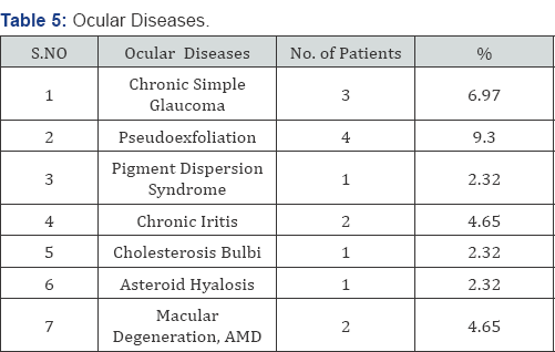
Material and Methods: This study in which only 50 eyes of 43 patients were included to treat by phaco emulsification was conducted from 1st January to 30th June 2016. 31 patients were male whereas 12 were female. 20 were right eyes, 16 were left while 7 patients were undergone bilateral phaco surgeries within 7 to 16 days.1 patient was in age group C, another 1 was in group D, 8 were in group E, 12 were in group F and remaining 21 were in group G. 21 were suffering from diabetes, 12 were hypertensive, 2 were with cardiac problems using pace maker and 2 was involved with HCV infection. Patients suffering from ocular diseases: open angle glaucoma 3, pseudoexfoliation 4, pigment dispersion syndrome 1, chroniciritis 2, cholestrosisbulbi 1, asteroid hyalosis 1, age related macular degeneration 2 Eyes were dilated with mydriacil/phenylephrine eye drops, local anesthesia as retrobulbar as well as facial block (von lint technique) were given using 2% xylocaine inj without adrenaline. 2.8mm incision, capsulorexhsis with bent 27 gauge needle, followed by hydrodissection and in some hydrodelienation with small caliber irrigation cannula, copious 2% methylcellulose used to save endothelial cells as well as to maintain anterior chamber, all 4 steps of phaco followed with divide and conquer method and finally injectable intraocular lens implanted. Every operation ended with subconjunctival injection of dexamethasone 2mg plus gentamicin 20mg.
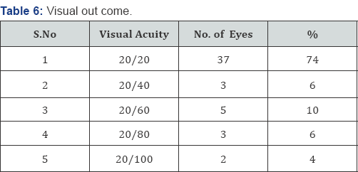
Results: 37 eyes gained 20/20 visual acquity on first post-operative day, 3 eyes gained 20/40, 5 gained 20/60 which over a period of five days improved to 20/20 after using topical prednisolone 1mg along with moxifloxacin eye drops, 3 gained 20/80 corrected with glasses, 2 were having 20/100 because of macular diseases.
Conclusion: In our experience phacoemulsification is an excellent technique which saves time, gives early rehabilitation depending upon the patience, experience and skill of surgeons.
Keywords: Cataract; Phacoemulsification results


Introduction

The term cataract is defined as an opacification of crystalline lens, having different morphological types; subcapsular; anterior and posterioir, cortical, nuclear opacification [1]. Cataract further divided according to density: grade 1 to grade 4. Cataract leads to decreased or blurring of vision.
Mostly senile [2] with other important causes like trauma, dibetes, myotonic dystrophy, atopic dermatitis, neurofibromatosis type 2, steroid induced, chronic iritis, high myopia, retinitis pigmentosa, gyrate atrophy, Stickler syndrome etc. [3].
With the passage of time lens increases in weight and thickness as new layers of cortical fibers are formed concentrically, the lens nucleus undergoes compression and hardening (nuclear sclerosis). Crystalline (lense proteins) are changed by chemical modifications and aggregation into high- molecular-weight proteins [4].
The only treatment for cataract is surgery either large incision ECCE or phacoemulsification, small incision early rehabilitation and with good visual outcome. The technique and results of cataract surgery have changed dramatically during the past three decades. In all over the world we have moved from intracapsular cataract extraction as the preferred technique to almost exclusively extracapsular techniques. Smaller incisions have become thestandard, with phacoemulsification now being the method of choice for most of surgeons [5].


Material and Methods

This study in which only 50 eyes of 43 patients were included to treat by phaco emulsification with injectable intraocular lens implantations at Indus Medical College Hospital Tando Mohammad Khan from 1st January to 30th June, 2016. All eyes were dilated prior surgery with mydriacil, phenylephrine eye drops, local anesthesia given using retrobalbur and facial (von lint) with 2% lidocaine (xylocain injections). Phaco done with infinity (Alcon) machine under Taggaki microscope.
Out of 43 patients 12 were females, 31 were males (Table 1), patients were divided into different age groups as: there was no patients in group A and B, group C and D have only one patient respectively, group E includes 8, group F includes 12 and group G includes 21 (Table 2).
Out of 50 eyes 20 were right, 16 left and 7 were both eyes (Table 3). 37 (86.04%) Patients Were suffering with systemic diseases like diabetes (48.83%), hypertention (27.90%), cardiac problem, using pacemaker (4.65%), HCV (4.65 %) underwent surgery after having fitness from their physicians (Table 4) 14 (32.55%). Patients were suffering from ocular diseases like Glaucoma (6.97%), pseudoexfoliation (9.30 %), Pigment dispersion syndrome (2.32%), old healed iritis with peripheral ant Synaechae (4.65%), Cholesterosisbulbi (2.32%), asteroid hylosis (2.32%), Age related macular degeneration (4.65%) (Table 5).
After asceptic techniques, drapping and using 2 drops of 10% povidine solution instilled into eye, after 1 minute copious irrigation done, incision started with 2.8mm phaco knife, capsulorexhsis done with 27 gauge bent needle, hydrodissection and in some hydrd ilea nation using small caliber irrigation cannula, copious use of 2% methylcellulose to save endothelium as well as to maintain anterior chamber. All 4 steps of phaco followed and finally injectable intraocular lens implanted. Wound closed with stromal hydration. Every operation finished with sub conjunctival injection of Dexamethasone 2mg plus gentamicin 20mg, and eye kept pached for 24 hours.


Results

50 eyes of 43 patients were undergone surgery by phacoemulsification with injectable intraocular lens implantation, 37 eyes (74%) improved visual acuity to 20/20 at first postoperative day, 3 eyes (6%) improved upto 20/40, 5 eyes (10%) upto 20/60, 3 eyes (6%) upto 20/80, and 2 eyes (4%) improved upto 20/100 (Table 6). Figure 1 a, b, c shows injectable lens implantation during one of our surgery.

Discussion

Phacoemulsification is a very safe and less time taking technique depending upon good dilation of pupil pre- operative and during surgery as well as the patience, experience and skill of surgeons. It is established that the smaller phacoemulsification wound gives less induced astigmatism, faster visual rehabilitation and improved wound security than ECCE [6-11]. Smaller wound heals more rapidly with less risk of leakage, viscoelastic do not leave the eye through small incision [12]. 37 (74 %) of my patient improved visual acuity upto 20/20 on first and secondday, 3 (6%) developed striate keratitis and treated with topical steroid and regained 20/20 on 5th post operative day. In 5 (10%) visual acuity corrected.
With glasses with in -1.50 D sphere and 0.75 cylinder at 90degrees, 2 (4%) who were suffering with age related macular degeneration remained after BCVA at 20/100. In our study not a single case suffered with post operative endophthalmitis same as in a study done by Cooper et al. [13].
Out of these 50 eyes only 3 developed striate keratitis, reason was hard nucleus more than grade 3 density needed high phaco power and time by the technique divide and conquer same as described by Gimbel [14]. Topical steroids were being prescribed and on 5th postop day vision become 20/20. Though it was fairly high 20% in one study by Popiela G et al. [15] but in our experience it was only 6 %, a grade 3 nucleus (severely dense) and long absolute phaco time as independent predictors for endothelial cell loss [16]. Phacoemulsification in the capsular bag by directing probe away from the corneal endothelium and keeping the lens fragments at deeper plane are the measures which would be helpful in minimizing the chances of corneal edema and striate after phacoemulsification same as suggested by Zetterstrin C [17] and Pirazzoli G et al. [18].


Conclusion

Small incision surgery such as phacoemulsification with injectable intraocular lens implantation is a very safe and less time taking technique which depends upon the experience and skill of surgeon who strictly follows selection and exclusion criteria and with a good knowledge when to abandon or convert the technique and always keeps the lens fragments in the capsular bag with the phaco tip directed away from endothelium, do not follow the lens fragments near the posterior capsule, allow fragments' to follow the tip.


Acknowle dgement


I am thankful to Aftab Ahmed Khan assistant professor for his assistance, Aisha Khan (4th year MBBS) and Atiqa Khan (final year BDS) students for their help in compiling data. Special thanks to my assistant Moin Shaikh who assisted all cases. Jawaid Iqbal assistant professor who is always helpful.
For more Open Access Journals in Juniper Publishers please click on: https://juniperpublishers.com


Friday, 15 November 2019

The Gas Ionization by Plasma Technology for Noninvasive Techniques in Oculoplastic-Juniper Publishers

The Gas Ionization by Plasma Technology for Noninvasive Techniques in Oculoplastic

Purpose: The objective of the research is to highlight the new non -invasive techniques there are in oculoplastic using plasma technology (4th stage of matters, gas ionization) aimed at resolving multiple eyesores problems in the ophthalmic area. We will talk about how through new techniques in oculoplastic we can solve problems like, xanathelasmas, Hemangiomas, eyelidcysts, ektropion, entropion, scars, syrignomas, papillomas, benign tumors, chalazion and benign tumors in difficult areas such as gray line.
Method: The methodology that is followed to solve the above-mentioned cases is the use of Plasma Exeresis using PLEXR machine. After first diagnosis and analysis in each region, we proceed to address and resolve the musing the Plasma Exeresis. The techniques of Plasma Exeresis are either spot by spot or spray method, depending on the case. There is no use of searing iron, the area is not cauterized but sublimated without complications, without scarring, and without affecting the optic nerve.
Results: Complete resolution of problem areas from the first session without the phenomena of recurrence. In case such as ektropion and entropion resolve can occur after 2-3 sessions.
Conclusion: In the past the solution to such problems requires surgical rooms, microscopes, stitches, and long period of recuperation. Now not only the patient can bloodlessly, more economically, without surgery and rehabilitation to get rid of these specific problems but also the doctor to perform all these methods, without complications in his clinic.


Introduction

In 2017, due to the fact that the advancements in the medical industry are changing rapidly, the need for direct and non-painful solutions to problems patients is increasing. The Exeresis Plasma method comes to cover this need because the use of it becomes necessary at the Oculoplastic and at the ophthalmology field. Now the doctor can proposing outright solutions to the patient without mentioning the word surgery thus eliminate the fear of the patient for the surgery. Our aim is to showthe new bloodless techniques in oculoplastic that their effectiveness equals that of surgery but without the complications and discomfort that may occur from a surgery [1-3].


Diagnosis

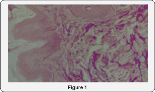
We examined and studied 347 patients who were experiencing problems as xanthelasmas (38), hemangiomas (4), eyelid cysts (20), entropion (2) ektropion (10), syrignomas (100), papillomas (100) benign nodules (45), chalazion (8) and benign tumors in difficult areas such as the gray line (20). The age range is from 25-78 years of age, smoking and non-smoking men and women. All incidents had as common fact the fear of surgery that is why the suggestive method was with Plasma Exeresis [3-6] (Figure 1).


Method

All patients accepted antisepsis in the areas that we would apply the Exeresis Plasma method. Then in all cases, except from those of ektropion, entropion, chalazion and benign tumors in the gray line, we applied numbing cream for 30 minutes. After the 30 minutes we removed the cream and started the process. The area was anesthetized and the patient did not felt anything during the treatment. In cases of ektropion, entropion, chalazion and benign tumors in the gray line, applied numbing injection with xylocaine [6-9].
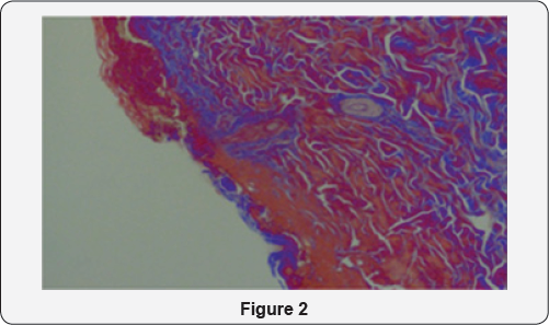
Once anesthetized the area either with anesthetic cream or anesthetic injection we started implementing Plasma Exeresis. For implementing the method we used Plexr machine which has three pistols with different intensity. Those pistols do not use electric power and they are not connected with cables. Depending on the site and the problem, we use the corresponding pistol. Sublimation methods are 2. Spot by spot method or Spray Method. All cases were treated with the two methods either separately or in combination [10,11] (Figure 2).


Analysis

  1. Xanthelasmas: Spray Method
  2. Hemangiomas: Spot by spot method
  3. Cysts: depending on the shape and size of the cyst, combination of 2 methods.
  4. Ektropion-Entropion : Spot by spot with specific direction
  5. Scars: Combination of 2 techniques depending on the scar
  6. Syrrigkoma: Spot by spot Method
  7. Papillomas: depending on the size either Spray Method or Spot by Spot Method
  8. Benign Tumors: Spot by spot around the tumor to anesthetize the area and then Spray method to sublimate the tumor
  9. Chalazio: If it’s in the gray line follow the spray method, if it is in tarsus use the spray method and the spot by spot method combined
  10. Benign tumors at the gray line: Spray Method


Results

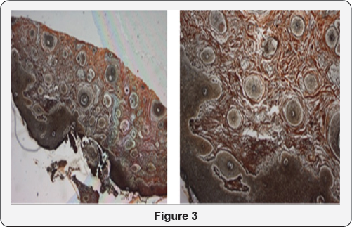
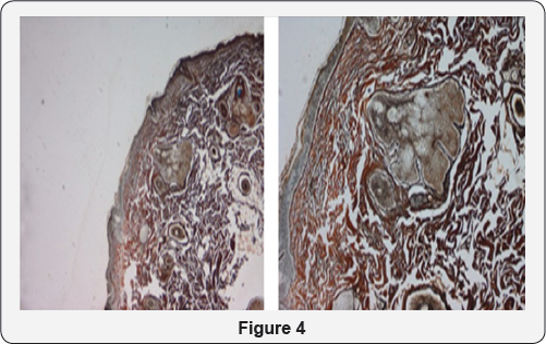
The result in all cases is the complete sublimation of the problems from the first session, without scarring, sutures, and long recovery periods. In cases of ectropion and entropion we may need a second session depending on the degree of ectropion / entropion (2nd or 3rd degree). Using Plasma Exeresis method is effective not only because the patient avoids surgery having the same effect with less cost but also for histological reasons. Histological analyzes before and after show that in regions that was applies Plasma Exeresis, the area not only reconstructed but created and type collagen III after application [12,13] (Figure 3 & 4).


Conclusion

After 347 successful cases in 1 year period, it is a fact that now every doctor can provide permanent and effective treatments to the patient using the Plasma technology (ionization of gas4th station of matter) in his clinic. The patient with low cost and without the fear of surgery solves his problem without blood, stitches and painful procedures. Finally it is worth to mention that the use of Plasma analysis is not only effective in ophthalmology and in other specialties as well.

Incidence of Myopia in Relation to Close Work at Indus Medical College Hospital, Tando Mohammad Khan-Juniper Publishers


JUNIPER PUBLISHERS- JOJ Ophthalmology


Incidence of Myopia in Relation to Close work at Indus Medical College Hospital, Tando Mohammad Khan
Purpose: To evaluate the incidence of myopia in relation with close near work particularly in those children involved in using small screen tablets and mobiles phones.
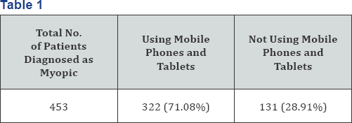
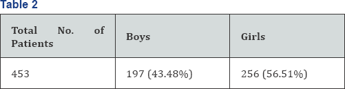
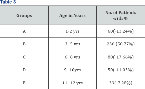
Material and methods: This study was conducted between January 2016 to June 2016 to know the incidence of myopic refractive error in children between 1 to 12 years in outpatient department of Indus medical college hospital and particularly the effect of close work like using tablets and mobile phones upon these children .We included 453 children who were diagnosed as myopic and having astigmatism , most of the children 322(71.08%) were using tablets and mobile while 131 (28.91%) were not involve in using mobile and tablets. 197 (43.48%) were boys while remaining 256(56.51%) were girls . They were divided into different age groups i.e. 60 (13.24%) children, were in age group A, majority of children 230(50.77%) belonged to group B, 80 (17.66%) were in age group C, 50 (11.03%) in group D and 33(7.28%) were in age group E. All of the patients in groups A to C and those non co-operative from group D and E were under gone cycloplegic refraction.
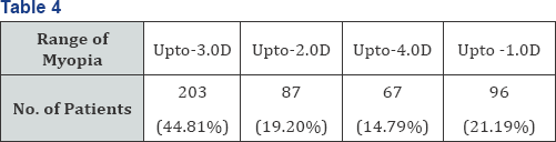
Results: Out of 453 children 203(44.81%) found to have myopia of up to -3.0D, 87(19.20%) have myopia of up to -2.0D, 67(14.79%) have myopia of up to -4.0D and 96(21.19%) having myopia of -1.0D. majority of patients i.e. 301(66.44%) having astigmatism of up to -1D, 83(18.32%) have astigmatism of up to -2.0D, 52(11.47%) were having astigmatism of up to - 3.0D and 17(3.75%) having astigmatism of up to -4.0D. Majority of patients (though female ratio higher than male ratio) were having myopic astigmatism of up to -1.0D , in the age group B i.e. between ages of 3 to 5 years and were involved in using mobiles and tablets.
Conclusion: Myopia is the more prevalent refractive error all over the world .in our study it is more prevalent in children between the ages 2 to 5 years of age particularly involved in close near work like apart from studying they were using mobiles and tablets particularly small screen for longer time. So better to use large screen avoid reading in dim light, do not use these small screens in lying position. Parents must take care while allowing the children to use mobiles and tablets screens must not be less than 12 inches in size better to allow them computers (desk top) rather than these small screens.
Keywords: Khan NA; Khan AA; Memon JI; Incidence of myopia in relation to close work at indus medical college hospital; Tando- Mohammad Khan


Introduction

Uncorrected refractive errors are a common cause of preventable blindness worldwide [1] and myopia is the result of complex hereditary and environmental factors, [2] most prevalent refractive error [3] defined as near sightedness caused by an incongruity between the power of the optical elements of the eye and its axial length. The object image is projected in front of the retina, and corrective lenses are necessary to displace the image backward, thus producing a clear retinal image. Although the causes of myopia are unclear, evidence supports both genetic and environmental components, among which are higher amounts of near work [4,5] years of education [6] and intelligence[7]. The world-wide urban rural patterns derived from both incidence and prevalence data are consistent with the near work hypothesis that increased reading and computer use may be a risk factor for myopia [5]. Researchers point to rigorous schooling system and the long hours children spend studying as being responsible for the high rates of myopia in Asia, rates that may be on the increase [8,9]. As myopia has onset and progression in childhood, it is important to focus research on these age groups.


Material and Methods

This study was conducted between January 2016 to June 2016 at Indus Medical College Hospital to know the incidence of myopic refractive error in children between 1 to 12 years and particularly to know the effect of using tablets and mobile phones upon these children. We included 453 children who were diagnosed as myopic and having astigmatism , most of the children 322 (71.08%) were using tablets and mobile while 131 (28.91%) were not involve in using mobile and tablets, (Table1). 197 (43.48%) were boys while remaining 256 (56.51%) were girls, (Table 2). They were divided into different age groups i.e. 60 (13.24%) children were in age group A, majority of children 230 (50.77%) belonged to group B, 80 (17.66%) were in age group C, 50 (11.03%) in group D and 33 (7.28%) were in age group E, (Table 3). All of the patients in groups A to C and those non co-operative from group D and E were under gone cycloplegic refraction.


Results

Out of 453 children 203 (44.81%) found to have myopia of up to -3.0D, 87 (19.20%) have myopia of up to -2.0D, 67 (14.79%) have myopia of up to -4.0D and 96 (21.19%) having myopia of -1.0D (Table 4). Majority of patients i.e. 301 (66.44%) having astigmatism of up to -1D, 83 (18.32%) have astigmatism of up to -2.0D, 52 (11.47%) were having astigmatism of up to -3.0D and 17 (3.75%) having astigmatism of up to -4.0D (Table 5).
Majority of patients (though female ratio higher than male ratio) were having myopic astigmatism of up to -3.0D, in the age group B i. e. between ages of 3 to 5 years and were involved in using mobile phones and tablets.


Discussion

In our study we included only patients between ages 1 to 12 years, were involved in close near work for long time particularly spending more time on mobile phones and tablets apart from their study time. We found that out of 453 myopic patients the highest rate of myopia, 44.81% was in the age group B (3-5yrs) and particularly these children were involved in using mobile phone and tablets for longer time, in a study done in 2011 at Dow University Hospital Karachi Pakistan by Rasheed et al. [10] shows rate of myopia was 26.6% while a multicenter study done in China, Chile and Nepal that shows the prevalence rates of myopia were16.2%, 5.8% and 0.3% in China.
Chile and Nepal respectively it is less than our study in another study the rate was almost same as our study that was conducted in Singapore and china, the prevalence rate of myopia in Singapore children was 36.7% compared to Xiamen (China) which was 18.5%. Singapore has highly competitive educational system, whereas Xiamen school system is not so demanding, more near work activity may explain the difference in the prevalence rates [11,12]. An epidemiological study, concerning the prevalence of myopia among the student population (15-18 years old) though the age is high as compare to our study of Northern Greece, myopia prevalence was 36.8% same as our results, It was found that myopia correlates strongly with near work and school performance [13]. Boys in orthodox Jewish schools were found to have higher rates of myopia (81.3%) that is too high ratio as compared to our study while boys in general Jewish schools have only (27.4%) myopia. Orthodox schooling is characterized by sustained near vision139and frequent change in accommodation due to the swaying habit during study [14]. In Xiamen, China the prevalence of myopia in urban school children was 19.3% and in rural school children was 6.6%. The average hours per day children spent in reading and writing outside of school was 2.2 hours in the city compared with 1.6 hours in is higher in the city than in the country side.These data suggest the prevalence of myopia is higher in the city than in the country side. One possible explanation for these different rates could be that school children in the city spend more time reading and writing outside of school compared with children in the countryside. Myopic children in both the city and the countryside spent more time reading and writing compared with non myopic children. This increased near-work activity may contribute to the prevalence of myopia [15]. So the above studies prove same as ours that the time spent for close near work is directly related to the increase in prevalence of myopia with only change that children of this era are using mobile phones and tablets rather than only studying.


Conclusion


Myopia is the more prevalent refractive error all over the world in our study it is more prevalent in children between the ages 2 to 5 years of age particularly involved in close near work like, apart from studying they were using mobiles and tablets particularly small screen for longer time. So better to use large screen avoid reading in dim light, do not use these small screens in lying position. Parents must take care while allowing the children to use mobiles and tablets, screens must not be less than 12 inches in size better to allow them computers (desk top) rather than these small screens.

For more Open Access Journals in Juniper Publishers please click on: https://juniperpublishers.com