Showing posts with label Strabismus. Show all posts

Saturday, 12 October 2019

Juniper Publishers: Diplopia and Torticollis in Adult Strabismus


JUNIPER PUBLISHERS- JOJ Ophthalmology


Abstract

Objective: To expose the diagnosis of an adult patient with vertical and unilateral divergent strabismus and to approach the surgical treatment in order to remove the symptoms of diplopia and signs of torticollis.
Methods: The measurement of the angle of deviation, the Lancaster test, the Bielchowsky test and the study of ocular motility, both in examination room (versions) and in the surgery room (test of ductions), demonstrated the diagnosis: right eye hipertropia (HTR) and right eye exotropia (XTR). Surgical intervention was performed on the right upper rectus muscle and on the right inferior oblique muscle, with the aim of resolving the deviation of both directions in only one surgical intervention.
Results: The intervention was satisfactory for both the patient and the medical team, because we were minimized the signs and symptoms. After 4 months the residual deviation angle was minimal and allowed the ability of fusion.
Conclusion: The diagnostic and treatment protocol performed in this case show the optimal resolution of the diplopia and torticollis that the patient suffered.
Keywords: Strabismus; Torticollis; Diplopia; Lancaster test


Introduction

We believe that, in some cases, in strabismus surgery there may be more than one option in the choice of surgical protocol, and each professional and his experience sets a more personalized pattern of action. Our objective is showing through this case, for ophthalmologists and other professionals who visit adult patients with strabismus, our experience in the surgical treatment of vertical and divergent deviation with torticollis and an associated diplopia [1-5].


History, Data and Method of Diagnostic Exploration

Patient 41 years old. The patient refers diplopia in increase for a year and torticollis for years. Tilt the head to the left shoulder to try to compensate the diplopia. Ocular antecedents: Lasik refractive surgery (myopia -5.00 both eyes).

Family history: Glaucoma.

Pre-surgery exploration

  1. Visual acuity without correction right eye = 1
  2. Visual acuity without correction left eye = 1
  3. Refraction (cycloplegic) right eye = +1.50-0.50x170°
  4. Refraction (cycloplegic) left eye = +0.50-0.75x145°
  5. Corneal topography: Central ablation in both eyes.
  6. Normal ophthalmic examination.
  7. . Synoptophore without correction in superaversion = 22Δ XTR / 28 Δ HTR. Primary position of gaze = 17Δ XTR / 28Δ HTR. In Infraversion = 9 Δ XTR / 30Δ HTR.
  8. Left eye dominance
  9. Stereopsis test (Titmus / Fly): There is no stereopsis.


Method Surgical

The day of the surgery is performed a tests of duction: right upper oblique muscle (- - -)

Surgical protocol

Backward movement of right upper rectus muscle 3mm. Backward movement of right inferior oblique muscle 6mm.

Post-surgery exploration

The synoptophore after the intervention in the primary position was 3Δ HTR. There is no existence of stereopsis, but it can even fusion the images. The patient is subjectively satisfied. It is proposed, 4 months after the surgery, to begin orthoptic treatment to strengthen the ability of fusion images [5-10].


Discussion

The deviation of an adult’s strabismus is diagnosed and resolved the vertical and divergent strabismus in only one surgical intervention. It was discarded the differential diagnosis of nonocular torticollis by the examination tests. It was considered, for the good maintenance of the obtained result and maintenance of the ability of fusion, to perform an orthoptic treatment after surgery. We consider that the patient will have more visual quality in their daily life when resolving their symptoms of diplopia, signs of torticollis and ocular asymmetry. We emphasize the exploration by the Lancaster test in patients with diplopia for its ease of use, graphical representation and its diagnostic ability and the following of the case over time.


Conclusion


The diagnostic and treatment protocol performed in this case show the optimal resolution of the diplopia and torticollis that the patient suffered.

For more Open Access Journals in Juniper Publishers please click on: https://juniperpublishers.com


Juniper Publishers: Comparative Study between Synoptophore and Cover Test with Prisms at Far Vision


JUNIPER PUBLISHERS- JOJ Ophthalmology


Abstract

Objective: Based on the measurement of strabismus angle with prisms at far vision, which is the most widely used method, we compare it with the measurement performed with synoptophore. We evaluate which factors influence the measurement with synoptophore and with Cover Test with prisms at far vision.
Method: In a sample of 64 patients with a mean age of 27.6 years, we consider two common clinical methods of measuring the angle of ocular deviation: synoptophore and cover test with prisms. Given the differences, we find between the measurement of the angle of ocular deviation and what factors may influence the variability with both methods.
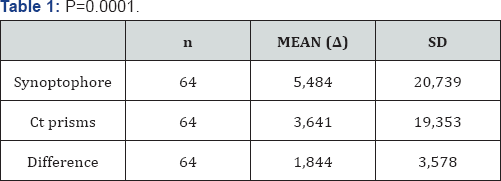
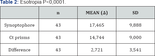
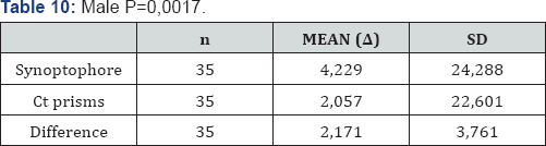
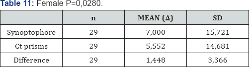
Results: Comparing the measured deviation angle in 64 patients with both methods at far vision, we found a statistically significant difference, with an average of 1,844 prism dioptres. If we compare both methods in esotropia or exotropia, in the 43 patients with esotropia found a statistically significant difference (mean difference 2.721Dp). On the other hand, in the 21 patients with exotropia there was no a statistically significant difference (mean difference 0.048Dp.). Considering gender, 35 men of the sample, we find statistically significant differences (mean difference 2.171Dp), and women, 29 patients of the sample, we also found statistically significant differences (mean difference 1.448Dp).
Conclusion: In esotropia we find that there are statistically significant differences in cover test with prisms at far vision. On the other hand, in exotropia we don’t find statistically significant differences. In both the groups of patients with previous strabismus surgery and non-surgery there are no statistically significant differences. The fact that we find a greater deflection angle with the synoptophore at convergent strabismus confirms the ability of dissociation.
Keywords: Synoptophore; Cover test; Dissociate prism dioptre; Strabismus; Exotropia; Esotropia


Introduction

The stereoscope invented by Charles Wheatstone in 1830 used for oculomotoras and sensory evaluation. Its main use is to assess and treat strabismus, also used in vision therapy for the treatment of amblyopia [1]. It simulates far vision conditions and presents separate images for each eye. It studies oculomotoras and sensory capabilities mainly in strabismus patients [2]. Although the measure of the deviation angle is usually performed with prism bar or loose prisms, it is worth considering the synoptophore, given its characteristics.
Compared with measurement with cover test (CT) and prisms, in synoptophore we obtain:
  1.  Ahigher value of deviation as it is more dissociating.
  2.  More simplicity and comfort in the measurement of the 9 positions of gaze thanks to its mechanical structure.
  3.  The values are more reproducible, making it easier for monitoring the case.
  4.  More accurate values in high-angle strabismus, since in these cases the measure is not performed in leaps.
  5.  More objective measurement


Materials and Methods

In a sample of 64 patients, with a mean age of 27.6 years, we consider two common clinical methods of measuring the angle of ocular deviation: synoptophore and Cover Test with prisms at far vision. We study which differences we find between both methods and what factors may influence the variability of the results. We studied the influence of factors such as the direction of the horizontal deviation (esotropia/ exotropia), if they had vertical deviation or some sort of alphabetical syndrome, if they had undergone strabismus surgery previously and finally the patient’s gender.


Results

Comparing the measurement of the deviation angle in 64 patients with synoptophore and cover test with prisms at far vision we find:

Comparison in factors (Table 1):

Type of deviation

  1.  Esotropia: P<0,0001 (Table 2)
  2.  Exotropia: P=0,9426 (Table 3)

History of strabismus surgery

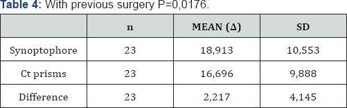
  1.  With previous surgery: P=0,0176 (Table 4)
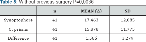
  2.  Without previous surgery: P=0,0036 (Table 5)

With previous surgery

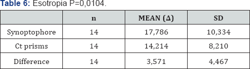
  1.  Esotropia: P=0,0104 (Table 6)
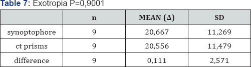
  2.  Exotropia: P=0,9001 (Table 7)

Without previous surgery

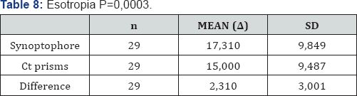
  1.  Esotropia: P=0,0003 (Table 8)
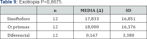
  2.  Exotropia: P=0,8675 (Table 9)

Gender

  1.  Male: P=0,0017 (Table 10)
  2.  Female: P=0,0280 (Table 11)
We also assessed whether other factors such as vertical deviations or alphabetic syndromes could influence the comparison. Given the low percentage of patients who showed vertical strabismus or alphabetical syndrome, comparative under these factors was dismissed [3-8].


Discussion

Despite significant statistical differences in convergent strabismus, in regular motor examination we always include the measurement with synoptophore as well as the measurement with prism at far vision.
We consider that the greater accommodative capability that convergent strabismus present, both at far and near vision, allows the synoptophore, as being more dissociating, to find greater deviation angles. We believe that although with synoptophore there is a risk of causing greater dissociation in some type of strabismus, for us its use for justified the measure, because it is more accurate, objective and reliable. Anyway we still complete the study with the measurement with prisms. In our protocol for measuring ocular alignment we consider the measurement with synoptophore, objectively, a complementary study to consider.


Conclusion


In esotropia we found that there was statistically significant differences respect to the measurement at distance with prisms at far vision. In exotropia we didn’t find statistically significant differences. Both in patients with previous strabismus surgery and non-surgery there are statistically significant differences. The fact that we find a greater deflection angle with the synoptophore at convergent strabismus confirms the ability of dissociation that we initially suspected occurs with Synoptophore [9-12].

For more Open Access Journals in Juniper Publishers please click on: https://juniperpublishers.com