Showing posts with label Syndrome. Show all posts

Friday, 31 January 2020

Schwartz Jampel Syndrome- A Case Report- Juniper Publishers


Juniper Publishers- JOJ Ophthalmology

Itroduction

Blepharophimosis is a general diminution of palpebral fissure in all its dimensions. The lids usually show ptosis, dystopia canthorum, lateral displacement of the lateral puncti, or abnormalities of the lashes such as ditichiasis or misdirected and stiff lashes. The other ocular defects associated with congenital blepharophimosis include strabismus, nystagmus, amblyopia, microphthalmus, anophthalmus, epicanthus inversus, microcornea and hypermetropia [1,2]. Schwartz-Jampel syndrome, an autosomal recessively transmitted disease, is a rare presentation of blepharophimosis.

Case History

A 2 year old male child, having dysmorphic features was referred from the department of Pediatrics for Ophthalmic assessment. The child was the first born of healthy non- consanguineous parents after an uneventful pregnancy. His mental and motor development was normal and he acquired independent walking at 16 months. Fine pincer grasp developed by 9 months of age. At the age of 2 years the child could talk only two words with meaning. Social development of the child was poor because of his abnormal appearance and poor language development. The parents noted the abnormal facial expression at the age of 18 months.
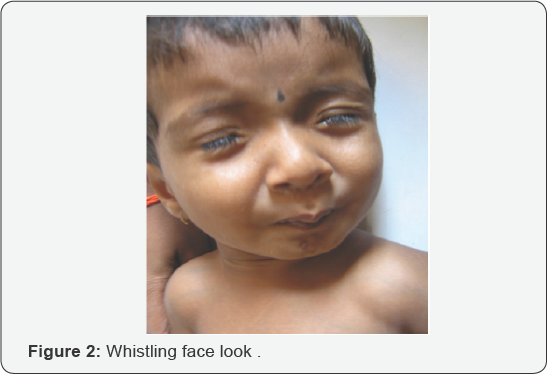
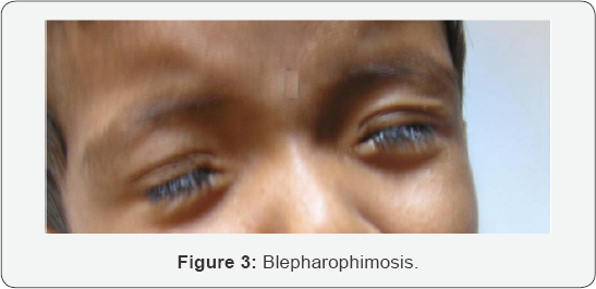
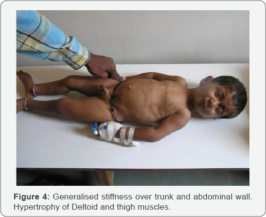
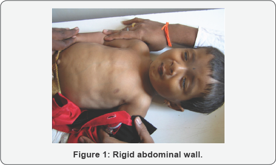
On examination the child had a short stature. The head posture was normal. Forehead did not show excessive wrinkling. The child had blepharophimosis (Figure 1). Lid crease was present. The child also had hypertrichosis. The globe examination was normal. The extraocular movements were normal. There was no refractive error. Fundus was normal. The child demonstrated pursing of lips giving him a 'whistling face' appearance and restricting his mouth opening (Figure 2). The shape of the chest was abnormal with sternal protrusion and sub-costal retraction (Figure 3). There was stiffness of his abdominal wall. The upper and lower limbs demonstrated hypertonia. The deep tendon reflexes were exaggerated. He had a waddling gait. The child had a high pitched voice (Figure 4).

Discussion

Schwartz-Jampel syndrome is a rare autosomal recessively transmitted disease, characterized by generalized myotonic myopathy, typical facial features, skeletal dysplasia, contracture of joints, growth retardation and bone maturation delay [3]. However a few cases showing dominant inheritance have also been reported. It is classified into 3 types based on age and severity
  1. Type 1A
  2. Type 1B
  3. Type 2

Type 1A

The type 1A disease is diagnosed in mid-childhood with recognition of myotonic facies with convex profile, short palpebral fissure, telecanthus, dimpling or quivering of the chin, prominent eyebrows, low hairline, low-set ears, flat base of the nose, micrognathia, microstomia, sometimes high-arched palate. The child exhibits progressive myotonia, muscle wasting and orthopaedic problems with decreased linear growth myotonia plateus by mid childhood. Additional findings reported in a few cases are myopia, hypertrichosis, and strabismus. The continuous myotonia is probably responsible for both muscular hypertrophy and peculiar facial appearance.

Type 1B

Type 1B is more severe than 1A, Bone dysplasia is present at birth. Long bones are shortened, femurs are dumbbell shaped. Bone epiphyses are large and vertebral bodies are flat.

Type 2

Type2 disease is more severe. Onset is neonatal, there is short limb dysplasia and long bones are bowed. Early death is frequent [4].
The diagnosis is predominantly on the basis of the typical dysmorphic facies [5]. EMG showing continuous discharges further supports the diagnosis. The gene defect in SJS type 1 is located in the 1p34-p36 of chromosome 1, whereas it is different in type 2 [6,7]. Perlecan the major proteoglycan of basement membranes is altered in patients with Schwartz- jampel syndrome disease [8]. However, a significant amount of molecular heterogeneity exists, genomically and proteomically, within SJS type 1. Currently no known correlation exists between the specific mutations found and the specific features of a given case However, the new mutations found by Stum et al. In 2006 have been discovered so recently that not enough time has elapsed to explore such possibilities. The new findings should be important tools to help find correlations among genetic variants, perlecan forms and levels, and clinical subtypes. Other facts yet unknown also may influence the severity and the specific characteristics of the disease [5]. The genetic tests for perlecan gene are not easily available in the commercial laboratories.
The child was diagnosed as having type 1A type of Schwartz- Jampel syndrome since the typical facial features became manifest at the age of 18 months. The old pictures of the child taken on his first birthday showed normal facial features. Medications that have been found useful in myotonic disorders such as phenytoin and carbamazipene may help to reduce the abnormal muscle activity. Warm baths are helpful in reducing stiffness. Botox injections are reportedly found useful to relieve blepharospsm.
Patients are generally treated with Carbamizipene 2030mg/kg body weight and most of them show improvement. Carbamazipene probably works by inhibiting neuronal sodium channels and may have direct effects on neurotransmitter systems. Orbicularis oculi myectomy, levator aponeurosis resection and lateral canthopexy are some surgical procedures which may be tried if the response to carbamazipene or botox is not adequate. The parents of the child were educated regarding the genetic nature of the disease and were referred to the geneticist. This particular child has not reported for follow-up as he belongs to a remote village far from our hospital and is probably reporting for follow-up at a nearby city.


Conclusion


Schwartz-Jampelsyndromeisararecauseofblepharophimosis. The condition can be managed with medications in most of the? cases. Surgery may be required if the condition does not improve with drugs.

For more Open Access Journals in Juniper Publishers please click on: https://juniperpublishers.com


Monday, 27 January 2020

Hay-Wells Syndrome: Case Report- Juniper Publishers


Juniper Publishers- JOJ Ophthalmology


Abstract

Introduction: Hay-Wells Syndromeis a rare disease with an autosomal dominant transmission. It is a type of ectodermal dysplasia, leading to impaired development of hair, nails, teeth and glands, usually in association with cleft palate and/or lip and ankyloblepharon. This syndrome is present at birth and diagnosis is made based on child phenotypic features. This study aims to describe a case of this syndrome.
Case report: Authors describe a case of a newborn with no history of parental consanguinity or other similar cases in the family. At birth alopecia, ankyloblepharon, cleftpalate, micrognatia and absent nails were detected. Surgical correction of ankyloblepharon was done. Postoperative period had no complications and after 12 months of follow-up lid edges are completely free.
Discussion: As a genetic disease, treatment is symptom-oriented. Antibiotic ointments can be used in skin lesions. Surgery is reserved for correction of cleft palate/lip and ankyloblepharon. Genetic counseling is recommended.
Keywords: Syndrome; Ectodermal dysplasia; Ankyloblepharon cleft palate and lip


Introduction

Hay-Wells Syndrome, also known as AEC syndrome (ankyloblepharon-ectodermal dysplasia- clefting syndrome), is a rare genetic disease [1], with an autosomal dominant transmission. Sporadic cases have, however, also been described. First described by Hay and Wells in 1976 [2], it was later found that it is caused by a p63 gene mutation, a known regulator of epithelial development/differentiation and homologous of the TP53 oncosupressor gene [3,4]. It is a type of ectodermal dysplasia, leading to impaired development of hair, nails, teeth and glands, usually in association with cleft palate and/or lip and ankyloblepharon. These are considered the cardinal signs of the syndrome by most authors. This syndrome is present at birth and diagnosis is made based on the child's phenotypic features. The authors aim to describe a case of a male newborn with the cardinal signs of this syndrome at birth, as it is a very rare syndrome with just a few cases reported worldwide.


Case Report

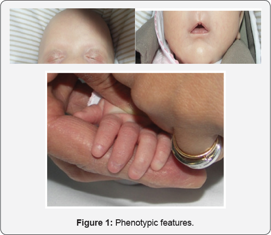
Male white newborn with no history ofparental consanguinity or other similar cases in the family. The baby was born by spontaneous vaginal delivery after an uneventful pregnancy with good prenatal care. At birth alopecia, ankyloblepharon, cleft palate, micrognathia and absent nails were detected (Figure 1a,1b & 1c- Phenotypic features). Anterior segment was normal, with no detected anomalies in the lacrimal apparatus. The rest of the physical examination was normal.
Ophthalmic ultrasound revealed a normal posterior segment. Abdominal and pelvic ultrasound were normal. Head ultrasound showed a small cyst in the pellucid septum and an apparent thickening in optic chiasmal area. Further study with Cranial MRI was normal, thus excluding a septum-optic dysplasia. Blood tests (CBS, electrolytes, hepatic function, kidney function, imunophenotyping and immunoglobulin levels) revealed a decrease in immunoglobulin A (X; normal range X), with no other changes. Hearing tests were normal.
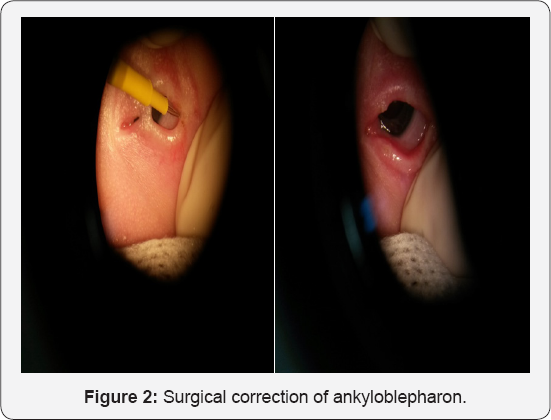
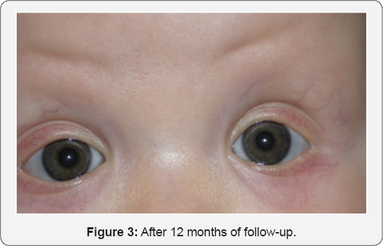
Dermatology consultation recommended topical streoids in scarceeczematous areas and daily mineral ointments. Later hair prosthesis was discussed with the parents. Surgical correction of ankyloblepharon was carried out by ophthalmologists. Tearing of lid bridges was done in the operating room, under general anesthesia, using an electric scalpel in both eyes (Figure 2- Surgical correction of ankyloblepharon). Careful tearing with gentle pulling of the eyelids was carried out in order not to damage the underlying cornea. Post-operative period had no complications and after 12 months of follow-up lid edges are completely free (Figure 3 - After 12 months of follow-up).
Cleft palate was complete, type III. Surgical correction was performed according to Furlow and Von Langenbeck procedures by pediatric surgeons. Blood molecular analysis of TP63 mutations and deletions/duplications was normal; we are still waiting for molecular analysis of mouth mucosa specimen. Unfortunately, it was not possible to have parental authorization to carry DNA analysis in their cells. Speech therapy was also prescribed.


Discussion

Hay-Wells Syndrome, also known as AEC Syndrome (ankyloblepharon-ectodermal dysplasia-clefting), is a rare autosomal dominant disease [1]. Ankyloblepharon, ectodermal dysplasia signs and cleft palate are considered cardinal signs of this syndrome [5]. All of these features were present in our case.
Ankyloblepharon results from fusion, partial or total, of superior and inferior lid edges. In general, eyelids remain closed until the 5th week of gestation, when they open spontaneously. The mechanism underlying this phenomenon is still on debate, but many author point out keratinization as the key [6]. Accordingly, any kind of anomaly occurring between 7th and 15th week of gestation can result in eyelid anatomy changes [6]. Ankyblepharon can also be present in Trisomy 18 and CHAND Syndrome (Curly hair-Ankyblepharon-Nail Dystrophy), being associated with heart malformations, hydrocephaly, imperforated anus and glaucoma. Ankyblepharon should then be a warning sign for the possibility of other important simultaneous diseases [7].
Ectodermal dysplasias are a group of diseases in which there is impaired development of hair, teeth, nails, sweet glands and other structures originating from ectoderm [8,9]. These anomalies, when associated with other malformations, correspond to a group of ectodermal dysplasia syndromes that includes EEC Syndrome (ectodactilia-ectodermal dysplasia- clefting), Rapp-Hodgkin Syndrome and Chand Syndrome, the most important differential diagnosis of Hay-Wells Syndrome [9]. In our case, none of the other malformations were identified, so that Hay-Wells Syndrome diagnosis was straight forward.
Hay-Wells Syndrome patients can have various degrees of alopecia, thin and scarce hair, onicodystrophies, palmoplantar hyperkeratosis, cutaneous pigment changes [10], hypohidrosis, hypodontia, teeth malformations and ear anomalies. Lacrimal canal obstruction is common. Other described features include super numerary nipples, otitis media, hypospadia, middle-facial hypoplasia, hypertelorism, low height, mental retardation, deafness and other ocular deformities.
At birth, a descamative erythrodemia can be seen, with superficial erosion and crusts. Scalp area usually shows an erosive dermatitis, which is a common source of infection, putting these patients at an increased risk of bacterial superinfection and sepsis; this leads to an increase in mortality and morbidity rate in newborns with this syndrome. A lot of Hay-Wells Syndrome cases are erroneously diagnosed as bullous epidermolysis due to the presence of erythrodermia and extensive areas of erosion. It is thought that scalp lesions can disappear with age or lead to alopecia [3].
Although concerns exist about wound healing in patients with this syndrome, there have been no reported instances of wound healing complications. Cleft lip and palate repair can then be performed safely in patients with Hay-Wells syndrome.
This syndrome is caused by a p63 gene mutation, an homologous of p53 oncosupressor gene [3,4], which has a central role in epidermal stratification process, regulating basal keratocyts proliferative capacity. Evidence showing that changes in this gene can be associated with other diseases like EEC and Rapp-Hodgkins syndrome reveal the high pleomorphic effect of p63 gene mutations. Hay-Wells Syndrome results specifically from amino-acid substitution in SAM domain (sterile alpha motif) [3,4].
Timely diagnosis is essential. As a genetic disease with multiple phenotypic features, multidisciplinary action is of main importance and treatment is symptom-oriented. Antibiotic ointments can be used in skin lesions. Surgical treatment is reserved for correction of cleft palate/lip and ankyloblepharon. Genetic counseling is recommended.


Disclosure statement


No sponsorship or funding arrangements relating to research and no conflicts of interest to declare.
For more Open Access Journals in Juniper Publishers please click on: https://juniperpublishers.com