JUNIPER PUBLISHERS- JOJ Ophthalmology
Aetiology
The most accepted theories until now are: Agenesis or partial development of the 6th cranial nerve nucleus in the brain stem and Branches from the 3rd nerve are redirected to innervate the lateral rectus. So lateral rectus has both Subnormal or no innervations from the 6th cranial nerve and anomalous innervations from the 3rd
nerve. According to the amount of anomalous innervations going to the
lateral rectus; different presentations and patterns of Duane’s syndrome
results (Figure1).

Duane Type 1
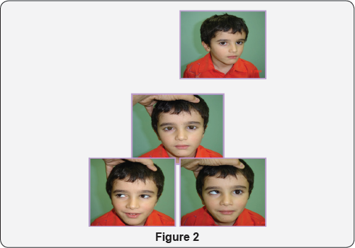
No innervations from the 6th nerve. Mild anomalous innervations from the 3rd nerve so the clinical picture is that limited abduction and mild narrowing of the palpebral fissure in adduction (Figure 2).

Duane Type 2
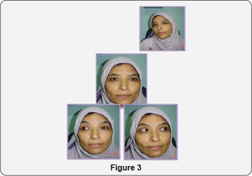
Normal innervations from the 6th nerve. Moderate
anomalous innervations from the 3rd nerve so the clinical picture
includes normal abduction and limited adduction together with narrowing
of the palpebral fissure in adduction (Figure 3).

Duane’s Type 3
No innervations from the 6th nerve. Nerve
fibers to the medial rectus is divided 50/50 between medial rectus and
lateral rectus so; the clinical picture is that limited abduction (no
innervations from the 6th), limited adduction due to equal contraction of both medial and lateral recti on adduction (Figure 4).

Duane’s Type 4
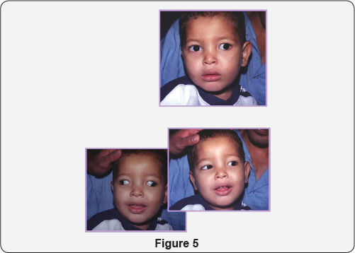
No innervations from the 6th nerve. Most
of the fibers of the medial rectus go to the lateral rectus so that with
attempted adduction, abduction occur (simultaneous divergence) (Figure 5).

For more articles in JOJ Ophthalmology (JOJO) please click on: https://juniperpublishers.com/jojo/index.php
No comments:
Post a Comment