Showing posts with label juniper publishers publons. Show all posts

Friday, 16 August 2019

Orbital Miasis: Complicating Squamous Cell Carcinoma Conjunctiva A Rare Case Report-Juniper Publishers


Juniper Publishers-Journal of Ophthalmology


Abstract
Miasis refers to invasion into the living tissues of humans and other mammals by the eggs or larvae of flies from the order of Dipteria. The invasive parasitic larvae almost invariably cause massive destruction of orbital tissue especially in neglected patients accompanied by marked inflammatory reactions and secondary bacterial infections. Infestation can be caused by multiple or single larvae. We report a case of 50 yr post menopausal women who was having squamous cell carcinoma of conjunctiva and was a neglected and belonged to low socioeconomic status. The patient ultimately ended with massive orbital miasis.
Keywords:Orbital Miasis; Carcinoma

Introduction

Orbital miasis is very rare worldwide. Miasis is a term first introduced by F. W. Hope. Miai is a Greek word meaning ‘fly’. Miasis refers to invasion into the living tissues of humans and other mammals by the eggs or larvae of flies from the order of Dipteria [1,2]. The invasive parasitic larvae almost invariably cause massive destruction of orbital tissue especially in neglected patients accompanied by marked inflammatory reactions and secondary bacterial infections. Infestation can be caused by multiple or single larvae [3]. Nursing staff, medical and paramedic staff should be aware of this condition, particularly in high risk cases like immobile, open wound and skin malignancy patients.

Case Report

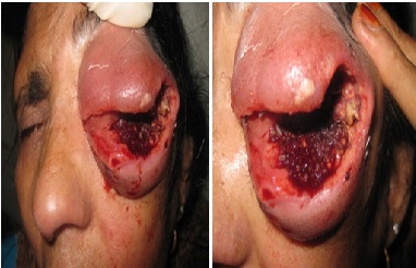
50 year old postmenopausal, normotensive, non diabetic, chronic hukka smoker patient comes to the accident and emergency department of SMHS Hospital with a complaint of blood stained discharge from the left eye and worms seen in the same eye (Figure 1). On examination the upper and the lower lids of the same eye were swollen, thickened and hard and could not be closed. The eye ball and the ocular tissue could not be seen. There was blood stained discharge and numerous live active worms in the left orbit. There was active oozing of blood on the left side. Orbital miasis was diagnosed. After going through the records of the patient, we came to know that patient was already diagnosed as a case of left poorly differentiated squamous cell carcinoma of the conjunctiva. CECT head showed soft tissue density mass with no bone invasion. MRI shows 5x4cm extracranial intraorbital mass with intraorbital component adherent to left lateral rectus muscle with left preauricular lymphadenopathy. The staging of tumor was T4 N1 M0. After cleaning and debridement of left orbital cavity the patient was referred back to the parent institution for emergency exentration and radiotherapy.
The maggots were identified as chyrosoma bezziana.

Discussion

Ophthalmomiasis is a rare form of eye disease and massive orbital or ocular miasis is even rarer [1,3-6]. Flies are small vingid orthopods of the class insecta. Some are biological or mechanical vectors of protozoal viral, bacterial for helminthic disease. Miasis in humans may be benign to asymptomatic or may result in mild to violent disturbances even death. Orbital miasis is the least common form of ophthalmomiasis.
By going through the literature we found very less cases of orbital miasis. Children and elderly persons or dementia patients who could not adequately take care of themselves are usually the victims. Most reported cases have belonged to the lower class of society being farmers, labours or beggars. In children, gonococcal conjunctivitis and in adult’s periocular ulcerated skin cancers may predispose to larval infestation3. Orbital miasis occurs when large number of diptherous larvae known as flies invades and destroy orbital contents. Ophthalmo-miasis may result in complications ranged from minor ocular ulceration to complete blindness and even death [7]. Common ophthalmomiasis inducing agents are cochhomyia hominivorax, hypoderma bovis, caliphora vomitoria, wohlfartia magnifia and chyrosoma. Bezziana the maggets identified in our patients belonged to C. Bezziana. Diagnosis is based on microscopic examination of the sliced caudal ends of the larvae (preserved in 70% alcohol or formalin). A dichomatous key is used to identify the number of interior spiracet opening and shape of body part [8]. Discharge associated with chyrosoma Bezziana foul smells and bloody.
The main predisposing factor for infestation in our patient was bed of necrotic tissue, provided by squamous cell carcinoma. Other important factors were lack of self care, general ignorance, low socioeconomic status and poor hygiene.
Any skin ulceration like diabetic, neurotrophic and malignant ulcers in the exposed areas in these patients are a risk factor and should be treated [9].
Miasis is not a common disorder in humans and it affects primarily wounds of skin, infection with purulent secretion and blood and body secretions are most common factors that attract flies.

Conclusion

Orbital miasis is a rare disease, which is marked by inflammatory reactions and secondary bacterial infections. Life threatening consequences like intracranial invasion may also occur. Old, debilitated and neglected patients are more prone to such disease. Malignant growths often form scaffolding for such infections to occur.

#
Figure 1: The maggots were identified as chyrosoma bezziana.

For more Open Access Journals in Juniper Publishers please click on: https://juniperpublishers.com


Monday, 12 August 2019

Micro Perforating Deep Sclerectomy: A New Modification of Non Penetrating surgeries to Improve results and Flatten learning Curve-Juniper Publishers


Juniper Publishers-Journal of Ophthalmology


Introduction

The High intra ocular pressure (IOP) is the only risk factor that we can control in glaucoma treatment, and it has been shown that reducing this pressure will stop or reduce glaucoma progression. Usually, when the patients diagnosed, wesill start treatment with drops that reduce aqueous humor production or increase its out flow in the trabecular mesh work in order to reduce the pressure inside the eye. But sometimes, this reduction is not enough and we can´t reach to our planified Target Intraocular pressure and it´s necessary to plan a surgical procedure that could reduce this pressure even more too finally achieve our intended IOP.
Glaucoma surgery has multiple variations but the gold standard procedure is still the trabeculectomy, creating a pathway from the anterior chamber, to the subconjunctival space by a scleral ostium and and peripheraliridotomy. This surgical technique is usually not very difficult to perform, but the chances of having surgery or post operation complications are very high, specially, those related to ocular hypotension due to unexaggerated and not controlled out flow. Another usual problem is when we try to control or reduce the out flow, and we perform really small ostiums or very tight sutures in the scleral flap and we get a poor result in the reduction of the intraocular pressure.
So there are many options to develop a surgery that can provide good intraocular pressure reduction with fewer complications than the penetrating procedures like trabeculectomy or valve implants. The development of Non penetrating Deep Sclerectomy became one of the most promising of this new surgical techniques. It is described as a good option for open angle glaucoma, especially if the target IOP, is not very low, or when the risk of hypotonic is high (phakic, nanoftalmos, Hypermetropia). This technique consist carving a first superficial scleral flap of about 4 or 5 mm wide and 1/3 of scleral thickness, then its carved a second Deep scleral flap is about 3 to 4 mm and almost 2/3 of scleral thickness arriving just choroid darkness. This Deep flap goes forward until we see the change of the scleral fibers when the scleral spur is located, then we find the external Wall of Schlemm's canal, Schwalbe´s line and continue forward at least one or two millimeters into clear corneal stroma. At this moment an anterior chamber paracentesis can be done to avoid prolapse of schlemm´s cannel tissue or the perforation of this very thin tissue that remains between anterior chamber and the outside, that we call trabeculo-Descemet membrane. These cond deepest flap is cutted and then next step is to remove that schlemm´s cannel outer Wall and at that moment we will see a continue controlled filtration or aqueous humor, without collapsing or abrupt compression of the anterior chamber.
This technique has shown good results in intraocular pressure reduction, compared to trabeculectomy, with an important difference in the incidence of complications related with hypotonic (atalamy, cataracts, endothelial damage, hypotonic aculophaty, retinal detachment, choroidal effusion or detachment) or to the communication of the outside environment with the inside of the eye (Endophthalmitis).
This is a safe, effective technique but with a very high learning curve, especially at the moment of the second Deep flap carving when the trabeculo-descemetic membrane is created, the perforation of this very thin tissue is a common complication and usually learning specialists try to avoid it by not going very Deep, over the choroidal plane and that is a reason, why the surgeons can´t get the correct plane, that will guarantee and adequate aqueous filtration trough the trabeculode scemtic membrane into the scleral lake as a decompression chamber, sub conjunctival space. The use of laser goniopunctures were needed to get the IOP lowering effects pected.
This was our main problem when we tried to learn and teach this technique in Bolivia, we had to convertion to trabeculectomy due to perforation or not enough IOP lowering effect.
The solution was to perform the surgery with the same steps as the original but when we found the remaining tissue was too thick to achieve a good filtering function, the risk of perforation trying to go deeper, we used a 30 gauge needle to perform about 5-10 micro perforations in the trabeculode scemetic like membrane we created, turning this membrane into a net like tissue that kept the resistance of the aqueous out flow, avoiding fast decompression problems. And hypotony problems in the post operation period without requering an extra procedure like yag laser goniopunctures or surgery revision.

Results

66 cases have been conducted from which (9.09%) should have been turned into trabeculectomy by perforation of the trabecular descemetic membrane. On a 3-year average follow-up, a 17+-3 IOP was obtained during the first year in 54 patients (81,8%) in 50 (75,7%) after 2 years, and in 48 patients (72,7%) after 3 years of control, and them education had to be restarted in the rest. Out of the operated patients, only 7required a new surgical procedure (Ahmed valve implant) on the 3rd year of the follow-up. No cases of hypothonia, at halamia, choroid detachment or end ophthalmitis were reported 3 years after the control.

Discussion

On our first years of experience with the non penetrating Deep sclerectomy, we found our selves continuosly with the situation of membrane perforation, or a thickker, non functioning membrane created. We need to turn many surgeries into big trabeculectomies, that increased the incidence of hypontony related complications in the post operatory time or if we were conservative, filtering surgeries that worked bad or didn´t work at all, needing new medications, new surgeries, oryag laser retreatment.
Turning Non Penetrating Deep sclerectomy surgeries into Micro Perforation Deep Sclerectomy give us a safe, effective option, that we used a lot in the beginning, with practice and gaining experience. We need to use less over time, once having dominated the surgery planes we needed to reach, when the learning curve was passed.
Its is a good option for learning residents or experienced surgeons who want to start with the Non penetrating surgeries to reduce the chances of complications and increse effectiveness of the filtering technique, until they achieve the needed practice to perform a perfect flaps carving and reaching a trabeculo-decement level needed to really reduce IOP.
Learning non penetrating surgeries, allows Glaucoma surgeons to identify and reach schlemm´s channel, and that opens options for other Glaucoma surgeries, like visco canalostomy, canaloplasty, ortrabeculotomy.

For more Open Access Journals in Juniper Publishers please click on: https://juniperpublishers.com


Tuesday, 28 May 2019

Juniper Publishers - Open Access Journals

Juniper Publishers - Engineering Technology Open Access Journal 


The Engineering Technology Open Access Journal is vast journal that deals with all types of Engineering Technology. The journal includes electronic communication, industrial engineering, Management in Engineering, design, manufacture, performance validation, and application of high precision machines, instruments, and components, including fundamental and applied research and development in manufacturing processes, fabrication technology, technology, innovation, kinematics, dynamics, control, and simulation of robots and intelligent machines and systems; design of robotic mechanisms, robotics-related computer hardware, software, and architectures, transistor-level design of integrated circuits, device modeling, technology, systems design, layout, and testing. Materials Science & Engineering, Mechanics of processes involved in material removal, accretion and deformation, in solid, semi-solid or in particulate form, Electro-Physical and Chemical Processes, Engineering and Metrology, Life-Cycle Engineering, innovation of elegant, effective, and economic engineering designs. Our journal welcomes all types of papers like Research, Review, Mini-Review, Opinion, Short Communication, Case Report, Proceedings, Editorials, Letter to Editors, Perspective etc.

To know more about Juniper Publishers click on https://juniperpublishers.com/


Friday, 10 May 2019

Juniper Publishers

Juniper Publishers


The main epigram of Juniper Publishers is to spread scientific knowledge globally. The credibility of published articles completely depends on the effective peer reviewing process; Hence, editors are the chief support for Juniper Publishers. The Editorial board members of Juniper Publishers are responsible for publishing quality manuscripts received from authors on concerned subjects.

To know more about Juniper Publishers, click on https://juniperpublishers.com/