Showing posts with label juniper publishers in california usa. Show all posts

Thursday, 12 August 2021

Juniper Publishers- JOJ Ophthalmology

Monocular Optic Neuropathy: Case Unsolved-JOJ Ophthalmology

Case Presentation

Female patient, 58 years old, unremarkable general medical and ophthalmological history, complains about pulsatile headache, with no irradiation, no specific location, and absence of nausea or vomiting. The headache would not aggravate after Valsalva maneuver and faded without medication. Also, refers vision loss in the right eye (RE) since the beginning of the headache, 10 days before. The best corrected visual acuity (BCVA) was hand movement in the (RE) and 9/10 Snellen scale in the left eye (LE). It was observed a relative afferent pupillary defect (RAPD) graded 3+, no color perception on the RE, Ishihara plates fully read in the LE, no eye movement alterations, and no facial proprioception alterations. The anterior segment had no inflammation signs and phacosclerosis was present in both eyes. Posteriorly, therewas no signs of disc inflammation, vitritis, macular or vascular alterations. Intra-ocular pressure (IOP) was 15 mmHg bilaterally.

Investigations

Emergency head Computorized Tomography (CT) and Magnetic Resonance Imaging (MRI) with gadolinium were performed to rule out optic pathway lesions or neuritis. ESR, CRP, blood count were performed to exclude Giant Cell Arteritis, wich all came negative [1]. Furthermore, testing for HIV, Syphilis, Hepatitis B and C, IGRA Quantiferon-TB (Tuberculosis), ACE and urinary calcium (Sarcoidosis), ANCA PR3 and MPO (Autoimunity), HLA-B51 (Behçet's disease) and Anti-NMO Aquaporin 4 antibodies (Devic's disease) was completed, with no positive results [2]. Optic disc and macular spectral domain Ocular Computorized Tomography (sdOCT) had no acute alterations. Computorized static perimetry (CSP) was unreliable due to a high percentage of false positive and negative responses, but revealed a deep and general loss of visual sensitivity across all right visual field.

Differential Diagnosis

At this time, the main differential diagnosis were:

Posterior optic neuropathy (PON)

Although there was no big abdominal or heart surgery previously, an RAPD graded 3+ and deep visual loss were present [3], without optic disc edema.

Optic neuritis (ON)

The patient was not young enough to fit the diagnosis of multiple sclerosis or Devic's disease, plus, there were no lesions revealed by the MRI or neurological symptoms, but due to the profundity of visual loss [4], these options had to be considered.

Anterior Ischemic Optic Neuropathy (AION)

No disc edema was observed, so by definition, this option was excluded [5]. The patient had no risk factors.

Treatment

After exclusion of infectious possibilities, a 1Gr metilprednisone/day was administered for 3 days. After this period [6], she was medicated with oral prednisone 1mgr/Kg/ day for 2 months, and then started to halt it in a slow manner.

Outcome

The patient improved greatly after 2 months. RE BCVA was 9/10, no RAPD was present, Ishihara plates were completely read and equally fast [7]. RE CSP, wich had initially a Visual Function Index of 2%, presented lastly 65%, with a medium deviation of -15.42dB and pattern stand deviation of 8.45dB. LE was considered unremarkable. sdOCT revealed RE optic disc atrophy, as the peripapillary nerve fiber layer had a thickness of 60|im, and an apparent loss of macular nerve fiber layer thickness [8]. The optic atrophy was observed at the slit lamp.

Discussion

Initially, due to the absence of optic disc edema, the options considered were posterior to the optic disc, which lead us to think of Multiple Sclerosis and Devic's disease, but the neuro imaging and blood testing ruled out these possibilities. After 2 months, although RE vision had improved, an optic atrophy was evident. It's curious as there was no optic disc edema observed throughout the follow-up, and no occupying-space lesion was identified. Since there was a clinical response to the steroids, ON is a possible diagnosis, although the study was negative.

For more Open Access Journals in Juniper Publishers please click on: https://juniperpublishers.com

For more articles in Journal of Physical Fitness, Medicine & Treatment in Sports

please click on: https://juniperpublishers.com/jpfmts/index.php

For more about juniper publishers  please click on: https://www.juniperpublishersgroup.com/

Wednesday, 4 August 2021

Juniper Publishers- JOJ Ophthalmology

Hemi-Central Retinal Vein Occlusion Secondary to the Habit of Forceful Sneeze Prevention-Juniper Publishers

Introduction

31 year-old man applied to the eye clinic with a complaint of silhouette in the lower paracentral visual field on his left eye. His central visual acuity was 10/10 both on his eyes. Fundus examination revealed hemi-central vein occlusion sparing the macula. A few days later widespread intra-retinal hemorhages fell into macula and visual acuity reduced dramatically. Detailed story of the patient pointed out that he had a lomber disc herniation 3 months ago at L3-L4 levels. Detailed history also showed that the patient had hypertrophic adenoids and hypertrophic nasal conc has which are under treatment by nasal decongestants. The patient did not have a heavy conditional work. But it had been speculated that powerful prevention and keeping the sneeze resulted both the herniation and hemi-central vein occlusion. Vein occlusions are common among olders but systemic disorders must be ruled out in youngers. We intended to share acute intra-ocular pressure increase as an uncommon cause for vein occlusion in this dramatic young man because systemic disease research came clean. Lomber herniation usually occurs secondary to forcing the inter-vertebral discs to high pressures that they can endure. So they can rupture and cause disc herniation. Performing valsalva maneuver by coughing, sneezing or sneeze prevention may increase pressure inside the medulla spinalis so it can result as disc herniation. This maneuver not only affects on the intervertebral discs, but also on all intra- and extra-dural structures and nerves [1]. So optic nerve head may be affected as an intra-dural component.

Hemi-central retinal vein occlusions which are classified in central retinal vein occlusion may result from hypertension, diabetes, atherosclerosis, hyper coagulable states and hypercholesterolemia. Younger patients with hemi-central retinal vein occlusion must be ruled out from diseases mentioned above. Valsalva manoeuver is the forced exhalation against a closed glottis. First described by an Italian anatomist in 17th century Antonio Maria Valsalva, it is now a well known entity amongst medical practitioners. It leads to increased intra- thoracic and intra-abdominal pressure and raised central venous pressure. It is an uncommon condition and has been reported to occur with various forms of Valsalva stress such as weight lifting, physical exercise, balloon blowing, birth labour [2]. Sneezing prevention may be accepted as a rare cause of valsalva stress. Preretinal haemorrhage caused by this sudden rise in venous pressure was first termed as Valsalva haemorrhagic retinopathy by Duane TD in 1972 [3]. Valsalva retinopathy is typically seen in young males. Sometimes it may make a fluid level which's location can be subinternal limiting membrane (ILM) or subhyaloid. Duane [3] first postulated the mechanisms for retinopathies due to distal trauma as alteration in arterial circulation (forward retinopathy), venous circulation (backward retinopathy) or to changes occurring in both systems (mixed retinopathy). Valsalva retinopathy may look like vein occlusions in appearance as spreaded intraretinal haemorrhages but transformation of valsalva retinopathy to vein occlusion related macular edema is a rare antity. Here i report a case about valsalva retinopathy which transformed to complete hemi-central vein occlusion in a week.

Case Presentation

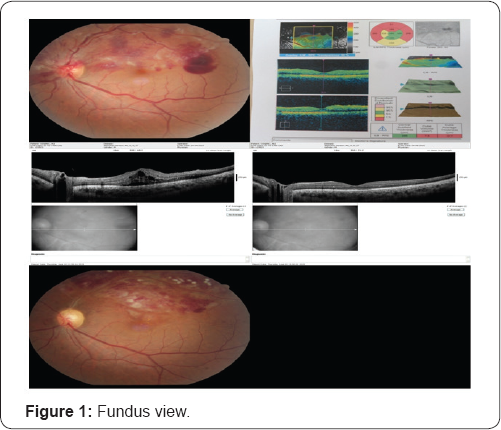
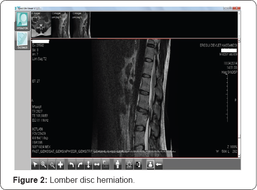
A 31 year-old healthy man complained about a blurry silhouette in the lower para-central visual field in his left eye after a powerfull sneeze prevention. Ophthalmic examination revealed bilateral 10/10 visual acuities and normal ocular tensions. Detailed examination showed a hemi-central vein occlusion sparing macula in the left fundus. There were widespread intra-retinal haemorrhages diffused in the upper hemi- central retina and a pre-retinal and subhyaloid haemorrhage just near the temporal macula. The fundus view is seen in Figure 1. Questioning about heavy work conditions for probable valsalva maneuver indicated a sneeze prevention habit. He had a lomber disc herniation at the L3-L4 levels 3 months ago and hypertrophic adenoids and nasal conchas which were still being treated by nasal decongestants. Lomber disc herniation is seen in Figure 2. Sneeze prevention habit was accused for lomber disc herniation by neuro-surgeon. After a week of rest recommendation and conservative management for valsalva retinopathy, the haemorrhages came to the bottom of macula and clinical macular edema settled upon as his visual acuity decreased to 3/10. Intravitreal bevacizumab injection was performed in other eye center at second week and he had full visual recovery again in two weeks time after the injection.

There were no retinal holes, tears, detachment, perivascular sheathing or snow banking at parsplana. Vein occlusions were not common among youngers so investigations including complete blood counts, clotting parameters such as antithrombin, protein c, protein s, d-dimer, factor 5, biochemical parameters such as urea, creatinine and glucose, crp, sedimentation were normal. Systemic evaluation was carried out by the physician and no abnormality was detected. We diagnosed him as hemi-central vein occlusion secondary to valsalva retinopathy. Systemic clotting abnormalities must be ruled out in hemi-central retinal vein occlusion among youngers. Disc herniation without predisposing factor and vein occlusion after sneeze prevention steered us to think about valsalva retinopathy. Intravitreal bevacizumab injection was performed and laser photocoagulation was performed for the macular edema secondary to hemi-central retinal vein occlusion. Because of the valsalva retinopathy was not limited to the pre-retinal and sub-hyaloid area, a week later the haemorrhages were spreaded widely to the macula and macular edema was settled. The visual acuity of the patient decreased to 3/10 snellen on the left and conservative treatment modality for the valsalva haemorrhagic retinopathy was replaced by the treatment of macular edema secondary to hemi-central retinal vein occlusion.

Results

The visual acuity of the patient is 10/10 on both eyes with complete resolution of macular edema for 2 months. Ghost vessels are seen on fundus examination and retinal haemorrhages are just about to resolve. These are seen in Figure 1. Bed rest and avoiding from valsalva maneuvers were adviced for lumbar disc herniation.

Discussion

Valsalva retinopathy is a unilateral or bilateral condition which occurs by rupture of superficial retinal capillaries. Heavy lifting, straining, coughing or vomiting may cause it. Valsalva maneuvers typically result in superficial retinal haemorrhages(sub-internal limiting membrane, subhyaloid) with a predilection for the macula, but subretinal, retinal and vitreous haemorrhages have been reported. The prognosis is variable, however complete visual recovery is possible [4] Some patients may develop a poor visual outcome due to the presumed toxic effects of dense premacular haemorrhages on the retinal pigment epithelium and/or epiretinal membrane formation. Nd-YAG laser puncture of the posterior aspect of the vitreous may allow drainage of premacular haemorrhage or vitrectomy may be performed in selected cases [4]. It may also managed conservatively with good outcomes. It may Valsalva retinopathy is a self-limited condition. Recurrence of valsalva retinopathy can be encountered if preventing any condition leading to valsalva like stress is not succeeded [2]. Our patient did not know about the harmful effects of sneeze prevention. A careful history is the key to the diagnosis. The prognosis is good and patient needs to be counselled to avoid any such Valsalva manoeuver like stress to prevent recurrence in future. Habits which are against the physiology such as sneeze prevention may be harmful. Patients should be careful in every physical activities which may induce valsalva maneuver. Valsalva retinopathy may be seen as central or hemi-central retinal vein occlusion.

For more Open Access Journals in Juniper Publishers please click on: https://juniperpublishers.com

For more articles in Journal of Physical Fitness, Medicine & Treatment in Sports

please click on: https://juniperpublishers.com/jpfmts/index.php

For more about juniper publishers  please click on: https://www.juniperpublishersgroup.com/
Monday, 10 February 2020

A Case of Temporal Arteritis Associated with Atypical Clinical Findings- Juniper Publishers


Juniper Publishers- JOJ Ophthalmology


Introduction

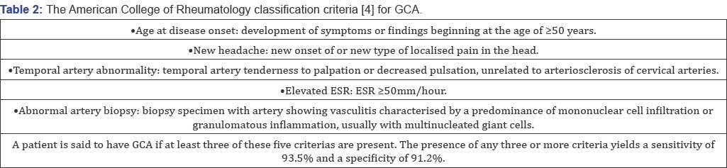
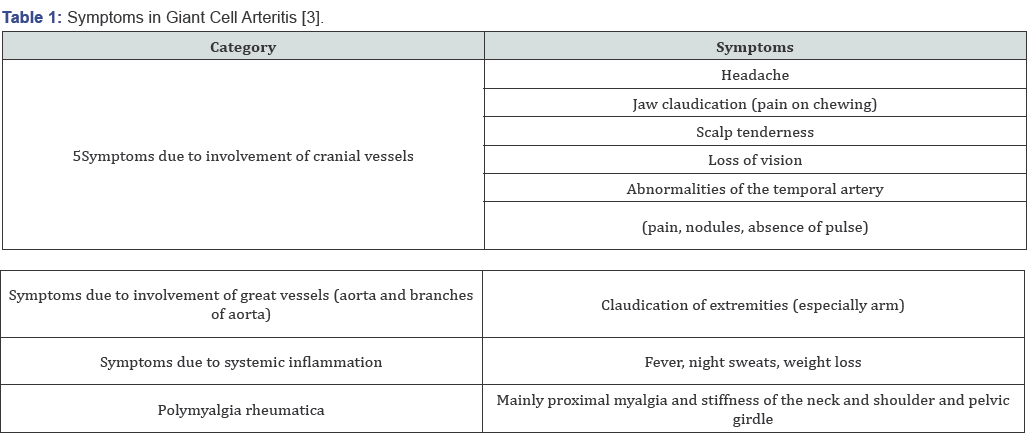
Giant cell arteritis (GCA), is a systemic vasculitis, affects medium and large sized cranial arteries [1], it can cause serious morbidities. Giant cell arteritis (GCA) predominantly affects elderly females [2]. The typical symptoms of new-onset GCA are bitemporal headaches, jaw claudiacation, scalp tenderness, visual disturbances, systemic symptoms such as fever and weight loss, and polymyalgia [3]. The diagnostic assessment comprises laboratory testing (erythrocyte sedimentation rate, C-reactive protein), temporal artery biopsy and imaging studies. The most important complication is permenant and deep vision loss. Diagnosis of TA is difficult if typical symptoms other than visual loss are absent. It can be difficult to diagnose this disease in the absence of typical clinical signs and labaratuary findings. Here we report a silent and atypical temporal arteritis case (Table 1& 2).


Case Report

63 year old hypertensive male patient applied to our hospital with complaint of vision loss in his right eye for two
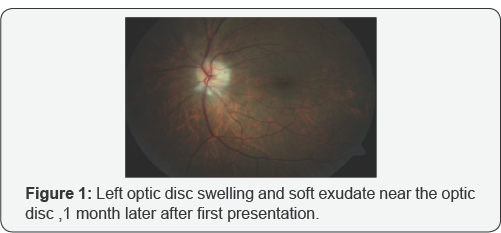
weeks. His visual acquity was perception of hand movements in right eye and 8/10 according to Snellen Chart in left eye. While anterior segment evaluation revealed bilateral nuclear sclerosis, fundoscopic examination revealed papiledema in his right eye. He did not complain of any associated headache, scalp tenderness, jaw claudication or constitutional symptoms such as weight loss, fever, malaise or sweats. We consulted the patient to internal medicine, rheumotology and hematology departments. His clinical and labaratuary findings were unremarkable except thrombocytosis. Following a presumptive diagnosis of non- arteritic ischemic optic neuropathy the patient was hospitalized and treated with intravenous 1000mg methylprednisolone for 3 days. After 3 days, oral prednisolone therapy was started 60mg per day. Oral steroid therapy was tapered 10mg with 3 days interval. However at 10-days' examination visual acquity in his right eye was reduced to loss of light perception. 1 month later, patient presented with sudden vision loss in his left eye, headache and malaise. Fundus examination revealed left optic disc swelling and soft exudates near the optic disc (Figure 1). Floroscein anjiography showed delayed filling at arterial phase and late staining of the optic disc. When labaratuary tests were repeated, erythrocyte sedimentation rate (ESR) was 65mm/ h(normal range: 1-15mm/h) and creactive protein (CRP) level was 15mg/L (normal range: <5 mg/L). According to these findings, with the diagnosis of arteritic ischemic optic neuropathy, intravenous steroid therapy was initiated. Rheumatology department evaluated the patient again and a temporal artery biopsy was performed; focal chronic inflammation and elastic fiber degeneration areas were identified at histologic sections (Figure 2). At the fifth day of intravenous steroid therapy visual acquity in left eye was 7/10 according to the Snellen Chart. The Humphrey Perimetry test revealed a paracentral island in his left eye.Since the visual acquity was at the level of loss of light perception at the right eye we didn't perform the Humphrey Perimetry test for the right eye.


Discussion

Patients with GCA typically present headache, jaw claudication, fever, weight loss, myalgia, arthralgia or malaise. Conversely, patients with occult GCA, first described by Simmons & Cogan [5], present with sudden visual loss without systemic symptoms and signs. Thus, due to the lack of symptoms, diagnosis and treatment of silent GCA may be considerably delayed when compared to typical GCA. Occult GCA , a potential cause of blindness, is defined as ocular involvement of GCA without any systemic symptoms and signs [6]. Ocular symptoms include visual loss, amaurosis fugax, diplopia and eye pain Hayreh et al. [6], described cotton wool spots in up to one third of eyes with visual loss dring the early stages of occult GCA [6]. Scalp abcess, chronic earache, bilateral central retinal artery occlusion, mydriaitic pupil, dry cough, aort anevrism , bilateral submandibular lymphadenopathy, hepatosplenomegaly are some atypic symptoms of GCA. The occult GCA group had higher C-reactive protein levels, a higher platelet count , and lower serum albumin levels [7]. Ophthalmic involvement can occur in up to 50% to 70% of the GCA patients, and this represents an ocular emergency. Arteritic anterior ischemic optic neuropathy (AAION) is the most common type of ophthalmic involvement in GCA and can cause permanent visual loss. Therefore, prompt diagnosis and treatment with a high dose of steroids is essential for these patients. In our patient at the first presentation labaratuary findings were unremarkable and there were no systemic symptoms; therefore at the beginning, our presumptive diagnosis was non-arteritic ischemic optic neuropathy of right eye. 1 month later, sudden vision loss appeared at left eye and he was complaining from headache, scalp tenderness ; ESR and CRP levels were elevated on labaratuary examination at that time. Hayreh et al. [6] found that ESR and CRP levels were relatively lower in patients with occult GCA compared to cranial GCA. On the other hand, Hamidou et al. [7] found higher CRP levels, a higher platelet count, and lower serum albumin levels in occult GCA group. In our patient's first presentation, ESR and CRP levels were within the normal limits. One month later when the other eye was effected ESR and CRP levels were remarkably high (Figure 3].
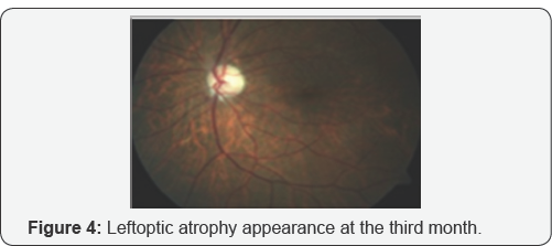
Although Liozon et al. [8] describes occult GCA as a protracted inflammatory response and a relatively benign short term outcome, in our case despite the long term corticosteroid and immunsupresive treatment, visual acquity was loss of light perception level in the right eye. Tan et al. [9] reported a case of GCA involving only the occipital artery which was revealed by magnetic resonance angiography. Chomlak et al. [10] reported a case of GCA with vertebral artery involvement, which was refractory to immunosuppressives. Hocevar et al. [11] claimed that, even early diagnosis and a prompt initiation of steroid did not prevent relapses in GCA. In our case after first attack despite the prompt intravenous steroid therapy one month later the patient consulted with visual loss at his other eye this verified Hocevar’s hypothesis. Cullen et al. [12] reported an occult GCA case in Singapore. Papakostas et al. [13] reported a case of GCA that presented with cotton wool spots and retinal vasculitis affecting small-size retinal arterioles. Shambhu et al. [14] reported an atypical case of GCA presenting as mild upper abdominal pain and generalized weakness in the context of hyponatremia as the presenting manifestation of vasculitis that was subsequently diagnosed by MRI scanning. Cheema et al. [15] described a case had the signs and symptoms consistent with GCA but who had an ESR within the normal limits, 27mm/h. In our case ESR at the first presenting ESR was within the normal limits,too. Labarca et al. [16] reported that patients with hypertension or diabetes at GCA diagnosis have more relapses during follow-up. Kermani et al. [17] reported seven patients with a positive temporal arteritis for GCA had a normal ESR and CRP at diagnosis (Figure 4].
In people older than 55 years, amaurosis fugax or visual loss, development of an acute ocular ischemic lesion with or without elevated erythrocyte sedimentation rate and systemic symptoms, should raise suspicion for giant cell arteritis. The diagnosis of temporal arteritis is easily made when sudden loss is contemporary with other symptoms and raised inflammatory markers. In the absence of known symptoms, in a patient diagnosed as ischemic optic neuropathy, occult temporal arteritis shouldn’t be out of mind. Despite the early diagnosis and treatment final visual acquity may not be satisfactory.


Conflict of Interest

Asfuroglu declares that he has no conflict of interest. Author Koz declares that she has no conflict of interest. Author Ozbalkan declares that she has no conflict of interest. Author Sandikci declares that she has no conflict of interest. Author Ciftci declares that she has no conflict of interest. Author Ozdemir declares that she has no conflict of interest.
This article does not contain any studies with human participant performed by any of the authors.

Informed consent was obtained from all individual participants included in the study.

For more Open Access Journals in Juniper Publishers please click on: https://juniperpublishers.com


Monday, 17 June 2019

Juniper Publishers Reviews


Juniper Publishers Reviews - 5 Reviews of Juniperpublishers.com


Open Access facilitates free and unrestricted access to the data for everyone to read and download. We believe that the Promotion of Open Access is essential to encourage innovation, socio-economic development and flow of knowledge around the world. It allows the permanent storage of scholarly journal articles and scientific data through digital copies. Abstracts and full texts (HTML, XML and PDF format) of all articles published by Juniper Publishers are freely accessible to everyone immediately after publication. The benefits of implementing Open Access are reaped by many end users such as students, researchers, clinicians, patients, policymakers and journalists. Our content is universally available in an easily readable format on the internet and strictly follows the open access policies.

For more details Open access journals please click on: Juniper publishers


For more details please click on: Peer reviewed Journal of Ophthalmology


For more details open access journals review please click on: Juniper Publishers Reviews - 5 Reviews

Tuesday, 28 May 2019

Juniper Publishers - Open Access Journals

Juniper Publishers - Engineering Technology Open Access Journal 


The Engineering Technology Open Access Journal is vast journal that deals with all types of Engineering Technology. The journal includes electronic communication, industrial engineering, Management in Engineering, design, manufacture, performance validation, and application of high precision machines, instruments, and components, including fundamental and applied research and development in manufacturing processes, fabrication technology, technology, innovation, kinematics, dynamics, control, and simulation of robots and intelligent machines and systems; design of robotic mechanisms, robotics-related computer hardware, software, and architectures, transistor-level design of integrated circuits, device modeling, technology, systems design, layout, and testing. Materials Science & Engineering, Mechanics of processes involved in material removal, accretion and deformation, in solid, semi-solid or in particulate form, Electro-Physical and Chemical Processes, Engineering and Metrology, Life-Cycle Engineering, innovation of elegant, effective, and economic engineering designs. Our journal welcomes all types of papers like Research, Review, Mini-Review, Opinion, Short Communication, Case Report, Proceedings, Editorials, Letter to Editors, Perspective etc.

To know more about Juniper Publishers click on https://juniperpublishers.com/


Friday, 10 May 2019

Juniper Publishers

Juniper Publishers


The main epigram of Juniper Publishers is to spread scientific knowledge globally. The credibility of published articles completely depends on the effective peer reviewing process; Hence, editors are the chief support for Juniper Publishers. The Editorial board members of Juniper Publishers are responsible for publishing quality manuscripts received from authors on concerned subjects.

To know more about Juniper Publishers, click on https://juniperpublishers.com/