Monday, 12 August 2019

Micro Perforating Deep Sclerectomy: A New Modification of Non Penetrating surgeries to Improve results and Flatten learning Curve-Juniper Publishers


Juniper Publishers-Journal of Ophthalmology


Introduction

The High intra ocular pressure (IOP) is the only risk factor that we can control in glaucoma treatment, and it has been shown that reducing this pressure will stop or reduce glaucoma progression. Usually, when the patients diagnosed, wesill start treatment with drops that reduce aqueous humor production or increase its out flow in the trabecular mesh work in order to reduce the pressure inside the eye. But sometimes, this reduction is not enough and we can´t reach to our planified Target Intraocular pressure and it´s necessary to plan a surgical procedure that could reduce this pressure even more too finally achieve our intended IOP.
Glaucoma surgery has multiple variations but the gold standard procedure is still the trabeculectomy, creating a pathway from the anterior chamber, to the subconjunctival space by a scleral ostium and and peripheraliridotomy. This surgical technique is usually not very difficult to perform, but the chances of having surgery or post operation complications are very high, specially, those related to ocular hypotension due to unexaggerated and not controlled out flow. Another usual problem is when we try to control or reduce the out flow, and we perform really small ostiums or very tight sutures in the scleral flap and we get a poor result in the reduction of the intraocular pressure.
So there are many options to develop a surgery that can provide good intraocular pressure reduction with fewer complications than the penetrating procedures like trabeculectomy or valve implants. The development of Non penetrating Deep Sclerectomy became one of the most promising of this new surgical techniques. It is described as a good option for open angle glaucoma, especially if the target IOP, is not very low, or when the risk of hypotonic is high (phakic, nanoftalmos, Hypermetropia). This technique consist carving a first superficial scleral flap of about 4 or 5 mm wide and 1/3 of scleral thickness, then its carved a second Deep scleral flap is about 3 to 4 mm and almost 2/3 of scleral thickness arriving just choroid darkness. This Deep flap goes forward until we see the change of the scleral fibers when the scleral spur is located, then we find the external Wall of Schlemm's canal, Schwalbe´s line and continue forward at least one or two millimeters into clear corneal stroma. At this moment an anterior chamber paracentesis can be done to avoid prolapse of schlemm´s cannel tissue or the perforation of this very thin tissue that remains between anterior chamber and the outside, that we call trabeculo-Descemet membrane. These cond deepest flap is cutted and then next step is to remove that schlemm´s cannel outer Wall and at that moment we will see a continue controlled filtration or aqueous humor, without collapsing or abrupt compression of the anterior chamber.
This technique has shown good results in intraocular pressure reduction, compared to trabeculectomy, with an important difference in the incidence of complications related with hypotonic (atalamy, cataracts, endothelial damage, hypotonic aculophaty, retinal detachment, choroidal effusion or detachment) or to the communication of the outside environment with the inside of the eye (Endophthalmitis).
This is a safe, effective technique but with a very high learning curve, especially at the moment of the second Deep flap carving when the trabeculo-descemetic membrane is created, the perforation of this very thin tissue is a common complication and usually learning specialists try to avoid it by not going very Deep, over the choroidal plane and that is a reason, why the surgeons can´t get the correct plane, that will guarantee and adequate aqueous filtration trough the trabeculode scemtic membrane into the scleral lake as a decompression chamber, sub conjunctival space. The use of laser goniopunctures were needed to get the IOP lowering effects pected.
This was our main problem when we tried to learn and teach this technique in Bolivia, we had to convertion to trabeculectomy due to perforation or not enough IOP lowering effect.
The solution was to perform the surgery with the same steps as the original but when we found the remaining tissue was too thick to achieve a good filtering function, the risk of perforation trying to go deeper, we used a 30 gauge needle to perform about 5-10 micro perforations in the trabeculode scemetic like membrane we created, turning this membrane into a net like tissue that kept the resistance of the aqueous out flow, avoiding fast decompression problems. And hypotony problems in the post operation period without requering an extra procedure like yag laser goniopunctures or surgery revision.

Results

66 cases have been conducted from which (9.09%) should have been turned into trabeculectomy by perforation of the trabecular descemetic membrane. On a 3-year average follow-up, a 17+-3 IOP was obtained during the first year in 54 patients (81,8%) in 50 (75,7%) after 2 years, and in 48 patients (72,7%) after 3 years of control, and them education had to be restarted in the rest. Out of the operated patients, only 7required a new surgical procedure (Ahmed valve implant) on the 3rd year of the follow-up. No cases of hypothonia, at halamia, choroid detachment or end ophthalmitis were reported 3 years after the control.

Discussion

On our first years of experience with the non penetrating Deep sclerectomy, we found our selves continuosly with the situation of membrane perforation, or a thickker, non functioning membrane created. We need to turn many surgeries into big trabeculectomies, that increased the incidence of hypontony related complications in the post operatory time or if we were conservative, filtering surgeries that worked bad or didn´t work at all, needing new medications, new surgeries, oryag laser retreatment.
Turning Non Penetrating Deep sclerectomy surgeries into Micro Perforation Deep Sclerectomy give us a safe, effective option, that we used a lot in the beginning, with practice and gaining experience. We need to use less over time, once having dominated the surgery planes we needed to reach, when the learning curve was passed.
Its is a good option for learning residents or experienced surgeons who want to start with the Non penetrating surgeries to reduce the chances of complications and increse effectiveness of the filtering technique, until they achieve the needed practice to perform a perfect flaps carving and reaching a trabeculo-decement level needed to really reduce IOP.
Learning non penetrating surgeries, allows Glaucoma surgeons to identify and reach schlemm´s channel, and that opens options for other Glaucoma surgeries, like visco canalostomy, canaloplasty, ortrabeculotomy.

For more Open Access Journals in Juniper Publishers please click on: https://juniperpublishers.com


Friday, 9 August 2019

Juniper Publishers Contact Number

Unusual Visual Presentation from Optic Neuropathy Secondary to Recurrent Oral Squamous Cell Carcinoma-Juniper Publishers

Juniper Publishers-Journal of Ophthalmology


A number of medical conditions may present to the emergency department involving a change or loss of vision. Many of these conditions are sudden in onset and may result in complete or partial loss of vision, such as stroke or multiple sclerosis. A 53 year old male with a past history of oral squamous cell carcinoma presented with a headache and changes in color hue in his right eye. CT imaging revealed recurrent oral squamous cell carcinoma involving the right sphenoid and ethmoid sinuses with extension into the right middle cranial fossa and orbit. It is important to note that a change in color vision may be from optic nerve dysfunction, which was found to be the case for this patient. The patient was diagnosed with optic neuropathy secondary to recurrent oral squamous cell carcinoma, which represents the first case report of such condition.
Keywords: Human Papillomavirus
Abbreviations: OSCC : Oral Squamous Cell Carcinoma

Introduction

Oral squamous cell carcinoma (OSCC) represents the most frequent malignancy of the oral mucosa [1,2]. Tobacco and alcohol are strong risk factors [2], and a number of studies have commented on the association between human papilloma virus and OSCC [3,4]. It represents a significant clinical challenge, requiring aggressive surgical resection with staging of the tumor. Chemotherapy and radiation therapy are required to achieve remission [5] and management requires evaluation by MRI and PET [6]. The most frequent spread of OSCC is to the cervical lymph nodes and lung, followed by metastasis to bone and liver [7]. Nasopharyngeal spread may occur in up to 28% of cases; however secondary ocular spread is rare [8]. We report a patient who presented to the emergency room with symptoms of color vision changes which was found to be secondary to metastatic spread of OSCC.

Materials and Methods

Biomicroscopy, color vision testing, visual field testing, dilated indirect ophthalmoloscopy, optical coherent tomography, fundus photography, and fluorescein angiography was performed. Subsequent CT and PET imaging was also obtained.

Case Report

A 53 year old male presented to the emergency room with headaches and decreased contrast sensitivity in his right eye. He was a nonsmoker, but would occasionally drink alcohol and use recreational drugs. Family history was unremarkable for malignancy. Medical history included controlled hypertension and OSCC involving the tongue, with spread into the cervical lymph node. After undergoing surgical resection, he underwent chemotherapy and radiation therapy to the head, neck, and lungs. Two years later he was noted to have a recurrence of OSCC, which involved his left maxillary and ethmoid sinuses with extension into the left orbit. He underwent left orbital exenteration, left external sphenoidectomy, left ethmoidectomy including resection of tumor at cribriform plate, and left medial maxillectomy. The patient concurrently received an additional three month course of chemotherapy and radiation therapy.
The patient was referred for ophthalmologic consultation. Due to the patient’s unusual presentation, there was concern for recurrence of OSCC in the right orbit. The patient was sent for MRI imaging of the brain and orbit. Due to surgical clips from the previous orbital exenteration, a CT scan was performed instead. He was referred to oncology for further evaluation, and received a PET scan and another CT scan. The patient elected for hospice care rather than further surgical or medical treatment.

Results

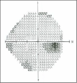
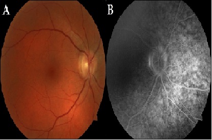
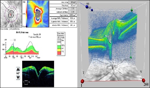
Ophthalmic examination disclosed a best corrected visual acuity of 20/25-2 in the right eye, and an intraocular pressure of 10 mmHg. There were no abnormalities of the right eyelids, conjunctiva, cornea, lens, pupillary response, or extraocular eye motility. There was no proptosis or pain or resistance on retropulsion. There were no abnormalities of color vision by Ishihara plate testing. Humphrey 24-2 visual field testing revealed an enlarged blind spot and small peripheral abnormalities (Figure 1). After dilation, the optic nerve showed mild hyperemia and mild retinal venous congestion (Figure 2A). No abnormalities of the macula, peripheral retina, or vitreous were noted. A fluorescein angiogram was performed and disclosed mild late staining of the optic nerve, with no other abnormalities (Figure 2B). Evaluation of the optic nerve by optical coherent tomography demonstrated mild optic nerve elevation, consistent with optic neuropathy (Figure 3).
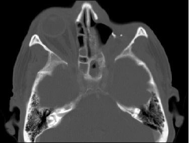
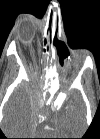
The CT scan disclosed sphenoid and posterior ethmoid sinus disease (Figure 4). PET scan imaging demonstrated progression of malignancy at the level of the right skull base with extension into the right cavernous sinus and extension to the posterior nasopharynx and adenoid region. Extensive new skeletal metastatic disease was also noted. Within a month, he presented with proptosis and an orbital apex syndrome. Extensive tumor invasion into the right orbit was noted by an additional CT (Figure 5). The patient succumbed to his illness within three weeks of this examination.

Discussion

Visual complaints can be a common presentation to the emergency department. This patient presented with a headache and changes in color hue, and was diagnosed with optic neuropathy secondary to recurrent OSCC. Given the ophthalmologic findings, CT and PET studies, it is likely the optic neuropathy was secondary to tumor spread in the cavernous sinus giving rise to the venous dilation and direct tumor involvement of the optic foramen from the sphenoid sinus. We suspect the patient’s headache was secondary to meningeal involvement. Macrophage polarization and regional spread to the cervical lymph nodes, both of which this patient demonstrated, have been indicators of a poor prognosis [9]. Fewer than 10 cases involving ocular spread have been reported, which also carries a poor prognosis [7]. None of the reported cases have presented as an optic neuropathy. In 2007, Feng et al. [10] reported the first case of acute visual loss in a head and neck cancer patient with ocular metastasis and sphenoid pyocele, however he points out that the visual loss occurred from compression of the optic nerve and the sphenoid pyocele.
This case serves to illustrate the unusual nature of visual complaints that may present to the emergency department and helps to illustrate the effectiveness of the multispecialty team approach in assessing complex patients.

Acknowledgments

The authors thank Dr. Vijay Suhag (Department of Oncology, Sutter Roseville Medical Center), Dr. Christopher Markus (Emergency Department, Sutter Roseville Medical Center), Dr. Randall Ow (Department of Otolaryngology, Sutter Roseville Medical Center), and Dr. Michael Kaplan (Department of Otolaryngology, Head and Neck Surgery, Stanford University Medical Center).
Figures and Tables
#
Figure 1: An enlarged blind spot and peripheral visual field defects in the right eye formally tested using the Humphrey 24-2 testing algorithm.
#
Figure 2: Fundus photography displaying mild hyperemia in the right optic nerve and retinal venous congestion (A), followed by fluorescein angiography displaying mild late staining of the optic nerve without other abnormalities of the macula or retinal vasculature (B).
#
Figure 3: Cirrus-HD optical coherence tomography demonstrating slight optic nerve elevation consistent with right optic neuropathy.
#
Figure 4: Computed tomography disclosing sphenoid and posterior ethmoid sinus disease.
#
Figure 5: Computed tomography demonstrating proptosis and an orbital apex syndrome with extensive tumor invasion into the right orbit.
For more Open Access Journals in Juniper Publishers please click on: https://juniperpublishers.com


Wednesday, 7 August 2019

Ocular Tissue Adhesive Application in DSAEK: a Comparative Study-Juniper Publishers


Juniper Publishers-Journal of Ophthalmology


Abstract

Purpose
To evaluate clinical safety and efficacy of a novel use of an ocular tissue adhesive in Descemet’s Stripping (Automated) Endothelial Keratoplasty (DSAEK).
Methods
35 consecutive DSAEK cases were evaluated retrospectively. In group-A (nA=15) the tissue adhesive (Re Sure Adherent Ocular Bandage, Ocular Therapeutix, Inc., Bedford, MA) had been used, prior to placement of one suture, while in group-B (nB=20), only nylon sutures were used for the closure of corneal incisions. Peri-operative complications were noted. Visual Acuity, refraction and topographic cylinder, Intraocular Pressure (IOP), and endothelial cell counts (ECC) were monitored long-term for up to two years.
Results
Follow-up time was 10.5±8.5 (8 to 29) months. No case from group-A required any additional air insertion following the tissue adhesive application and no case required additional intra operative surgical manipulation for further graft centration. In group-B eighteen (out of twenty) cases required intra-operative supplemental air insertion, and four of those intra-operative repositioning of the graft. The differences in visual acuity and IOP were not statistically significant; ECC change of -16% in group-A vs. -21% was noted in group- B (statistically significant, p =0.03). Hyperopic shift was noted in both groups; cylinder reduction was noted, too, with group-A performing better.
Conclusions
Tissue adhesive may be a valuable adjunct in clear-cornea DSEAK by stabilizing the potential of air escape from the main incision inadvertently occurring during suture placement.
Keywords: LED Cassini; multi-color LED topography; Acanthamoeba keratitis; Point-source topography; Pentacam HR; Placido topography; Scheimpflug topometry; Differential topography; Irregular corneal astigmatism; Stray light measurements; C-Quant; Anterior-Segment Optical Coherence Tomography

Introduction

Descemet’s Stripping (Automated) Endothelial Keratoplasty (DSAEK) surgery has become the standard of care worldwide for endothelial dysfunction management [1] superseding for this purpose Penetrating Keratoplasty (PK). PK involves a full corneal thickness, open-chamber procedure; among the disadvantages noted with this procedure are the prolonged visual rehabilitation, unpredictable cylindrical refractive changes (high postoperative astigmatism), susceptibility to ocular surface complications (wound dehiscence), and vulnerability to traumatic wound rupture [2,3].DSAEK, introduced 2002, [4] involves the posterior cornea lamellae in a closed-chamber procedure [5]. Because of this, it is considered safer, provides faster visual recovery, [6] usually requires only few sutures and causes less astigmatic change,[7] overcoming some of the limitations of PK.
The corneal incisions associated with ocular corneal surgery, such as cataract and lamellar keratoplasty, are becoming smaller, depending on surgical instrumentation and techniques, as well as on implantation materials and designs. There is, nevertheless, concern that closure of DSAEK incisions with nylon sutures may induce air-bubble escape and possible graft slippage. Additionally, astigmatic changes may affect visual function. We have observed during our experience with DSAEK [8] topographic and tomographic changes consistent with significant irregular astigmatism along the incision site.
Application of ocular tissue adhesives for the closure of corneal incisions in DSAEK is considered a novel approach. This technique carries the benefits of an absence of risk of intraoperative needle stick injury and later suture removal. The purpose of this study was to evaluate the clinical efficacy of ocular tissue adhesive application in DSAEK cases.

Materials and Methods

This retrospective case series study received approval by the Ethics Committee of our Institution, adherent to the tenets of the Declaration of Helsinki. Informed written consent for the anonymous use of data had been obtained from each subject at the time of the first clinical visit or prior to the operation.

Inclusion Criteria

All consecutive DSEAK cases in our institution were considered for this study. The decision to proceed with ocular tissue adhesive-assisted closure (group-A, nA= 15 eyes) or traditional nylon-suture closure (group-B, nB= 20 eyes) or was based on random choice (coin toss). No case included in the study involved concurrent cataract removal and/or intraocular lens replacement surgery, which in our clinical practice corresponds to near 1/3 of the DSAEK operations. All cases were performed by the same surgeon (AJK), as an ambulatory outpatient surgery procedure (not requiring hospital admission), and under monitored local anesthesia with peribulbar block [9].

Surgical Technique

We prepared the DSEAK grafts ourselves, with a Moria artificial chamber (Moria Surgical, Antony, France), an LSK (Gebauer Medizintechnik GmbH, Enzkreis, Germany) microkeratome (350 μm microkeratome head), and a Hanna suction punch block (Moria). Typical central graft thickness was of the order of 120 μm. A main clear cornea 4.50-mm incision at the 9th hour was employed for implantation using the singleuse Busin glide spatula #17300 (Moria SA, Antony, France). Additionally, two clear cornea paracenteses were performed, a 1-mm at the 6th hour for a Moria coaxial microforceps forceps insertion and a 1-mm at the 3rd hour for the anterior chamber maintainer insertion and infusion. The DSEAK procedure was otherwise standard, to include scoring the host Descemet’s with a reverse Sinskey hook, removal of the central hosts Descemet’s membrane, and bi-manual pull of the DSEAK graft through the Busin glide spatula with forceps placed through the anterior chamber. Following the graft lenticule insertion in the anterior chamber, the anterior chamber maintainer infusion was restarted and the graft was unfolded. Last, a large air bubble was introduced in the anterior chamber in order to secure superior tamponade of the graft, against the host cornea. Following this last step, the two groups had different completion.
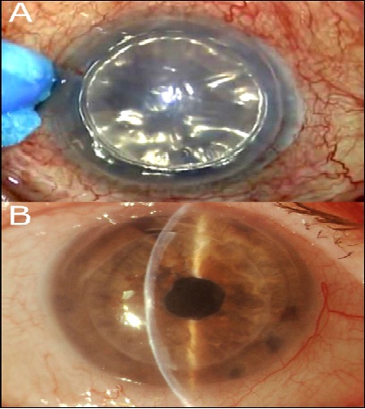
In group-A, the tissue adhesive (ReSure Adherent Ocular Bandage, Ocular Therapeutix, Inc., Bedford, MA) was mixed on the operating instrument stand, and applied in liquid form (painted) on the main incision borders with a special spear sponge following the absorption of any redundant surface fluid (Figure 1A). Within 3 to 5 seconds, the material was stable in soft form and any excess over the peripheral conjunctiva was removed with a dry spear sponge. The eye was observed for an additional 45 minutes for graft stability while the anterior chamber was filled with at least 75% with air and air tamponade, only the ‘matress’ 10-0 polypropylene sutures employing the CS 160-6 needle (Ethilon, Ethicon Inc, Somerville, NJ) were placed to secure the main incision. The sutures were removed typically at the one-month visit; in all cases the sutures had been removed well prior to the threemonth scheduled visit.

Data Collection and Analysis

All patients had been evaluated pre-operatively and at least one-year post-operatively for best-spectacle distance corrected visual acuity (CDVA) reported decimally, spherical and cylindrical error reported in diopters (D), Intraocular pressure (IOP) reported in mmHg, and endothelial cell density (ECC) reported in cells/mm2. In the case of pre-operative ECC, the data from the cornea bank certificates were used, while post-operatively, ECC was measured by specular microscopy (FA-3709, Konan Medical, Irvine, CA). Slit-lamp evaluation was also part of the complete ophthalmological evaluation performed. Figure 1B illustrates an example of slit lamp imaging from a sutureless DSEAK with the use of tissue adhesive, 1 week post-operatively.
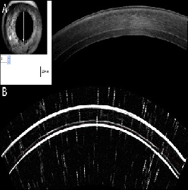
Spherical and cylindrical error corresponds to the refraction for which the CDVA was reported, and was based on phoropter manifest refraction examination. IOP values were not adjusted for corneal thickness changes. In addition, qualitative evaluation by means of corneal tomography (Pentacam, Oculus Optikgeräte GmbH, Wetzlar, Germany) and anterior-segment as well as retinal optical coherence tomography imaging (RtVue-100, Optovue, Fremont, CA) were part of the standard protocol performed during all visits [10]. Figure 2A presents an example of OCT imaging performed on DSAEK case two years postoperatively. Whenever possible, high-frequency scanning ultrasound imaging (Artemis ii+ superior, Artemis Medical Technologies Inc., Vancouver, British Columbia, Canada) was also employed for the imaging of the anterior segment. An example of Artemis cross-sectional imaging is illustrated in Figure 2B. Due to the nature of the condition, post-operative recovery was followed for at least once a year past their 12-month visit in all cases.

Results

The subject age in group-A (nA=15, 5 male and 10 female, 8 OD and 7 OS), at the time of the operation was 72.94±15.59 (36 to 90) years, while in group-B (nB=20, 6 male and 14 female, 9 OD and 11 OS) was 69.57±11.9 (50 to 88) years.
No case from group-A required any additional intra operative air insertion following the tissue adhesive application and no case required additional intra operative surgical manipulation for further graft centration. No case required post-operative rebubbling or had any graft rejection incidence.
In group-B, 18/20 cases required intra-operative supplemental air insertion (re-bubbling), and four of those, intra-operative graft repositioning. Additionally, five cases required post-operative re-bubbling (on average 5.7 months post-operatively), and one case lead to graft rejection, followed by penetrating keratoplasty 9 months from the initial DSAEK operation. The cases from group-B with post-operative rebubbling or graft failure were excluded from the subsequent data analysis, leaving thus 14 cases whose refractive data are reported in this study, of which 4 were female and 11 male; 7 belonged to right eyes (OD) and 8 to left eyes (OS). Average follow-up time for all cases was 10.5±8.5 (8 to 29) months.
In group-A preoperative CDVA was 0.14±0.17 (0.01 to 0.60) decimal, spherical error was -1.05±3.30 (-9.50 to +4.00) D, cylinder was -3.75±2.05 (-10.50 to -0.50) D, IOP was 17.25±5.60 (10 to 29) mmHg, and graft/donor ECC was 2,567±310 (1,790 to 2,935) cells/mm2.
In group-B pre-operative CDVA was 0.15±0.16 (0.01 to 0.50) decimal, spherical error was -0.94±3.60 (-12.50 to +5.00) D, cylinder was -3.24±2.44 (-10.00 to -0.25) D, IOP was 18.83±6.02 (8 to 32) mmHg. Graft (donor) ECC was 2,440±532 (1,635 to 2,850) cells/mm2.
Table 1 summarizes the pre-operative as well as the 3-month and 12-month refractive and corneal data pertaining the two groups of study. The two groups were matched on all aspects of the parameters involved in the study (age, gender laterality, eye laterality, visual acuity, sphero cylindrical error, IOP and graft ECC), as none of the paired-test p-values was less than 0.05.
The improvement in visual acuity within the same groups had a noted and statistically significant improvement at the 3-month interval (Δ = +0.22 and +0.10 for group -A and –B, respectively) as well as at the 12-month interval (Δ = +0.31 and +0.25). IOP was increased (Δ = +5.51 and +4.65 mmHg for group-A and –B, respectively) at 3-months as well as at 12-months (Δ = +5.77 and +5.22 mmHg). The increase in IOP between the two groups was rather similar, and not statistically significant. ECC change at 12-months of -16% was noted in group-A vs. -21% in group-B (statistically significant difference between the two groups, p =0.03).
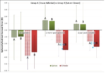
Figure 3 illustrates the sphero cylindrical error (sphere and cylinder) pre-operatively, as well as 3-months and at 12-months post-operatively. The spherical error indicated a hyperopic shift of +1.97 D in group-A and +1.79 D in group-B (not statistically significant difference between the two groups). Cylinder was improved (reduced), by 2.10 D in group-A, and by 0.81 D in group-B. The difference of cylinder improvement between the two groups was statistically significant (p =0.012).

Discussion

Management of surgical cornea incisions with nylon sutures may have been the acceptable standard in the past, but there are a number of complications associated with this technique [11]. Induction of astigmatism, potential to fluid ingress and egress from the ocular surface,[12] endophthalmitis, [13] and increase of surgical time and patient discomfort are some that may be listed. There is, therefore, interest in an adhesive to replace and/or supplement sutures in the repair of corneal wounds and improve corneal incision sealing [14]. The material must be biocompatible, self-dissolving, and safe (eg without affecting visual function).
Cyanoacrylate, a biocompatible material, has long been employed in surgical incisions [15]. The latest cyanoacrylate adhesive for medical use, FDA-approved in 2002, was n-butyl-2- cyanoacrylate (Indermil®, Vygon-Ecouen, Lansdale, PA). It has been used to close small skin wounds in pediatric patients, with successful results [16]. Applications in ocular surgery have also been reported [17,18].
Polyethylene glycol (PEG) polymers are also among the materials considered for corneal wound sealing [19]. Biocompatible PEG polymers form the same type of hydrogel used in contact lenses in wetting agent applications [20]. The liquid hydrogel compound is administered (painted) over the wound, and polymerizes fast (in approximately half a minute) into a soft form that adheres to the ocular surface, forming a bandage that leads to a watertight seal. Two such ocular tissue adhesive products are the ReSure (Ocular Therapeutix, Inc., Bedford, MA) [21] and the OcuSeal (BD Medical, Waltham, MA) [22].
Ocular tissue adhesives have been evaluated for their applicability in limbal-conjunctival wound after fornix-based trabeculectomy [23] and cataract surgery [24]. To the best of our knowledge there is no report in the peer-reviewed literature on the topic of ocular tissue adhesives in DSAEK.
The present work is to the best of our knowledge the first investigation that presents the clinical applicability of employment of ocular tissue adhesive in DSAEK. We evaluated comparatively two matched groups of study over a long followup period. The differences in regard to far fewer cases needing intra-operative re-bubbling have been compelling. Postoperatively, similar visual acuity and IOP changes are noted. CDVA was improved in both groups, while also IOP increase has been noted; the latter can be explained by the thicker cornea (as a result of the DSAEK procedure) and the known dependence of IOP readings on central corneal thickness [25].
We noted a statistically significant ECC loss in both groups (by -16% in group-A, and by -21% in group-B). These data are in accordance with published results in the literature. For example, ECC loss of -19% has been reported in DSAEK cases [26]. We note, however, that the ocular adhesive group-A appears to perform better in this aspect (p =0.045 between the two groups).
Regarding the refractive data, both groups indicate a significant hyperopic shift. The average increase in sphere was more than +2.50 D at the 3-month interval and near +2.00 D at the 12-month interval. This hyperopic shift has been modeled, and the average predicted hyperopic shift in the overall power of the eye was calculated to be +0.83 D [27]. It is explained by the fact that the graft is thinner centrally, as a result of the microkeratome pass creation procedure over the donor cornea, and the known increased corneal thickness peripherally. It appears that our technique introduces more, but predictable hyperopic shift. As a result the graft is thinner centrally and thicker peripherally, the ‘new’ posterior corneal surface has a smaller radius of curvature; our calculations based on application of the Gullstrand’s formula indicate that for approximately 1 mm change in the posterior curvature (e.g from 6.8 to 5.8 mm) there is a corresponding 1.00 D change in the posterior corneal refractive power (e.g. from -5.88 D to -6.90), resulting thus in 1.00 D of hyperopic shift.
A noted improvement in cylinder was noted in both groups, with the ocular adhesive group-B appears to perform better in this aspect (p =0.032). The sutureless group-A had 12-month improvement in cylinder by 2.1 D, while the suture group-B by 0.8 D. This may be explained by the reduced effect on cornea distortion by the ocular adhesive. Considering the nonsymmetrical nature of the suture placement in the traditional wound sealing, the noted improvement in surgically-induced astigmatism in the sutureless group-A may offer perhaps the clinical advantage over the traditionally applied technique.
Application of a material that seals wounds safely, effectively, and comfortably enable better suture placement and possibly improve corneal surgical outcomes. This procedure enables better suture placement and provides the benefit of the reduction of wound leak during suturing and possible graft slippage. This procedure may also be applied to DMEK cases. Further studies involving the clinical impact of the use of these new polymer corneal sealants may be warranted.

Conclusions

This novel tissue adhes ive may be a valuable adjunct in sutureless DSEAK clear cornea surgery in enhancing intra operative anterior chamber stability and possibly offering more secure wound closure.
Figures and Tables
#
Figure 1: A. Application of tissue adhesive in liquid form over the incision.
B. Slit-lamp picture of a sutureless DSEAK with the use of the tissue adhesive, 1 week post-operatively.

#

Figure 2: Cross-sectional imaging of a DSAEK case two years postoperatively: top, utilizing anterior-segment OCT, and bottom, utilizing high-frequency scanning ultrasound.

#

Figure 3: Comparative spherocylindrical error between the two groups, pre-operative, 3-months and 1-year post-operatively. *indicates statistically significant difference between the two groups.

For more Open Access Journals in Juniper Publishers please click on: https://juniperpublishers.com


Thursday, 1 August 2019

Multi-modality imaging of a Highly Irregular Cornea: comparison of findings to a Novel LED Multicolor-Spot Reflection Topography-Juniper Publishers


Juniper Publishers-Journal of Ophthalmology


Abstract

Background

This case report aims to evaluate safety, efficacy and feasibility of anterior corneal surface imaging by a novel multi-color Light-Emitting-Diode (LED) tear film-reflection topographer in comparison to several standard corneal imaging modalities in patient with severe ocular and corneal changes due to old acanthamoeba keratitis.

Case Description

An18-year old female patient, infected approximately two years ago with acanthamoeba in her right (OD) eye, was subjected to a multitude of anterior-segment and ocular imaging diagnostic devices. The imaging modalities included Placido topography, Scheimpflug tomography, Anterior-Segment Optical Coherence Tomography (AS-OCT), ocular stray-light measurement quantifier, and the Cassini, a novel multi-spot, multi-color LED reflection topographer.

Clinical Relevance

The ease of use and comparable results offered by the Cassini, in comparison to established Placido topography and Scheimpflug tomography, as well as the increased sensitivity that the novel topographer may offer new clinical diagnostic significance. Scheimpflug-imaging derived pachymetry has limitations in cases of partially opaque corneas.

Accurate

This case report, based on a novel corneal topography imaging in comparison to a variety of corneal imaging modalities, verifies the clinical applicability of the new technology in the imaging of disturbed and highly asymmetric cornea.

Keywords:

LED Cassini; multi-color LED topography; Acanthamoeba keratitis; Point-source topography; Pentacam HR; Placido topography; Scheimpflug topometry; Differential topography; Irregular corneal astigmatism; Stray light measurements; C-Quant; Anterior-Segment Optical Coherence Tomography

Introduction

The limitations of Placido-based topography have been identified in the past, particularly pertaining to imaging of radial, rather than only contour topographic changes [1]. Topography systems with color-coded LED reflection forward ray tracing have been proposed [2] as an alternative to Placido ring imaging [3]. The VU topographer (Vrije Universiteit Medical Center, Amsterdam, The Netherlands) [2] introduced a different approach, with a color-coded squares in a chess-pattern array projected on the cornea instead of the Placido rings [4]. The algorithm for the surface reconstruction employed data from the pattern crossing points to eliminate source-image mismatch, enabling one-to-one match. In principle, this color-coded topographer was found to be more efficient in reconstructing the non-rotationally symmetric anterior corneal surface [5].
The Cassini (i-Optics, The Hague, The Netherlands) topography system is a novel topographer employing multi spot (up to 700), multicolor (red, yellow and green) Light Emitting Diode (LED) tear film-reflection-imaging, following the steps of the VU topographer. The difference is instead of a limited number of color-coded squares, there are hundreds of LED spots on radial and contour arrangement imaged on the cornea. Image processing algorithm locates feature points in the reflection image and accounts for smearing and deformation in irregular corneas. The system has been recently introduced [6] and has received FDA approval for clinical use in corneal topography.
Due to the novelty, clinical validations of this novel topography system and its clinical implications have not yet been extensively investigated and reported. We have recently reported [7] clinical results of the Cassini in a case of forme fruste keratoconus (FFKC).
The scope of this manuscript is to examine the clinical feasibility of this newly-introduced corneal topographer in a case of a patient with old acanthamoeba infection. Very little has been documented in the last ten years in cases involving acanthamoeba keratitis – infected eyes, from the point-of-view by contemporary anterior segment imaging.

Case Report

Case Report

We present the case of an 18-year old female subject, diagnosed with acanthamoeba keratitis [8] on her right (OD) eye, affected at approximately two years ago. Informed consent was obtained from the subject at the time of the first clinical visit. This study adhered to the tenets of the Declaration of Helsinki and was approved by the Ethics Committee of our Institution.
The non-affected left eye (OS) had best-corrected distance visual acuity (CDVA) 20/20 and manifest refraction -7.00 S-0.25 C × 180°. The affected OD eye had CDVA 20/50 when wearing the manifest refraction of -3.50 S -2.00 C × 18°.
The Cassini (running on software version 1.2, updated September 2013) was employed to provide anterior cornea imaging. The system produces anterior elevation, tangential, axial curvature, and refractive power three-dimensional maps covering approximately a 7.5-mm diameter area. The report provides keratometry data (steep and flat K, simulated astigmatism), and computerized topographic and keratoconus indices. In addition, aberrations report is provided, according to the Zernike nomenclature. Four additional ocular imaging modalities were employed in this work. Placido imaging was provided by the Wave Light Allegro Topolyzer (Alcon Surgical, Ft Worth, and TX). The Topolyzer is a wide-cone corneal topographic Placido system with 22 concentric rings for the detection of up to 22,000 elevation points. Scheimpflug topometry was provided by the Wave Light Oculyzer II (Alcon Surgical, Ft Worth, TX), a Pentacam HR (high-resolution) camera providing corneal pachymetry and tomography imaging [9] was also included in this study.
The Fourier-domain Anterior-Segment Optical Coherence Tomography (AS-OCT) system RTVue-100 (Optovue Inc., Fremont, CA), running on analysis and report software version A6 (9,0,27) was employed in the study to provide cornea cross-section, and corneal and epithelial thickness 3-dimensional mapping [10]. Finally, the C-Quant (Oculus Optikgeräte GmbH, Wetzlar, Germany), a system that measures stray-light scattering in the eye [11] was employed to provide quantitative data of scatter in relation to the identified corneal opacity.
Figure 1 presents OCT high-resolution meridian scan of the affected eye, corneal and epithelial pachymetry 3-dimensional maps. We observed the deep stromal scar, appearing as opacity associated with the residual stromal scarring, located in the near inferior-nasal area. Local corneal thinnest point and epithelial hyperplasia were also indicated in the same affected area. Central corneal thickness and minimum corneal thickness, as measured by the OCT were 381 and 318 μm respectively. A thinner epithelium (56 μm) over the thinnest, most ectatic region of the cornea also noted; overall, however, the epithelium was thick, in the vicinity of 70 – 74 μm, associated with large topographic variability (standard deviation of pachymetry) computed from the Scheimpflug system. The imaging successfully depicts the corneal opacity. The thinnest point also located inferior-nasally; however we note that the Scheimpflug imaging-derived central and minimum corneal thickness were 438 μm and 167 μm, a distinct disparity when compared to the OCT measurements (381 μm and 318 μm, as noted above). As it can be noticed in Figure 2a, the posterior surface was poorly identified at the lesion, a possible consequence of the corneal opacity. This is a clinical example of a cornea with considerable opacity that alters the ‘normal’ densitometry interpolation of the Scheimpflug algorithm and can possibly be associated with the reported cornea thickness results by the device.
Figure 3 presents stray-light measurement report by the C-Quant. The measurement, based on the compensation method, [12] reported a stray-light logarithm value of 1.43±0.05. When comparing this result to the average value of 0.80±0.05, obtained from a large number of healthy subjects of the same age (reference light curve in the upper graph), [13] it is clear that the measurement suggests that the affected eye has a substantially ‘abnormal’ ocular scatter.
Figure 4 presents Placido ring imaging raw data and the corresponding refractive map. We observed the highly distorted ring pattern along the vertical direction. The refractive map indicated very large variation of the cornea refraction, ranging from a localized 33.5 D inferiorly, to more than 48 D superiorly, and a distinct ‘astigmatic’ – like pattern in the mid-superior cornea. Figure 5A presents Cassini imaging raw data imaging on the affected eye. Anterior surface axial curvature and refractive map (C) computerized with the above data are reported in Figures 5B and 5C, respectively.

Discussion

Acanthamoeba keratitis is a rare disease in which amoebae invade the cornea [14-16]. The disease [17] as well as it sequalae is a common cause of corneal blindness in the United States, often a result of improper disinfection of contact lens use [17-19]. If it is not diagnosed early and not treated aggressively, infection by trophozoites [20] may result in extensive ocular damage, and enucleating may be required [21]. Even when cured, the disease may result in permanent irregular corneal surface and extensive central opacities,[22] thus rendering accurate imaging of the cornea is challenging. Subsequent corneal scarring may have severe impact on the anterior surface regularity, tear film regularity, as well as stromal density, as indicated by the OCT imaging, renders Placido and Scheimpflug imaging in this case challenging, as expected.
In this work we examined the clinical feasibility of the Cassini, the newly-introduced topographer in a case of Acanthamoeba keratitis in comparison to the established methods of clinical imaging, namely Placido topography, Scheimpflug topometry, and AS-OCT.
Thinnest cornea pachymetry, measured with Scheimpflug topometry (167μm) was in large disparity with the OCT-measured value (318μm). We believe this is a disadvantage of the Scheimpflug imaging principle, which assumes clear cornea interpolation of the posterior surface.
When correlating the novel Cassini to the established Placido topographer, it is clear from this case that the increased sensitivity, which includes both radial and contour differences, based on differential spot-imaging, enabled proper imaging of this highly irregular cornea. Particularly considering the extent of the abnormality presented in this case, and the fact that significant distortion was also present at the corneal center, the Placido device was not successful in imaging this case. The Scheimpflug topometry, on the other hand, was successful in imaging the anterior surface, but not the posterior.

Discussion

The facility and comparable results offered by the novel Cassini topographer, in comparison to established Placido and Scheimpflug imaging, as well as the possibility of increased predictability that may be offered by the novel corneal imaging system in highly irregular corneas, may hold promise for wider clinical applications, such as screening of highly asymmetric and corneas with highly coinciding opacities.

For more Open Access Journals in Juniper Publishers please click on: https://juniperpublishers.com