Monday, 16 December 2019

Juniper Publishers PubMed Indexed Articles


Juniper Publishers PubMed Indexed Articles

Author Information : Louise EJ Thomson and C Noel Bairey Merz1*
PubMed ID : PMID: 30976755
ISSN: 2573-2609


Author Information : Mark R Burge1*
PubMed ID : PMID: 30828700
ISSN: 2573-2188
     
Author Information : Rupak Datta1
PubMed ID : PMID: 30465048
ISSN: 2575-8543

Author Information : Eliane Thaines Bodah1
PubMed ID :  PMID: 30345411
ISSN: 2471-6774

Author Information :
PubMed ID :  PMID: 30148262
ISSN: 2575-8594
Author Information : Ishtiaq Qadri1
PubMed ID : PMID: 29863159
ISSN: 2472-6400

Author Information : Qiuqian Wu
PubMed ID : PMID: 29911686
ISSN:  2577-2864

Author Information :  Beibei Guo1
PubMed ID : PMID: 29645007
ISSN: 2573-2633
Author Information : Kenneth Blum
PubMed ID : PMID: 29057394
ISSN: 2573-2447

Author Information :  Huang J
PubMed ID : PMID: 28825056
Journal Name :
ISSN:
Full Article Link :

Author Information :  Zaina P Qureshi
PubMed ID : PMID: 28845476
ISSN: 2473-554X

Author Information :   Laura Duque-Serrano and Jeff C Huffman
PubMed ID : PMID: 29276801
ISSN: 2476-1435

Author Information :  Lalit P Singh
PubMed ID :  PMID: 29376145
ISSN: 2473-5477

Author Information :  Hemachandra Reddy P
PubMed ID : PMID: 29399663
ISSN: 2476-1435
Author Information :   
PubMed ID :  PMID: 29450408
Journal Name :   
ISSN:
Full Article Link :

Author Information :   
PubMed ID :  PMID: 29399668
Journal Name :  Journal of Addiction
ISSN: 2573-2447


juniper publishers PubMed indexed articles


Tuesday, 10 December 2019

Case Report: Lifting A Femtoflap first time after 2 days of Flap Creation... Opening the Unopened?- Juniper Publishers


JUNIPER PUBLISHERS- JOJ Ophthalmology


Purpose

To report an uncommon refractive incident where a patient was scheduled for a routine Femto LASIK procedure using Intralasae IFS and excimer ablation with Wave Light® Allegretto Wave® Eye-Q machines, and an unfortunate technical error of the Allegretto machine delayed the ablation for two days after successful flap creation with the Intralasae with no flap raising at the incident.


Method

A 27 year old male myopic patient came to the clinic seeking refractive surgery. Full opthalmological examination was done for him, which was all normal. He had BCVA 20/200U corrected with refraction of-3.25/-2.50*250D and -2.00/-2.25*160S. Corneal assessement with Pentacam was performed revealing a prolate normal cornea, upon which the decision was for a Femtolasik procedure for refractive correction was decided, and for a wavefront optimized Excimer Laser ablation using the Allegretto machine [1].
The procedure started with a successful complete bilateral femto flap creation of 100um thickness using the INTRALASE IFS machine. Proceeding to the excimer ablation, an unexpected sudden technical error, due to administrative misreporting about it, occurred to the Allegretto machine before starting .After asking for technical support, and tremendous efforts from the maintenance team to fix the problem for three hours with no thread of hope to solve the problem that day ,the decision was taken to postpone the case for another day. Two days later, the problem was totally solved, and the machine was ready to use. The patient was informed and consented to continue the procedure. The patient was examined on the slit lamp before the procedure to determine the flap borders [2].
The procedure was performed in right eye first by entering the flap elevator from the edge of the flap obliquely to make an opening point, then circumfrentially opening the gutter 270 degrees. Afterwards, the superior hinge flap was totally elevated with the elevator smoothly, and wave front optimized excimer laser ablation was applied, returning the flap at the end. The procedure was successively repeated in the left eye as well. Topical antibiotics, steroids and lubricant eye drops were prescribed postoperatively [3].


Results

First day postoperative the patient was examined. The flaps were coapted and cornea clear both eyes, and mild subconjuntival hemorrhage. One month later postoperative UCVA was 20/20 OU.


Conclusion


Succesful femtoflap creation makes flap lifting possible even after two days of its creation. The potential space created is still there, and facilitated flap elevation and continuation of the procedure with no mentioned difficulty. The question that arises to be further evaluated is the maximum duration primary f lap elevation can be done after flap creation without facing wound healing issues.

For more Open Access Journals in Juniper Publishers please click on: https://juniperpublishers.com


Monday, 9 December 2019

A Comparison of Automated Refractions Using Plusoptix S04 Photoscreener, Nidek AR-20 Handheld Autorefractor and Nidek ARK-510A Auto Ref-Keratometer- Juniper Publishers


JUNIPER PUBLISHERS- JOJ Ophthalmology


Abstract

Purpose: To evaluate automated refraction measurements using the Plusopti X S04 photo screener (OP), Nidek AR-20 hand held auto refractor (NAR) and compare them to the more traditional Nidek ARK-510A (ARK).
Methods: 102 patients aged 3 to 81 years were included. All patients underwent on-cycloplegic automated refraction using the PlusoptiX S04 photo screener, Nidek AR-20 handheld auto refractor and the Nidek ARK-510A. This was followed by measuring Cyclo plegic auto refraction using the Nidek AR-20 handheld auto refractor and Nidek ARK-510A.
Results: Without Cyclo plegia, the mean sphere values were significantly different for both the OP (-0.41 D, SD±2.39D) versus ARK (-0.60D, SD±2.24) groups with a p=0. 008, and for NAR (-0.48, SD±2.01) Versus ARK groups with a p=0.029. With cycloplegia, there was no significant difference in the mean sphere values between the OP versus ARK groups as compared to the OP versus NAR and NAR versus ARK groups (p=0.049 and p=0.001 respectively).
Conclusion: Our study indicates similarities between the OP photo screener and the traditional table-mounted ARK results after cycle plegia. These results were not reflected when comparing the NAR handheld auto refractor to the ARK. Although efficient as screening tools, we recommend caution when using handheld instruments.
Keywords: Refractometer; Refractive errors; Myopia; Hyperopia; Astigmatism; Cycloplegia; Middle east; Lebanon


Introduction

Refraction is a clinical test used to determine the refractive state of the eye and to assess the individual's need for visual aid. Although hretinoscopy is still considered the gold standard for determining the refractive state of the eye, it is subject to interobserver variability [1,2]. Over the past few decades, new advancements have introduced the auto refractors which have gained a popular routine use among ophthalmologists [3,4]. Auto refractors are easy to use, time saving and relatively accurate in measuring the amount and type of refractive error. Today, many auto refractors are available in the market (Canon, Nikon, Topcon, Carl Zeiss Meditec, Nidek etc.). A disadvantage of most auto refractors is they need to be mounted on a table due to their relatively large size. As such, ametropia measurements may therefore be difficult in bed ridden patients, disabled persons and very young or handicapped children. Handheld auto refractors or photo screener scans are useful in such scenarios.
Plusopti XS04 (OP) (Plusoptix GmbH; Nuremberg, Germany), is a third generation vision screener designed to screen for refractive errors, anisocoria, and strabismus in children starting at the age of 6 months [5]. Screening is quick with both eyes screened simultaneously and can be performed by non-medical personnel, with a screening distance of 1 meter [5]. The device measures refraction, pupil size, and corneal reflexes. Results with abnormal limits are displayed as "refer". Referral criteria include anisometropia, astigmatism, hyperopia, myopia, anisocoria and corneal reflexes [5].
To date, multiple studies show that the OP vision screener is useful when used in screening for amblyopia oramblyogenic risk factors [6-10] small-angle strabismus [11] is considered effective when compared to cycloplegic pediatric ophthalmic evaluation [9,10] and can be used to screen patients with intellectual disability foramblyogenic risk factors with 95% sensitivity and 50% specificity [12].
In our manuscript, we explore the accuracy of both the OP vision screener when used as suggested in noncyclopleged patients, the Nidek AR-20 (NAR) (NidekCo. Ltd, Gammagori; Aichi, Japan) handheld auto refractor on non cyclopleged and cyclopleged patients and compare the results with those obtained before and after cycloplegia using a more traditional table- mounted NidekARK-510A (ARK) autorefractor/keratometer (NidekCo. Ltd, Gammagori, Aichi, Japan).


Materials and Methods

We prospectively evaluated 102 consecutive patients presenting to the outpatient clinic during August 2013, without any bias to their age, gender, diagnosis or previous medical history. Patients were excluded if they could not be refracted because of poor cooperation or any ocular pathology. After the initial history was taken, a trained ophthalmology fellow (H.B) and professional optometrist (J.H) used all 3 instruments to obtain non Cyclo plegic automated refraction. This was followed bycycloplegia using 1 drop of Mydriacyl (tropicamide 1%, Alcon, Puurs, Belgium) in each eye twice (10 minute interval). Twenty to 30 minutes later, Cyclo plegic auto refraction was obtained using the ARK, and NAR auto refractors.
Data entry and statistical analysis were performed using SPSS 22.0 for Windows (SPSS Inc, Chicago, IL, USA). Analysis variables included:gender, age (years), auto refraction (sphere, cylinder, axis) and spherical equivalent for both eyes. For each of the 3instruments, the data were divided into 4 main categories: sphere, cylinder, axis and spherical equivalent. Spherical equivalent (sphere power+(0.5 x cylinder power)) measured in diopters was calculated from the auto refraction measurements. For the ARK and NAR handheld auto refractor, data were further divided into non cycloplegic and Cyclo plegic auto refractiCases with missing variables were excluded from the analysis. Two types of statistical testing were used to determine differences or similarities between the OP, NAR handheld auto refractor, and the ARK. Analysis of variance (ANOVA) testing was performed to compare the 3 groups. A variable ratio (F) was calculated to determine overall statistical differences. Paired-samples t-tests were performed to compare means between 2 groups. Significance was defined as p values less than or equal to 0.05.


Results

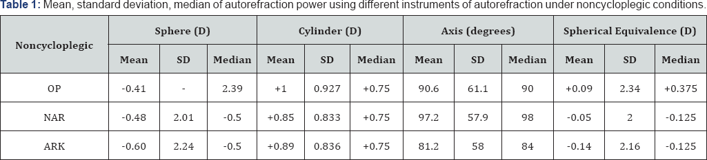
One hundred two patients (204 eyes) were initially recruited. Nineteen eyes (9.3%) could not be refracted by OP (one patient could not be refracted for both eyes by neither OP nor the NAR) and were excluded from the analysis. A total of 185 eyes completed the study on all 3 instruments and were included in the analysis (45.7% males, 54.3% females). The patients ranged in age from 3 years to 81 years (mean 41.02±19.7years, median 43 years).Using OP, the mean recorded sphere on 185 eyes was -0.41 diopters, mean cylinder was +1 diopters, and mean axis were measured at 90.6° (Table 1). The median spherical value was Plano (range -6.75 to +10). The mean spherical equivalent determination of the OP group was +0.09 diopters (range -6.125 to +10.5). A myopic spherical value was found in 47.6% of the eyes 42.7%were hyperopic and 9.7% were plano. Astigmatism was identified in 96.8%
OP:PlusoptiX S04 (Plusoptix GmbH; Nuremberg, Germany); NAR: Nidek AR-20 (Nidek Co.Ltd, Gammagori; Aichi, Japan); ARK: Nidek ARK-510A (NidekCo.Ltd, Gammagori; Aichi, Japan).

Non cycloplegic auto refraction results for nidek AR-20 handheld autorefractor and nidek ARK-510A

The mean sphere value recorded by the NAR for 185 eyes was -0.48 diopters, mean cylinder was+0.85 diopters, and mean axis were 97.16°. The median spherical value was -0.5 diopters (range -6.75 to+8.5). The mean spherical equivalence for the NAR group -0.05 diopters (range -6.125 to+9.12). A myopic spherical value was identified in 60.5%; 33.5% were hyperopic, and 5.9% were Plano. Astigmatism was identified in 91.9% (Table 3). The mean spherical value recorded using the ARK on 185 eyes was -0.604 diopters, mean cylinder was +0.89 diopters, and mean axis measurement was 81.2° (Table 1). The median spherical value was -0.5 diopters (range -6.25 to+10.5). The mean spherical equivalence measured in this group was -0.136 diopters (range -4.875 to+1G.875). A myopic spherical value was diagnosed in 61.1%, 3G.3% were hyperopic, and 8.6% were Plano. Astigmatism was identified in 95.7% of 185 eyes (Table 3).

Cycloplegicautorefraction results for nidek AR-20 handheld autorefractor and nidek ARK-510A

The mean sphere value recorded by the NAR for 185 eyes was -0.266 diopters, mean cylinder was +0.837 diopters, and mean axis were 91.3° (Table 2). The median spherical value was Plano (range -5.75 to+10.5). The mean spherical equivalence for the NAR group +0.145 diopters (range -4.75 to+10.875). A myopic spherical value was diagnosed in 48.6%, 41.6% were hyperopic, and 9.7% were Plano. Astigmatism was identified in 97.3%. The mean spherical value recorded using the ARK on 185eyes was -0.40 diopters, mean cylinder was +0.86 diopters, and mean axis measurement was 79.95° (Table 2). The median spherical value was -0.25 diopters (range -6.25 to+10.5). The mean spherical equivalence measured in this group was +0.05 diopters (range -4.875 to+10.875). A myopic spherical measurement was diagnosed in 52.4% of eyes, 35.1% were hyperopic, and 12.4% were Plano. Astigmatism was identified in 95.7% of eyes (Table 4).
OP: PlusoptiX S04 (Plusoptix GmbH; Nuremberg, Germany); NAR: Nidek AR-20 (NidekCo.Ltd, Gammagori; Aichi, Japan); ARK: Nidek ARK-510A (NidekCo.Ltd, Gammagori; Aichi, Japan).
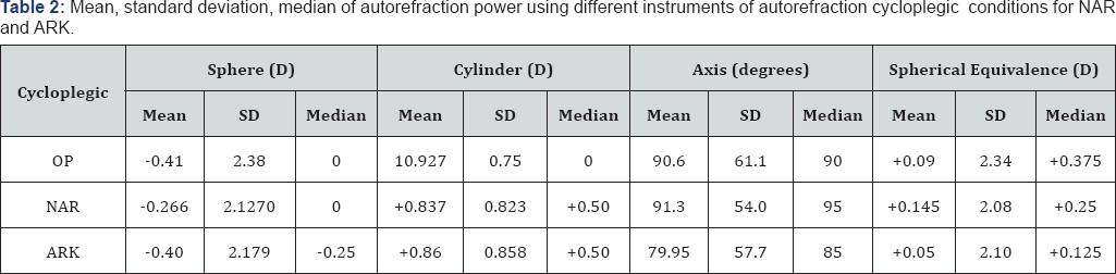
Optix: PlusoptiX S04 (Plusoptix GmbH; Nuremberg, Germany); C-NAR: cycloplegic-Nidek AR-20 (NidekCo.Ltd, Gammagori; Aichi, Japan); C-ARK: cycloplegic- Nidek ARK-510A (NidekCo.Ltd, Gammagori; Aichi, Japan).

Optix: PlusoptiX S04 (Plusoptix GmbH; Nuremberg, Germany); C-NAR: Cycloplegic- Nidek AR-20 (NidekCo.Ltd, Gammagori; Aichi, Japan); C-ARK: Cycloplegic- Nidek ARK-510A (NidekCo.Ltd, Gammagori; Aichi, Japan).
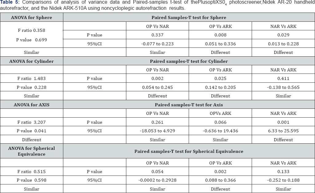
Except, of the axis value, ANOVA testing for auto refraction parameters of all instruments before cycle plegia revealed no significant difference among the 3 groups (Table 5). While paired-samples t-test comparisons of OP and NAR sphere values showed no statistical significance (p=0.34). Comparisons of sphere values for OPversus ARK and NAR versus ARKwere statistically significant (p=0.008 and 0.03 respectively) (Table 5) . Paired-samples t-test comparisons of cylinder values of NAR versus ARK showed similar results. However, the OP versus NAR and OP versus ARK groups showed a significant difference (p= 0.002 and p=0.025 respectively) (Table 5). The axis value analysis by ANOVA revealed a significant p-value (p= 0.034), indicating an overall significant difference between the 3 groups. Conversely, paired-samples t-test results were only significant between the NAR and ARK groups (p=0.001) (Table 5).
Calculated spherical equivalence measurements did correlate with our initial sphere measurements in the NAR versus ARK groups. Paired-samples t-test comparisons between the OP versus ARK demonstrated a difference with a p= 0.002. When comparing the OP versus NAR and the NAR versus ARK groups, no statistical difference was observed (Table 5).
ANOVA: analysis of variance test; CI: confidence interval; F: Variance ratio; OP: PlusoptiX S04 (Plusoptix GmbH; Nuremberg, Germany); NAR: Nidek AR-20 (NidekCo.Ltd, Gammagori; Aichi, Japan); ARK: Nidek ARK-510A (NidekCo.Ltd, Gammagori; Aichi, Japan).
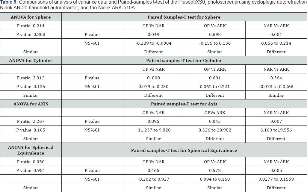
Data from the NAR and ARK instruments were reassessed using Cyclo pelagic autorefractions (C-NAR and C-ARK); ANOVA testing for all refraction parameters did not indicate any statistical differences (Table 6). Similarly, paired-samples t-test comparisons showed comparable results among the OP versus C-ARK groups. However, there was a statistical significance in OP versus C-NAR and C-NAR versus C-ARK groups (p= 0.049 and 0.001 respectively) (Table 6). Paired-samples t-test for the cylinder values of the OP versus the C - NAR group and OP versus C-ARK indicated statistical differences (p=0. 000 and p =0. 001) (Table 6).
For Axis values, paired-samples t-test comparisons of OPversus C-NAR values were statistically similar, whereas those of OP versus C-ARK showed a significant difference (p= 0.04). The C-NAR axis value compared to that of the C-ARK also showed a significant difference (p= 0.07) (Table 6). Finally, when analyzing spherical equivalence, paired-samples t-test comparisons showed similar results except for the C-NAR versus C-ARK group which showed a significant difference with a p=0. 005 (Table 6).


Discussion

Although considered as the gold standard for measuring refractive status, Cyclo pelagic refractions can be time consuming, cause patients discomfort and adds additional costs. As amblyopia is one of the leading causes of visual impairments in adults [13] developing accurate, easy to use, friendly autorefractors that do not require the use of cytoplegic agents could help in identifying significant refractive errors more rapidly and efficiently while avoiding the drawbacks of cycloplegia.As new instruments for refractive error measurements become available, it is of critical importance to evaluate and compare their validity to that of existing reliable technology. The OP photo screener has been marketed toward health care providers as a tool to assess refractive errors in young children without cycloplegia [5,9,10]. Although many studies showed that the OP vision screener is useful as a screening tool for amblyopia or amblyogenic risk factors [6-10] Dahlmann-Noor et al argue that the use of OP as a single screening test in children may miss a significant number of children with amblyopia or amblyogenic risk factor [14].
As in other studies, we found that both the OP vision screener and NAR handheld auto refractor were simple and user friendly, as well as quick in reporting refraction result without the need for direct contact or patient compliance. However, limitations that we encountered while using the OP included the need for a dimly lit room to obtain proper measurements, and its inability to obtain measurements in patients with a number of ophthalmologic conditions. Of the 19 eyes that failed autorefraction by the OP, 6 had a high refractive errors (more than -5), 4 had a history of cataract surgery, 2 had a history of cataract surgery and corneal rings for keratoconus (also failed autorefraction by NAR), 2 had a history of diabetic retinopathy, 1 had a history of a corneal ring for keratoconus, while the remaining 4 had no known ocular problems but were read as "pupil undetected". Disadvantages similar to those claimed by Rajavi et al which may have limited our use included: pupil size, fixation problems and posterior segment pathologies [15]. Limitations with the NAR were mostly a failure of patient cooperation resulting in no measurements being taken especially in children. A child's ability to hold the head still limited our ability to produce repeatable measurements.
The ARK table-mounted auto refractor readings were more frequently myopic compared to the OP and NAR (Table 3 & 4). This may be explained by the fact that it is well known that auto refractive instruments tend to overestimate myopia and underestimate hyperopia [10]. Except for the axis value prior to cycloplegia, when comparing all 3 instruments using ANOVA, our data indicated that there was no significant difference in auto refraction results before and after cyloplegia (Table 5 & 6). Comparing the OP spherical values with values obtained before cycloplegia using the NAR and the ARK indicated that there was a significant difference between the OP and the ARK groups as well as the NAR and ARK groups. This may lead us to conclude that the two handheld instruments may not be as reliable as the well established ARK for diagnosing refractive errors. In contrast, however, the results from both handheld instruments were similar (Table 5). Although the above mentioned results were reflected in the calculated spherical values for both the OP versus ARK and OP versus NAR groups, the NAR versus the ARK group showed similar results.
After cycloplegia on NAR and ARK, our spherical values indicated a difference between OP versus C-NAR and the C-NAR versus C-ARK groups (Tables 6). However, there was no difference between the OP and C-ARK values. Our study results are consistent with data from other studies that claim the OP to be a screening tool to assess refractive without the need cycloplegia [5,9,10]. While in both the cyclopleged and non cyclopleged groups, the OP recorded myopia less often than those recorded by the NAR and the ARK, astigmatism recordings were more comparable in all 3 groups (Table 3 & 4). The astigmatic (cylinder) readings were statistically similar between all 3 instruments by ANOVA however, paired samples-T test detected a difference between the OP and both the NAR and ARK groups before and after cycloplegia (Table 5 & 6). These results could mean that with regards to diagnosing astigmatism (measuring cylinder values) the OP may not be as reliable as the ARK as compared to the NAR.
Although handheld refractors are proving to be more practical, time efficient and provide a value as screening tools, we suggest that they should be used with caution when determining automated refractions especially in children. In the era of refractive surgery, it is important to assess the reliability and reproducibility of newer instruments when compared to existing technology and should not compromise the accuracy of the refractive state of a patient at the expense of practicality and time saving. We do not recommend the use of the hand held instruments for diagnostic purposes, with the exception of remote or inaccessible areas where portable instruments are essential and cycloplegia is difficult.

A positive aspect of our study setup is that our study population was non bias to age during recruitment. To our knowledge the Optixphotoscreener has only been extensively studied in screening younger population groups. This study may give us a better analysis of the use of the open adult patients. Several limitations are inherent in our study. In view of the time constraints and the difficulty of getting young children and elderly patients to cooperate and concentrate for long durations, repeated measurements on all 3 instruments were not documented. Furthermore, and for the same reason of time constraint, two separate individuals obtaining measurements on the handheld instruments may have introduced inter-observer bias. Finally, the same observer documenting measurements from both the handheld and table-mounted auto refractor was not masked to subjective refraction readings, introducing possible observer bias.

For more Open Access Journals in Juniper Publishers please click on: https://juniperpublishers.com


Wednesday, 4 December 2019

Inferior Rectus Myositis after an Uneventful Repair of Blowout Fracture- Juniper Publishers


JUNIPER PUBLISHERS- JOJ Ophthalmology


Summary

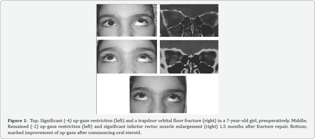
PA 7-year-old girl who had undergone a successful orbital floor blow-out fracture repair continued to have up-gaze and down gaze restriction post-operatively. She was observed for 6 weeks when an orbital imaging showed inferior rectus enlargement. Enlarged IR muscle associated with pain and restriction of up and down gaze led to a provisional diagnosed of myositis to which oral steroid was commenced and resulted in full recovery of up-gaze in 2 months.
Keywords: Blowout fracture; Inferior rectus; Myositis; Orbital fracture


Background

Early repair of a white-eyed blowout orbital floor fracture has been recommended in order to avoid permanent ischemic damage to the entrapped inferior rectus (IR) muscl0e [1]. However, even after a proper surgical repair of orbital floor fracture, up-gaze restriction may persist predominantly in children [1,2]. This may result from necrosis of muscle [2], IR muscle fibrosis [1], residual entrapped IR muscle sheath or peri- muscular tissue [3], and preoperative severe injury and swelling of the IR muscle [4].
This is, to the best of our knowledge, the first report of IR myositis after an uneventful white eyed blow out fracture repair. Iran University Eye Research Center ethic approval and patient’s parents’ consent were obtained.


Case Presentation

A 7 year-old girl was referred 2 days after a facial trauma. On examination, there was no or little eyelid swelling but marked painful restriction of up-gaze (-4) and moderate restriction of down gaze (-2) on the right eye. There was also hypoesthesia on the right cheek area. Vision (20/20 on both eyes) and ocular examinations were otherwise normal. Coronal Computed tomography (CT) scan showed a trapdoor floor fracture with inferior rectus entrapment which was extended from mid-globe to mid-orbit sections (Figure 1). Forced duction test was performed just before starting the operation 4 days after trauma which was strongly positive. Using trans-conjunctival approach, the entrapped muscle and peri-orbital tissue were released and the fracture site was covered by a properly fashioned porous polyethylene (Medpor, USA) sheet (0.85mm). The IR muscle was found to be discolored but viable. Forced duction test showed no restriction at the end of operation. Postoperatively, she was instructed to take oral systemic antibiotic (Cephalexin 250 mg, 4 times daily for 5 days), topical antibiotic and topical steroid (4 times daily for a week). A day after operation, there was less limitation of motion in down gaze (-1) and up-gaze (-2). However, she continued to have the same degrees of restriction associated with mild pain throughout post-operative follow ups at 1, 4, and 6 weeks. Since the repair was uneventful and postoperative follow up did not show improvement of muscle restriction, an orbital CT scan was requested. It showed no residual entrapment but significantly enlarged IR muscle. Increased thickness of IR muscle associated with pain on movement led to a provisional diagnosis of post-operative IR myositis and or persistent intra-sheath hematoma. Therefore, oral prednisolone (1mg/kg/day) was commenced and tapered within 6 weeks time. Up- and down restriction improved a week after its commencement. Completely normal examination was observed 1 year afterward with no recurrence of restriction and pain.


Conclusion


Possible explanations for persistent up-gaze restriction after a successful blowout fracture repair are: residual entrapment of any part of orbital soft-tissue [3] especially in the presence of posterior floor fracture [5], strangulation and necrosis of IR muscle [2], IR muscle fibrosis [1] and preoperative severe injury and swelling of the IR muscle [4]. Younger patients seem to recover longer than adults and a satisfactory force duction test at the end of the operation does not guarantee free voluntary movement of the involved eye [1,2]. In the presenting case, up- and down gaze restriction mildly improved a day after operation, but remained the same up to 1.5 months then after. In order to assess the possibility of residual IR entrapment, an orbital CT scan was requested which showed no entrapment but significantly enlarged IR muscle. In the context of up and down gaze restriction, pain, and enlarged IR muscle an orbital inflammatory myositis and or persistent IR hematoma were the provisional diagnoses. A rapid response to oral steroid was in favor of post-operative IR myositis. Post strabismus surgery extraocular myositis has been previously reported [6]. Whereas, to the best of our knowledge, there has been no any report of IR myositis after orbital fracture repair. Preoperative IR muscle swelling was reported to be a useful indicator of longer recovery after orbital floor fracture repair [4]. On reviewing the case, there was no significant IR enlargement preoperatively. Since this patient was treated with systemic steroid, it is not possible to comment on whether post-operative myositis is self limited if left untreated. The myositis did not recur one year after steroid treatment which may imply that it was due to either trauma or intra-operative manipulation. In conclusion, post-operative IR myositis should be considered in cases with residual up-gaze limitation after an uneventful orbital floor fracture repair.

For more Open Access Journals in Juniper Publishers please click on: https://juniperpublishers.com


Tuesday, 3 December 2019

Long Term Evaluation of AGV Implantation in Post PK Glaucoma- Juniper Publishers


JUNIPER PUBLISHERS- JOJ Ophthalmology


Introduction

Glaucoma after penetrating keratoplasty (PK) is an important cause of graft failure besides loss of vision due to glaucomatous optic atrophy. Causes of post PK glaucoma are multifactorial [1] such as increased post-op inflammation due to synechiolysis/pupillopasty, combined with cataract extraction and or vitrectomy, retained viscoelastic, tight suturing causing tissue compression, large graft size, long suture bites, peripheral anterior synechiae and prolonged use of steroids. Management includes medical therapy and in refractory cases surgical interve. The present study aims to describe long-term outcomes of Ahmed Glaucoma Valve (AGV) implantation in Post Keratoplasty glaucoma performed at a Tertiary Eye Centre.
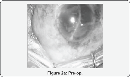
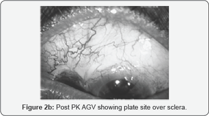
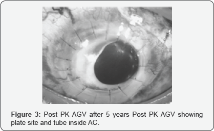
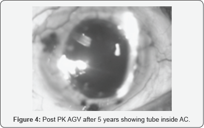
All subjects with Post PK Glaucoma who had undergone AGV implantation with a follow-up of minimum 5 years were evaluated. Preoperative antiglaucoma medication continued postoperatively and titrated according to the need. Surgical steps followed same in all except plate was covered either by conjunctiva or by scleral patch. Before starting the surgery, priming of the tube was done (Figure 1). After entering AC the tube was inserted and cut at desired length and the plate was covered. Clinical photographs were taken at different time period of time (Figure 2-4).
Of 32 eyes, the IOP decreased from a mean preoperative value of 32.8±3.6mmHg to 20.0±8.6mmHg at last visit. The number of medications decreased from 3.2±0.8 preoperatively to 2.2±1.2 postoperatively. Cumulative qualified success was achieved in 72% of eyes. The common complication was focal endothelial corneal decompensation at the site of tube-cornea touch.

Discussion

Management of Post PK glaucoma includes topical antiglaucoma medications and surgical options [1-3]. Medical management alone is not sufficient to control the Pressure besides its own hazards such as corneal decompensation, dry eye, CME in aphakic, psedophakic eyes, and recurrence of herpetic infection and depression. A result of Trabeculectomy in such eyes often fails. The valve helps to minimize postoperative hypotony and its associated complications.


Conclusion


AGV implantation with adjunctive topical anti-glaucoma drops controlled IOP in approximately 72% of eyes with Post PK glaucoma.

For more Open Access Journals in Juniper Publishers please click on: https://juniperpublishers.com


Monday, 2 December 2019

Recovery of Outer Retinal Laminations on Optical Coherence Tomography After Treatment of Cancer Associated Retinopathy- Juniper Publishers


JUNIPER PUBLISHERS- JOJ Ophthalmology


Abstract

Purpose: To report novel optical coherence tomography findings in a case of anti-α-enolasecancer associated retinopathy.
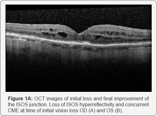
Observations: An elderly female presented with bilateral decreased vision and a recent diagnosis of ovarian carcinoma. Optical coherence tomography demonstrated bilateral loss of outer retinal structures and macular edema. Serum testing found antibodies against α-enolase and 82-84kDa proteins. Outer retinal structures showed recovery, macular edema resolved and repeat anti-retinalantibody testing became negative following cancer therapy and topical difluprednate treatment.
Conclusion and importance: Cancer associated retinopathy is a paraneoplastic disease that results in damage to retinal structures through an autoimmune response. The damage is generally considered to be irreversible however, in rare cases, such as observed here, retinal structures may demonstrate recovery after treatment.
Keywords: Cancer associated retinopathy; Optical coherence tomography


Introduction

Cancer associated retinopathy (CAR) is a paraneoplastic disease in which retinal degeneration occurs as an immune response to cancer antigens sharing homology with endogenous retinal proteins [1]. Past studies have found various retinal proteins to be antigenic, which include recoverin, α-enolase, arrestin, and transducin [https://www.ncbi.nlm.nih.gov/pubmed/245316532]. The inhibition of enolase, a glycolytic enzyme, results in metabolic disruption of retinal cells and the induction of apoptosis [3]. Anti-α-enolase autoantibodies are capable of accessing tissue and targeting ganglion cells, Muller cells, and photoreceptors. It is believed that death of retinal cells is an irreversible process. We report a patient with gynecologicalCAR who experienced objective improvement in photoreceptor architecture following treatment of her underlying malignancy.


Case Report

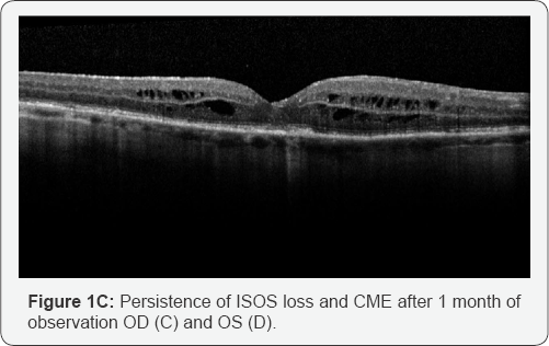
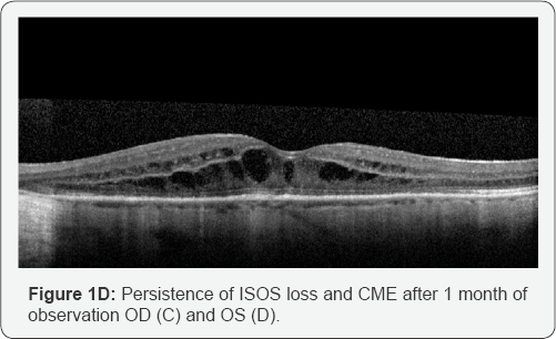
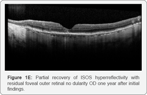
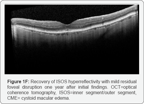
An 80 year Hispanic female with a history of chronic, bilateral Vogt-Koyanagi-Harada associated uveitis presented to the Casey Eye Institute Uveitis Clinic for a routine follow up visit. At that time, she reported a newdiagnosis of ovarian carcinoma and had started her first chemotherapy session consisting of carboplatin and paclitaxel. Due to severe aortic stenosis, the patient was not a candidate for surgical intervention. Her vision was 20/30 bilaterally without evidence of active uveitis. Four months later she returned with a bilateral decrease in vision to 20/50. The patient underwent imaging with macular volume scans centered on the fovea (Heidelberg Spectralis spectral domain ocular coherence tomography (OCT) with eye tracking software, Heidelberg, Germany) that demonstrated a disrupted inner segment/outer segment junction (ISOS) and cystoid macular edema (CME) bilaterally (Figure 1A & 1B). Clinical and OCT findings were suspicious for CAR and anti-retinal antibody testing was pursued. The patient declined local or systemic immunosuppression specifically for her ophthalmic diseaseand continued to undergo planned chemotherapy. One month later, her vision had dropped to 20/50OD and 20/100OS. Repeat OCT mapped to the original images continued to demonstrate loss of the ISOS junction and CME in both eyes (Figure 1C & 1D). Serum tested for the presence of anti-retinal autoantibodies showed antibodies against α-enolase and 82-84kDa proteins. Immunohistochemistry of the patient’s serum showed positive staining of the photoreceptor cell layer in human retina. The patient continued to decline periocular injection or systemic immunosuppression and was prescribed difluprednate drops three times daily. Two months later, there was partial return to normal reflectivity of the IS/OS junction on OCT and the CME had improved. Six months later, there was resolved CME on OCT and the IS/OS reflectivity returned to near normal in the subfoveal region. At this time, the vision was 20/40 bilaterally. The patient was instructed to stop difluprednate drops. Over the following six months, the patient’s visual acuity stabilized at 20/60OD and 20/50OS. There was no recurrence of CME and the OCT showed normalized IS/OS reflectivity except in the fovea where there was a stable elevated outer retinal lesion OD and near-normalized ISOS reflectivity in the left macula except in the fovea (Figure 1E & 1F). Serum was negative for anti-retinal autoantibodies on repeat testing.


Discussion

Anti-retinal autoantibodies can be detected in both retinopathy patients and healthy individuals. Individuals with gynecological CAR have a higher proportion of seropositivity than normal individuals [4,5]. Our patient became symptomatic after diagnosis of ovarian cancerand initiation of chemotherapy treatment. Autoantibodies may be present before the diagnosis of cancer, but it is not until they breach the blood retinal barrier that symptoms become evident [4]. Although the presence of anti-retinal autoantibodies can occur in normal patients, high antibody titers are a better indicator of retinopathy [5]. Anti-enolase autoantibodies affect the catalytic activity of the enzyme thus depleting glycolytic ATP, increasing levels of intracellular calcium which then induces mitochondrial-mediated apoptosis by the activation of its key elements [3].
The loss of outer retinal structures and retinal atrophy observed in autoimmune retinopathy arefrequently thought to be irreversible [5,6]. Partial recovery of SD-OCT outer retinal changes in a patient with CAR after treatment with rituximab has been reported, which suggests that therapy targeting B cells and consequently reducing production of anti-retinal antibodies may be beneficial [7]. Our patient showed improvement of the reflectivity of the photoreceptor IS/OS junctiondespite only local therapy with difluprednate, which was started to treat CME and reduce local inflammatory damage, but unlikely to significantly affect autoantibody production. We hypothesize that the prompt initiation of chemotherapy may have contributed to the patient’s improvement by possibly decreasing tumor expression of enolase and diminishing the production of anti-retinal autoantibodies or that chemotherapeutic treatment non-specifically immunosuppressed antibody production.The recovery of outer retinal structures in the present case corresponded to anti-retinal antibodies no longer being detected in the patient’s serum, supporting a pathologic role for autoantibodies in our patient.Treatments that may limit the production of anti-retinal antibodies such as rituximab should continue to be studied for efficacy in CAR.
The history of prior uveitis is a potential confounder to our findings; however, the patient did not demonstrate active inflammation during this follow up period. The presence of CME may also confound the ability to image the outer retina; however, we observed patchiness of the ISOS junction outside of regions of retinal edema indicating the outer retinal changes were not a sequel of CME alone. CME is not a common manifestation of CAR, more frequently observed in non-paraneoplastic autoimmune retinopathy [8], but previous case reports of CAR-related CME suggest it is responsive to steroids, as was observed in our patient [9]. Unfortunately, the patient declined additional objective testing (visual fields, electroretinography), which would have allowed further clinical correlation.
We report a patient with CAR who experienced objective improvement in photoreceptor architecture following treatment of her underlying malignancy, a novel observation previously reported only following rituximab therapy. We also note the successful treatment of CAR-associated CME with topical difluprednate, suggesting an alternative therapy to previously reported treatments with periocular or intravitreal steroids.


Conclusion

Damage to retinal structures from CAR can be objectively captured by OCT and may demonstrate recovery after treatment in rare cases.


Patient Consent

Consent to publish the case report was not obtained. This report does not contain any personal information that could lead to the identification of the patient.


Funding

P30 EY010572 from the National Institutes of Health (Bethesda, MD), unrestricted departmental funding to the Casey Eye Institute from Research to Prevent Blindness (New York, NY).


Conflict of Interest


JTR, PL, GA. The following authors have no financial disclosures FJI, LJK, SSS, MS, KB. All authors attest that they meet the current ICMJE criteria for Authorship.

For more Open Access Journals in Juniper Publishers please click on: https://juniperpublishers.com