JUNIPER
PUBLISHERS- JOJ Ophthalmology
Summary
PA 7-year-old girl who had undergone a successful
orbital floor blow-out fracture repair continued to have up-gaze and
down gaze restriction post-operatively. She was observed for 6 weeks
when an orbital imaging showed inferior rectus enlargement. Enlarged IR
muscle associated with pain and restriction of up and down gaze led to a
provisional diagnosed of myositis to which oral steroid was commenced
and resulted in full recovery of up-gaze in 2 months.
Keywords: Blowout fracture; Inferior rectus; Myositis; Orbital fractureBackground
Early repair of a white-eyed blowout orbital floor
fracture has been recommended in order to avoid permanent ischemic
damage to the entrapped inferior rectus (IR) muscl0e [1].
However, even after a proper surgical repair of orbital floor fracture,
up-gaze restriction may persist predominantly in children [1,2]. This may result from necrosis of muscle [2], IR muscle fibrosis [1], residual entrapped IR muscle sheath or peri- muscular tissue [3], and preoperative severe injury and swelling of the IR muscle [4].
This is, to the best of our knowledge, the first
report of IR myositis after an uneventful white eyed blow out fracture
repair. Iran University Eye Research Center ethic approval and patient’s
parents’ consent were obtained.
Case Presentation

A 7 year-old girl was referred 2 days after a facial
trauma. On examination, there was no or little eyelid swelling but
marked painful restriction of up-gaze (-4) and moderate restriction of
down gaze (-2) on the right eye. There was also hypoesthesia on the
right cheek area. Vision (20/20 on both eyes) and ocular examinations
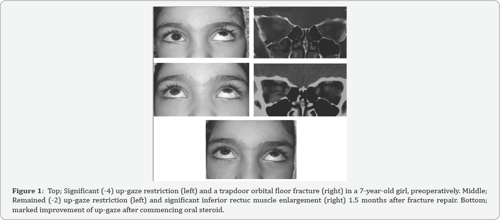
were otherwise normal. Coronal Computed tomography (CT) scan showed a
trapdoor floor fracture with inferior rectus entrapment which was
extended from mid-globe to mid-orbit sections (Figure 1).
Forced duction test was performed just before starting the operation 4
days after trauma which was strongly positive. Using trans-conjunctival
approach, the entrapped muscle and peri-orbital tissue were released and
the fracture site was covered by a properly fashioned porous
polyethylene (Medpor, USA) sheet (0.85mm). The IR muscle was found to be
discolored but viable. Forced duction test showed no restriction at the
end of operation. Postoperatively, she was instructed to take oral
systemic antibiotic (Cephalexin 250 mg, 4 times daily for 5 days),
topical antibiotic and topical steroid (4 times daily for a week). A day
after operation, there was less limitation of motion in down gaze (-1)
and up-gaze (-2). However, she continued to have the same degrees of
restriction associated with mild pain throughout post-operative follow
ups at 1, 4, and 6 weeks. Since the repair was uneventful and
postoperative follow up did not show improvement of muscle restriction,
an orbital CT scan was requested. It showed no residual entrapment but
significantly enlarged IR muscle. Increased thickness of IR muscle
associated with pain on movement led to a provisional diagnosis of
post-operative IR myositis and or persistent intra-sheath hematoma.
Therefore, oral prednisolone (1mg/kg/day) was commenced and tapered
within 6 weeks time. Up- and down restriction improved a week after its
commencement. Completely normal examination was observed 1 year
afterward with no recurrence of restriction and pain.
Conclusion
Possible explanations for persistent up-gaze
restriction after a successful blowout fracture repair are: residual
entrapment of any part of orbital soft-tissue [3] especially in the presence of posterior floor fracture [5], strangulation and necrosis of IR muscle [2], IR muscle fibrosis [1] and preoperative severe injury and swelling of the IR muscle [4].
Younger patients seem to recover longer than adults and a satisfactory
force duction test at the end of the operation does not guarantee free
voluntary movement of the involved eye [1,2].
In the presenting case, up- and down gaze restriction mildly improved a
day after operation, but remained the same up to 1.5 months then after.
In order to assess the possibility of residual IR entrapment, an
orbital CT scan was requested which showed no entrapment but
significantly enlarged IR muscle. In the context of up and down gaze
restriction, pain, and enlarged IR muscle an orbital inflammatory
myositis and or persistent IR hematoma were the provisional diagnoses. A
rapid response to oral steroid was in favor of post-operative IR
myositis. Post strabismus surgery extraocular myositis has been
previously reported [6].
Whereas, to the best of our knowledge, there has been no any report of
IR myositis after orbital fracture repair. Preoperative IR muscle
swelling was reported to be a useful indicator of longer recovery after
orbital floor fracture repair [4].
On reviewing the case, there was no significant IR enlargement
preoperatively. Since this patient was treated with systemic steroid, it
is not possible to comment on whether post-operative myositis is self
limited if left untreated. The myositis did not recur one year after
steroid treatment which may imply that it was due to either trauma or
intra-operative manipulation. In conclusion, post-operative IR myositis
should be considered in cases with residual up-gaze limitation after an
uneventful orbital floor fracture repair.
For more articles in JOJ Ophthalmology (JOJO) please click on: https://juniperpublishers.com/jojo/index.php
No comments:
Post a Comment