Friday, 7 February 2020

Efficacy of Amniotic Membrane Transplantation in Refractory Infective Keratitis Leading to Stromal Thinning, Descematocele and Perforations- Juniper Publishers


Juniper Publishers- JOJ Ophthalmology

Introduction

Diseases affecting the cornea are a major cause of blindness all over the world, second only to cataract in overall importance [1]. One of the commonest corneal causes is Infectious Keratitis. The prevalence of blindness directly resulting from complications of Infective Keratitis is estimated to be 5% [2]. Cases refractory to the medical therapy requires urgent surgical intervention to retrieve the vision and most importantly to salvage eye. Available surgical management in refractory keratitis cases include tissue adhesives, Bandage Contact Lenses (BCL), penetrating or lamellar keratoplasty [3] patch grafts, or conjunctival flaps. Unfortunately, these therapies are associated with a considerable number of complications and address only the tectonic problem, without solving the ongoing infection and inflammation. BCL and conjunctival flaps being a temporary measure does not provide with new collagen to improve corneal thickness and stabilize the cornea. For such situations Penetrating Keratoplasty (PK), Lamellar Keratoplasty (LK) or patch grafts was the only option and is still being used widely. PK and patch grafts performed to seal a corneal perforation may be complicated with synechiae, glaucoma, uveitis, and graft failure in the setting of an inflamed or infected eye [4]. Recurrence of infection in corneal grafts is also challenged. LK being difficult to perform may result in a double chamber between the donor and recipient cornea in some cases. Tissue adhesives may dislodge and are used as a temporary measure, obviating the need for a PK within a few days [5,6].
Preserved human amnion has been successfully used as a biological bandage, promoter of epithelialization, inhibitor of inflammation and angiogenesis, as well as a carrier for ex vivo cultured limbal stem cells [7]. Amniotic Membrane Transplant (AMT) offers the advantage of avoiding potential allograft rejection. Even if corneal transplantation is needed, the success rate is improved if performed on an eye that underwent AMT reducing inflammation [8,9]. Amniotic Membrane (AM) integrates in cornea and thus can be used as a treatment for corneal perforation by restoring corneal stromal thickness so that emergency PK can be avoided, as suitable donor corneal button availability is difficult in every place. Therefore, an alternative management for various stages of infectious keratitis including deep refractory stromal ulcers, descematocele and corneal perforations is reconstruction of the surface with AMT adjuvant with appropriate antimicrobials and supportive medications. In this prospective study AMT in various gravities refractory infective keratitis has been attempted to understand the efficiencies and limitations associated with it.


Methods

A prospective, interventional study was done on 150 eyes of 150 patients. All patients with refractory (unresponsive to conventional treatments significantly for more than 2 weeks) infective keratitis, advanced infectious keratitis with descematocele and corneal perforation requiring urgent concealment to salvage the eye, were treated with single or multi layered AMT. Patients with non-infective ulcers and perforations were excluded from the study. Corneal ulcer was graded 1-5 according to the depth of corneal involvement on slit lamp biomicroscopy (Table 1). Microbial investigations (staining for bacteria and fungus with culture-sensitivity) were done and antimicrobials started accordingly. B-scan ultra sonography was done in hazy media to rule out involvement of posterior segment. Any systemic (diabetes) or ocular (dacryocystitis) conditions hindering the healing of ulcer or triggering the infection were investigated and managed.
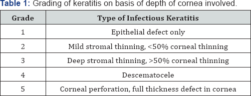
On basis of slit lamp examination at the site of most impact.


Technique

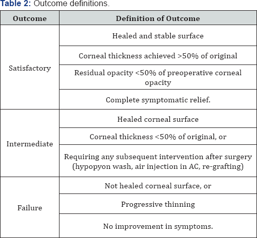
Surgery was performed preferably under sub conjunctival or peribulbar anesthesia. In children or uncooperative patients general anesthesia was used. Debridement of the necrotic tissue was done from and around the ulcer bed. Care was taken to remove the pseudo cornea over the perforation at the end of debridement to prevent leaking of aqueous and thus allowing proper keratectomy. Single layer preserved AM was used in cases of deep stromal ulcer. AM with epithelial side up was spread over the ulcer and trimmed to fit the ulcer. It was secured with continuous or interrupted 10-0 monofilament nylon suture. Descematocele and small corneal perforations up to 4mm were treated with multilayer AMT owing to deep corneal involvement. A sheet of AM, folded over it-self with epithelial side out, filled the ulcer crater and anchored to the healthy ulcer margin with interrupted 10-0 nylon suture. It was covered with a single sheet of AM similarly as in cases of deep stromal ulcers. In large corneal perforations of 4-6mm with extensive surrounding stromal necrosis, margins were not sturdy to hold the suture and there was a risk of cutting-off a corneal bite. In such cases single layer was sutured at limbus to at least provide tectonic support to the eyeball and delaying the need for PK. Side port or paracentasis was made in cases hypopyon and corneal perforation to reform the anterior chamber with air and reposit the prolapsed iris with help of spatula. Anterior synechiae if present were broken to prevent formation of adherent leucoma and thereby secondary glaucoma. Hypopyon if present was washed through the side port and intracameral antibiotic or antifungal was also injected according to sensitivity. At the end a BCL was placed over the cornea to prevent irritation from corneal sutures and maintaining AM in place. Antimicrobial, cycloplegics, ocular hypotensive and lubricating drops were continued along with systemic supportive therapy. Frequent follow-ups were done weekly for 1 month, biweekly till 3 months and monthly till 6 months. Efficacy was monitored on basis of improvement in symptoms and visual acuity, healing of the ulcer by re-epithelization and formation of anterior chamber, achievement of corneal transparency and corneal thickness. Accordingly patient's outcome was described as satisfactory, intermediate and failure (Table 2).

Observation and Results

Keratitis was classified (Table 1) according to the depth of the cornea involved into 5 grades. Grades 1 and 2 responded well with medical management, therefore did not require AMT. Grades 3-5 with deeper corneal penetration of infection did not heal merely with medical management, there was an apprehension of corneal thinning and progression of infection, which required AMT. Of the 150 patients who underwent AMT, 55 (36.67%) were deep stromal ulcers, 25 (16.66%) were descematocele and maximum 70 (46.67%) patients were of corneal perforation ranging from 1-6mm. There was no age group or gender preponderance. Symptoms of redness, pain, watering and foreign body sensation (FBS) were collectively present in all the cases. Lid oedema and photophobia were also present in majority of the cases (70.6% and 90% respectively).Presence of discharge was seen in moderately less cases (30%). ranging between 1-2mm and 10 cases (20%) had hypopyon of Hypopyon was present in total 50 (33.3 %) cases where 10 cases >2mm (Table 3).
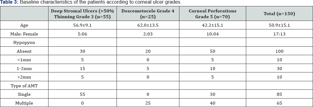
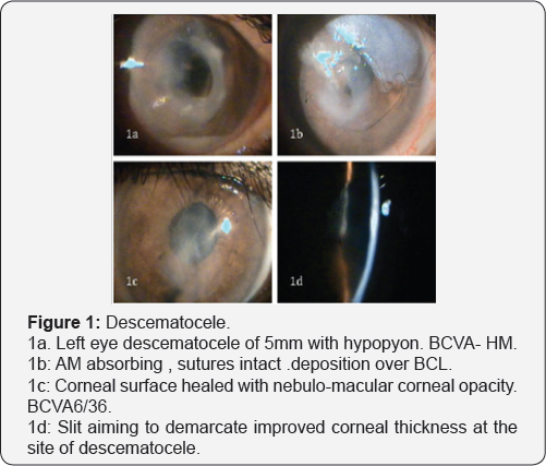
Single layer AMT was done in total 85 cases, all 55 cases of deep stromal ulcer and 30 cases of corneal perforation >4mm with extensive necrosis to provide tectonic support to maintain integrity to eyeball. Roofing with multilayer technique was done in 65 cases, all 25 cases of descematocele and 40 cases of corneal perforation >4mm in largest dimension where neighboring corneal tissue was healthy to hold the corneal sutures (Table 3). Patients were observed in repeated postoperative days. Rapid descent of symptoms was observed after the AMT. There was drastic improvement in pain, lid oedema, FBS and discharge in the first week. Symptoms were barely present in few cases by 1 month, which totally recovered by 3 months in all the cases (Figure 1).
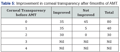
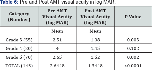
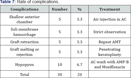
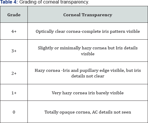
Corneal transparency graded from 0 (leucomatous opacity) to +4 (clear cornea, with no haze) was measured objectively at the site of most impact on slit lamp (Table 4). Improvement was seen in 105 of 150 cases and was statistically significant (p=0.016). However none of the cases improved to +4 transparency that is totally clear cornea (Table 5). Visual acuity was recorded before and after 6 months of treatment in 145 of 150 cases as 5 cases of fungal ulcer failed to heal with AMT (Table 6). Improvement in BCVA when taken collaborate, was extremely significant (p >0.0001). Mild to moderate complications were faced during the entire course of treatment. They were shallow anterior chamber in 5 cases in perforation which was tackled with air injection in anterior chamber and breaking anterior synechiae. Hemorrhage beneath AM in five cases which resolved spontaneously. Graft retraction was seen in five cases for which repeat AMT was done. Hypopyon developed in 10 cases and did not resolve with topical therapy was managed with anterior chamber wash and intracameral moxifloxacin and amphotericin-B respectively (Table 7). Hypopyon did not redevelop in these cases. All the complications were successfully managed with appropriate treatment with no recurrence and good results. Also no re-infection was noted. Graft melting and corneal perforation was seen in 5 cases of fulminant fungal ulcer and required urgent therapeutic PK.
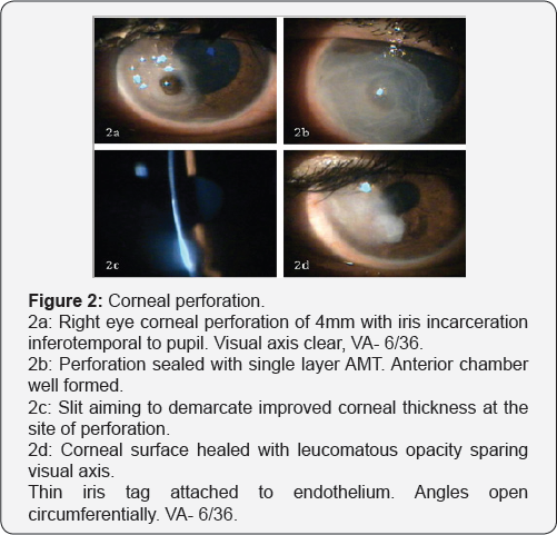
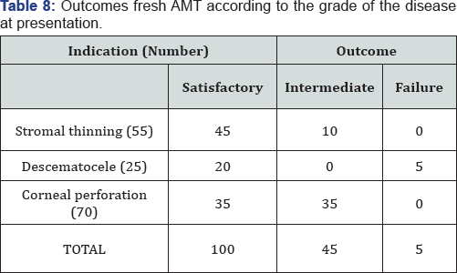
Satisfactory results were seen in 100 of 150 eyes (66.67%), intermediate results seen in 45 cases (23.33%). Failure was noted in 5 cases (3.33%) of fulminant fungal ulcers that showed subsequent corneal perforation requiring Therapeutic PK (Table 8). All the cases in intermediate category which also required subsequent intervention, healed with stable cornea. Thus, successful results were seen in 145 of 150 cases (96.67%) of which in 30 cases subsequent penetrating keratoplasty was done for leucomatous corneal opacity obscuring the visual axis left after healed ulcer (Figure 2).

Discussion

Approximately one-third of cases of infective keratitis require surgical interventions at the acute stage to prevent perforation or spreading of infection [10-14]. Keratoplasty being majorly followed in such situation faces a limitation of availability of good quality donor corneas, mainly in developing countries, recurrence of infection, difficulty in technique and graft rejection. Moreover, for fungal keratitis PK is technique dependent and may also carry a risk of recurrent infection [15].
Thus AMT is sought as an alternative, which has been extensively reported in ophthalmology literature [16-19]. AMT offers the advantage of stimulating re-epithelization, preventing neovascularization and scar formation and avoiding potential allograft rejection. Even if corneal transplantation is needed, the success rate is improved if performed on an eye with reduce infection and inflammation, this can be achieved with AMT [8,9]. In present study complete epithelization was noted in 145 of 150 cases, that is 96.67% success rate. Similar to our study, Chen et al. [20], showed 82.61% success rate, 4 of 23 cases in there study faced AM melting and graft failure requiring therapeutic PK in 3 and delayed healing with vascularization in the other. Kim et al. [21] used multilayer AM in cases of descematocele and corneal perforation. Corneal surface was healed successfully in all cases, and no recurrence of infection or rejection was experienced. Hanada et al. [22] used multiple layers of AM for deeper stromal ulcers down to descemetocele, to restore the normal corneal thickness as well as in corneal perforations from 0.5 to 3mm with or without additional tissue adhesive with high success rates (73-93%). In present study corneal perforations in cases of infectious keratitis up to 6mm have been treated successfully with AMT alone, and 100% corneal epithelization with more than 50% corneal thickness have been achieved in all 70 cases of perforation. In a series by Heiligenhaus et al. [23]. Seven patients with herpes simplex virus or varicella zoster- induced severe ulcerative keratitis, 5 of 7 eyes healed after first AMT [23]. In another study, stromal defect was filled up with multilayer technique proved to be better than monolayer procedure [22,24,25].
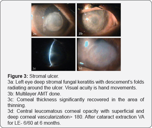
In present study 70% showed significant improvement in corneal transparency and increasing corneal transparency improves the best-corrected visual acuity further emphasizing the healing properties of AMT. Chen et al. [20], preserved useful vision after AMT in cases of fungal keratitis in 52.2% eyes. Kim et al. [21], reported 21 cases of successful AMT in infectious keratitis, in which visual acuity increased except for 5 cases because of irreversible corneal opacity. AMT has come up as a very effective managing technique for refractory ulcers. It aids in permanent healing of the refractory infective keratitis and prepares the cornea for definitive reconstructive procedure if required (Figure 3).

Conclusion


We have found that AMT represents a viable method of treatment to promote healing and prevent progressive melting of refractory infectious keratitis. Besides being cost-effective it’s easy to perform, with a short learning curve. Thus, it might be considered a first-line surgical technique when maximal medical treatment has failed.

For more Open Access Journals in Juniper Publishers please click on: https://juniperpublishers.com


Thursday, 6 February 2020

Medial Rectii Recessions a Surgical Procedure for Bil Alternating Infantile Esptropia in 6 Months Male Twins- Juniper Publishers


Juniper Publishers- JOJ Ophthalmology


Abstract

6 months un identical twins were seen by me in 2002 at qatif central hospital eastern province ks a with parents having noticed bill alternating inward ocular deviation since 2 monthsft normally delivered twins no history of exposure to oxygen birth trauma convulsions jaundice fever or any other cong ocular disorder like 1 sclera cornea 2 keratitis corneal dystophy peters corneal anomaly or limbal corneal dermoid on exam both twins dhad bil alt 15 degrees esotropia no limitation of ocular movements 2 no turning of head towards the direction of paralysed muscle 3 no diplopia 4 no abnormal head posture or false orientation under sedation mydriatic refraction and fundus examination was done ref was equal in both eyes in both eyes so were the normal fundii key words esotropia is inward ocular deviation 2 expotropia is outward ocular deviatin 3 hetrophoria is latenr ocular deviatin 4 infantile is age from birth to 3 years .


Introduction

squint is a common ocular disorder nowadays due to abuse of playing games on mobiles and i pads incidence of ocular deviation has increased recently I saw twins of 6 years of age parents complained that one twin had left eye ocular deviation since 3 months as this twin was constantly playing video games on exam this twin had plus 2d cylinder 130 axis other twin as he was not playing games was normal squints can be 1 esotopia 2 esotropia 3 heterotopias 4 paralytic 5 non paralytic 6 accommodative 7 non accommodative 8 pseudo squint in those who have broad bridge of nose epicanthal folds and wide inter canthal distance results of bill alt infantile esotropia with bill 5mm medial rectii recessions are very satisfactory both 1 from correction of angle of squint and reaction of eye which is minimal [1].


Discussion

Bill alt esotropia presents as 1 crossed fixation 2 uncrossed fixation 3 over action of inf oblique 4 a v patern 5 broad angle 6 covering the dominant eye will make p child to cry so after all investigations under g a both twins were operated under g a exposure of eye done with eye speculmm rotation of eye ensyred with 6 zero silk sutures 5mm recessions of medial rectii one and position of muscle secured on sclera using 6 zero vicoryl suture closure of conjuctival wound done with 8 zero silk sures next day children had very satisfactory correction of angle of deviation and most important thing was that eye reaction was minimal

Conclusion


Bill m rectii recessions is a very satisfactory procedure for cases of bill alt infantile esotropia as we do not cut muscle that is resection so the eye reaction is minimal and secondly correction of eye deviation is good I did this procedure in 2002 even today same procedures is done even after 17 years due to satisfactory modality of this surgical procedure however some complications r seen like 1 under correction 2 over action of inf oblique 3 amblyopia 4 d v d dissociated vertical deviation 5 accommodative element in my cases more than 1 buyers follow up of twins was normal.

For more Open Access Journals in Juniper Publishers please click on: https://juniperpublishers.com


Wednesday, 5 February 2020

Profile of Stereoscopic Acuity of School Children Aged 3 to 5 Years in the Yaounde 2 Sub-Division-


Juniper Publishers- JOJ Ophthalmology


Introduction

Binocular vision is the fundamental refinement of the visual function. It has 3 degrees: spontaneous vision, fusion, and stereoscopic vision. Stereopsis refers to the ability of the visual function to perceive depths and landscapes using binocular vision [1]. Stereoscopic acuity measured by the smallest detected retinal disparity [1], gives clear indication of the quality of binocular vision of an individual [2]. At adulthood, the absence of binocular vision can have an impact on professional orientation. People affected are excluded from certain professions as in aeronautics, marine and military careers: police, fire fighters. The very young child without binocular vision has at times difficulty learning how to walk, to read, or even how to write. A reduction in stereoscopic acuity can be associated to a number of vision problems as strabismus, amblyopia and anisometropia. As such, the measurement of stereoscopic acuity is frequently used to screen for visual dysfunction in children. It is more reliable than visual acuity in the screening of amblyopia [3].Stereoscopic vision continues to evolve after birth. According to a number of authors, the maturation of stereoscopic vision is almost complete in children between 3 and 5 years [4-6]. In view of the importance of this characteristic, we aimed at establishing the profile of stereoscopic acuity of schooling children aged 3 to 5 years in the Yaounde 2 sub-division.


Methodology

Participants selection

We conducted a cross-sectional descriptive study from March 13 to May 15, 2015 in 10 nursery schools of the Yaounde 2 subdivision. Were included all schooling children aged 3 to 5 years who obtained written informed consent from their parents. Were excluded from our study all non-cooperating children after numerous trials or sick on the day of the descent, or refusal from parents. To obtain our sample size, we used probabilistic sampling in 4 degrees. Raffle draw permitted to choose Yaounde 2 sub-division amongst the seven M foundi divisions. For the 2nd degree which concerned the choice of the 10 schools amongst the 86 schools of the Yaounde 2 sub-division, we used Excel 2010. By random draw, a class was chosen per section. As for the 4th degree, the choice of children in the classes. We agreed on a systematic draw of the first 20 pair numbers according to the order of the class list, which followed the order of registration in school. In total, we chose 600 children. We sent 600 questionnaires with notification letters and informed consent forms to the parents of the pupils chosen 3 days before our visit to the school.

Data collection procedure

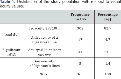
This was done in 4 steps. Firstly, hetero-anamnesis of children from their parents, using the pre-prepared questionnaire filled at home by themselves. We looked for neonatal and ophthalmological past medical history. Then was general inspection performed with emphasis on eyes, looking for malformation, torticollis, nystagmus, apparent strabismus, or an abnormality of the cornea. After this we undertook, measurement of distance visual acuity with and without optic correction using Pigassou's scale placed 5 metres in front of the child. At the end came evaluation of stereoscopic vision using Stereo test TNO. The child with duo chromic lenses (red/green), the test placed at 40cm away from his/her eyes, perpendicular to the visual axis, the different plates were moved in front of them so that they should identify. Significant reduction of distance visual acuity (RdVA) was defined by distance visual acuity, dVA≤6/10 in at least one eye or an anisoacuity≥2 line on Pigassou's scale stereoscopic acuity, SA≥240” corresponded to an abnormal or bad stereoscopic vision.


Statistical Analysis

Data collected was entered in a database created with CSPro 6.0 software and were exported to IBM SPSS 21 software. Tables and diagrams were designed using Excel 2010. Anova test was used to compare the stereo acuity between different age groups and Pearson's Chi2test and Fischer Exact test to look for associated factors to bad stereoscopic vision. Significance was set at 0.05.


Results

Participation rate

We sent 600 questionnaires together with informed consent forms to parents of selected pupils. We registered 374 forms amongst which 6 refusal and 3 absences. Our analysis was on 365 children in total; a participation rate of 60.8%.

Age distribution of the population

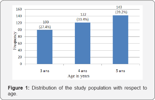
Among the 365 children examined, 175 were girls (47.9%) and 190 boys (52.1%), with a sex-ratio of 1.1 in favour of boys. Our population was divided in 03 groups: 100 (27.4%) children aged 3 years, 122 (33.4%) aged 4 years, and 143 (39.2%) aged 5 years (Figure 1). The most represented age group was that of 5 years. The mean age was 4.2±0.81 years.

Parental hetero-anamnesis of children

In the past-history mostly mentioned were prematurity 11cases, trauma 8 cases, and eye redness 6 cases.

Ophthalmological exam

At general examination, 4 children had strabismus, 2 others a vicious position of the head and one presented a nystagmus. We also found 2 children with optic corrections. Distance visual acuity was normal in 319 children (87.4%), (Table 1). The children had a poor distance visual acuity, represented 12.6% of the study population.

Stereoscopic vision evaluation

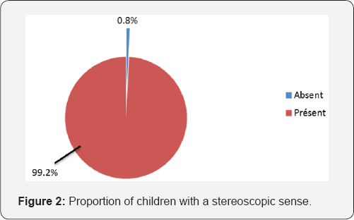
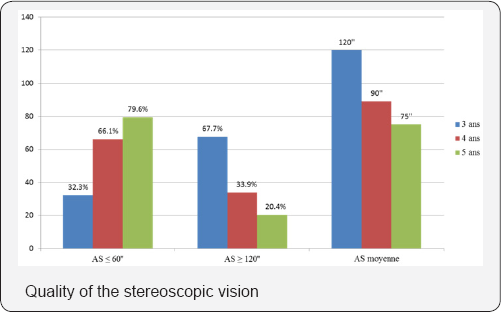
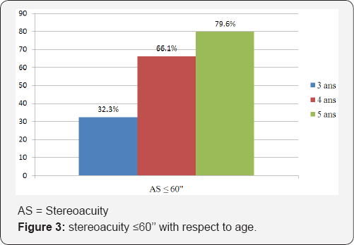
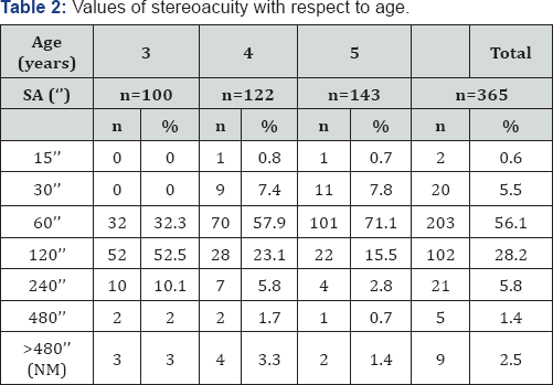
In our study population, 99.2% (362 children) had the stereoscopic sense as represented by (Figure 2). In 03 (0.8%) cases, we discovered an absence of stereoscopy; one in each age- group. Among the 3 children (0.8%), with ocular dominance,2 had left eye dominance. The value of stereoacuity (SA) with the largest proportion was 60"; 56.1% (203) of the study population. We counted 102 children (28.2%) with SA of 120" The least represented class was that of 15", 2 (0.6%) (Table 2). The proportion of children with SA <60" increased with age, 32 children (32.3%) amongst those aged 3 years, 80 (66.1%) in those aged 4 years, and 113 (79.6%) amongst the group of children aged 5 years. This increase in percentages with age was statistically significant (p=0.000) (Figure 3). The tendency inverted with values of 120" to 480", the proportions decreased with age. These differences were statistically significant with p=0.000. The median values of stereoacuity gradually sharpened with age. They varied between 120" at 3 year, to 60" at 4 and 5 years. This variation was statistically significant (p=0.000).

Factors associated to poor stereoscopic vision

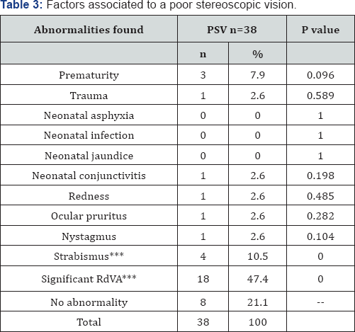
In total, we found poor stereoscopic vision in 38 children, 10.4% of the study population (Table 2). According to the fact that age increased, the percentage of children with a normal stereoscopic vision increased and inversely, the proportion of children with poor stereoscopic vision decreased, significantly (p=0.000). The significant Reduction in distance visual acuity (RdVA) and strabismus were the most encountered abnormalities in the group of children with poor stereoscopic vision (>240"), with respective percentages of 47.4% and 10.5% (Table 3). After bivariate analysis, these two abnormalities were identified as being associated to poor stereoscopic vision with a p=0.000 (Table 3).


Discussion

The principal objective of this study was to establish the profile of stereoscopic vision of schooling children aged 3 to 5 years of the Yaounde 2 sub-division. Specifically, it aimed at determining the proportion of children with the stereoscopic sense, to measure the median values of stereoacuity and finally, to distinguish the factors associated to poor stereoscopic vision in our study population.

Study Population

A total of 600 forms were distributed and only 365 children were examined, giving a participation rate of 60.8%. This rate was less than that obtained in the North department of France, during the 2011-2012 vision campaign organized by APESAL (Association de Prévention Et de Dépistage de troubles visuels Actions Locales) [7]. During this campaign in favour of children aged 2 years and a half to 3 years and a half, the participation rate was 80.05%. This difference could be explained by the fact that the French health system encourages screening of childhood visual disorders. It could also be due to the level of alphabetisation of Cameroon (71.3% according to the 3rd global population census and of habitat of Cameroon) [8], which is less than that of France (99%, according to the Institute national de la statistique et des étudeséconomiques). Moreover, screening campaigns in France are scheduled well ahead of time and introduced in the calendar of targeted schools. Parents are informed many times for their participation. On the other hand, our study took place on a short period. The delay between distribution of forms and the field work was just 02 days, and no reminder was sent to parents. Also, we can add the skepticism of certain parents.

Distribution of the study population according to age

We targeted children aged 3 to 5 years. Given that the schooling rate of the populations of Yaounde is 88.8% according to the results of the Demographic and health investigation and having multiple indicators done in 2011 (EDS 2011) [9], the majority of children aged 3 to 5 years of Yaounde are in school at least 5 hours per day. That is why the site of recruitment chosen was nursery schools. In nursery schools, we generally find children from 3 years. However, many are those who will celebrate their birthday during the school year. Thus, in the small section, we can find children of 4 years, same for the midsection with children of 5 years. This could explain why in our study population, children aged 5 years were more represented (39.2%), whereas the least represented were those aged 3 years (27.4%), with a mean age of 4.12±0.81 years.

Stereoscopic vision evaluation/ Profile of stereoacuity Proportion of children with the stereoscopic sense in our study population

According to our results, 0.8% of children didn't have the stereoscopic sense. The study Vision in Preschoolers (VIP), Ciner et al. [5] in the USA, on the stereoscopic acuity of children of 3 to 5 years, reported that 1.0% of children of the age range did not have the stereoscopic sense [5], results with corroborates ours. Moreover, he mentions that the proportion of children without the stereoscopic sense increased with age. Our sample respected this finding but this tendency was not statistically significant (p=0.800), which could be due to our sample which is smaller than that of Ciner et al.

Progression of SA with age/ quality of stereoscopic vision

In our sample, measured with the stereo test TNO, the cumulative percentages of children with stereo acuity<60" was 32.3% at 3 years, 66.1% at 4 years and 79.6% at 5 years. This increasing tendency with age was statistically significant (p=0.000). The TNO measures disparities till 15". Our results are similar to those of Ciner et al. [5] who using the Stereo Smile II whose finest measured disparity is 60". He compared the stereo acuity among age groups (3, 4 and 5 years) and between the group of children with and without any disorder. His study population was made up of children from the Vision in Pre-schoolers programme. He reports that the proportions of children having reached that disparity increased significantly with age. Thus, 52.2% of the 3 years, 64.9% of the 4 years, and 71.4% of the 5 years were able to see in landscape with an SA ≤60" [5].The median SA of schooling children aged 3 to 5 years in Yaounde 2 varied from 120" to 60", respectively from 3 years, to 4 and 5 years. This improvement with age was statistically significant (p=0.000). In 1975, Romano conducted his study using Titmus stereotest. He found median disparities from 200", 90" and 40" respectively for 3 years, 4 years and 5 years [6]. Likewise, Birch et al. [10] in 2008, using the Randot stereo acuity test observed that the median values of SA sharpened with age, going from 100" at 3 years, to 60" at 5 years [10]. Thus, the median of SA with respect to age that we calculated sharpened with age, as described by the previous authors irrespective of the test used. Thus, the thresholds of SA for children of 3 to 5 years are very close to those found in adults suggesting therefore the maturation of stereoscopic vision is almost complete in children of that age range.

Proportion of children with poor stereoscopic vision

The number of children per age group, with poor stereoscopic vision, progressively significantly decreased with age increase. Fifteen percent of the population aged 3 years presented with a poor stereoscopic vision, 10.7% of 4 years, and 4.9% of 5 years (p=0.000). Ciner suggested the same variation in 2014: 29.6% at 3 years, 22.5% at 4 years, and 19.2% at 5 years [5]. The greatest proportions registered by his group could be due to the fact that his study population was far greater than ours, and had a large number of children suffering from at least one vision disorder which could hamper stereopsis.

Factors associated to abnormal stereoscopic vision

We detected 4 cases of strabismus. All of them had a poor stereoscopic vision, representing 10.5% of children with abnormal stereoscopic vision. An association was established between strabismus and abnormal stereoscopic vision (p=0.000). This corroborates what many authors have described [5,11]. Sharma et al conducted a case-control study. They compared the SA of strabismus patients to that of a control. They showed a significant (p<0.001) poor stereopsis in the strabismus patients [11].The presence of a reduction in distance visual acuity (RdVA) was an associated factor to poor stereoscopic vision (p=0.000). This association is in the same line with reports from Ciner in 2014. Indeed, he found values of SA≥240" in 40.7% of children with a reduction in distance visual acuity (p<0.05) [5]. Amongst the children with poor stereo acuity, we identified 3 premature births (7.9%). According to our findings, prematurity does not influence stereoscopic vision (p>0.05). In 2000, Hard et al. [12] worked on a population of 51 premature children with mean age of 7.2±2.1 years. He showed that this population had an important prevalence of vision disorders, such as altered stereoscopic vision, associated or not to strabismus. Hard counted 7 children with a pathological stereoacuity, among the 31-extreme premature (GA<28 weeks), giving a prevalence of 22.6% [11]. This disagreement between the two studies could be explained by our sample size. It is small and the risk groups, notably the premature children, were not sufficiently represented.


Conclusion


The profile of stereoacuity in Cameroonian children does not present any specificity with respect to literature. Stereoacuity continues to sharpen between 3 and 5 years to reach adult threshold values. This test is recommended for the screening of childhood vision disorders for it is more reliable than the measurement of visual acuity in the screening of morbidities such as strabismus and amblyopia.

For more Open Access Journals in Juniper Publishers please click on: https://juniperpublishers.com


Juniper Publishers: Virtuous Engineering is the Need of Hour: A Perspe...

Juniper Publishers: Virtuous Engineering is the Need of Hour: A Perspe...: Civil Engineering Research Journal Abstract The best quality of someone or something that creates something valuable can be con...

Tuesday, 4 February 2020

Juniper Publishers: Responses of Four Sweet Potato (Ipomoea Batatas L....

Juniper Publishers: Responses of Four Sweet Potato (Ipomoea Batatas L....: Agricultural Research

Long Term Follow-Up of Persistent Outer Retinal Defects following Macular Hole Surgery- Juniper Publishers


Juniper Publishers- JOJ Ophthalmology

Introduction

Pars plana vitrectomy, peeling of internal limiting membrane and gas tamponade is currently the standard treatment for macular hole with high success rate and generally favorable visual outcomes. Previous studies have indicated that a significant number of surgically repaired macular hole patients may have persistent outer retinal defects (microholes) detected with OCT. These are associated with lower best corrected visual acuity (BCVA) following surgery [1-4]. Previous studies have also reported that such outer retinal defects represent discontinuities in the ellipsoid zone (EZ) and the external limiting membrane (ELM) and that there was increased normalization of the EZ and ELM over the first 12 months of post-op follow-up associated with improved visual acuity [5-6]. The purpose of this study is to report a series of such cases with long (greater than 1 year) follow up.


Methods

Retrospective case series reviewing medical records of adults undergoing pars plana vitrectomy (PPV) and gas tamponade for repair of stage 2 to 4 idiopathic macular holes from 2006 through 2009 at Casey Eye Institute and Devers Eye Institute was performed. Peeling of internal limiting membrane (ILM) was not performed in any of the cases. Demographic data, visual acuity, clinical course and optical coherence tomography (OCT) in post-surgical follow-up visits were reviewed and recorded. Time domain (TD) Stratus OCT was available and performed in the early postoperative period followed by spectral domain (SD) OCT in the later follow up visits. The study was approved by the institutional review board at Oregon Health and Science University and was conducted in accordance with the Helsinki Declaration.


Results

During the study period, 80 eyes of 73 patients (25 males and 58 females, mean patient age 67.4 years) with idiopathic macular holes underwent macular hole surgery with standard pars plana vitrectomy without Internal limiting membrane peeling, gas tamponade with either short-acting non-expansile concentrations of sulfur hexafluoride (SF6) or longer-acting non-expansile perfluoropropane (C3F8) or hexafluoroethane (C2F6). Macular hole closure was achieved in 61 eyes (76.3%). Post-operative OCT data was available in 43 of these eyes (70%). Of these 43 eyes, 11 cases (25.5%) presented with persistent outer retinal defects on OCT at least three months after surgery. The group consisted of nine females and two males with mean age of 66.8±8.2 years at the time of surgery. Analysis of right versus left eye and short versus long-acting gas tamponade failed to show any statistically significant difference among eyes who had persistent outer retinal defect.
Mean follow up time of these 11 cases was 60.5±43.2 months, with a range of 3 to 118 months. Of these eleven cases, three had less than one year of follow up due to death (one) or decision not to follow up further (two), during which time the outer retinal defect persisted in all three. Another patient had a re-opened macular hole at approximately a year after surgery but did not undergo further treatment. Seven eyes had longer follow-up with a minimum of 6 years (mean 89.8±18.6 months, ranging 72 to 118) (Table 1). Of these eyes, three (42.8%) developed spontaneous late closure of the outer retinal defects (Figure 1). All three had improved visual acuity after the outer defect was closed (from average 20/50 to 20/25). The final visual acuity in patients without a persistent outer retinal defect at the last follow-up was better than those with a defect, but the difference was not statistically significant due to small sample size.


Discussion

In our study, the overall rate of macular hole closure after single primary PPV was similar to previously contemporaneous reported closure rates using the same technique without ILM peeling [7-14]. Of note, the routine ILM peel and other advances have increased the anatomical success rate of MH surgery to more than 90% hole closure. Successful macular hole closure after surgery is determined clinically by bio microscopy and a negative Watzke-Allen test and is often verified with OCT. Several previous studies have evaluated ultra structural OCT imaging of the foveal region in an attempt to correlate this anatomy with visual outcomes and have suggested that outer retinal features are more important than inner retinal features in determining visual acuity [1,2,15-18]. Several studies have reported similar rates of outer retinal defects following successful macular hole surgery [1,2,17,18]. Histopathologic examination of eyes after macular hole surgery has demonstrated that hole closure start by re-approximation of the edges of the hole to the retinal pigment epithelium followed by growth of Müller cells and astrocytes into the hole to fill in the photoreceptor cell layer [19-24]. This is followed by circumferential and radial contraction of the glial plug pulling the photoreceptor cell layer toward the center of the hole. During this process, there is a potential for anterior displacement of tissue and therefore the presence of a space between migrating glial and photoreceptor cells from the retinal pigment epithelium in the central area. This can be viewed in OCT as persistent outer layer defect and can be seen as focal foveal detachment, ellipsoid zone disruption or both. Other possible explanations for such defects include outer retina (Müller cells/ photoreceptor or RPE) damage from long standing macular hole or surgical trauma during vitrectomy. These defects can also be associated with decreased visual acuity, although case reports of outer retinal defects with good visual acuity are present in the literature [15,19]. Further migration of the glial cell (and/ or photoreceptors) and their adherence to the retinal pigment epithelium centrally is probably responsible for healing of such defects and disappearance of the central OCT defect.
To our knowledge, no other study has investigated the long term outcome (to the extent of average 60 months post operatively) of such defects after successful macular hole surgery. A variety of lesions with similar morphology have been described and referred to as macular microholes [25]. They are small lamellar defects in the outer retina or retinal pigment epithelium that occur through a variety of mechanisms, including spontaneous vitre oretinal interface changes, trauma, photo toxicity, abortive macular hole formation and other unrecognized causes. The condition is non progressive, occurs in patients of all ages, and is compatible with good visual acuity. The retinal changes described in our study can provide a presumptive patho physiological mechanism and natural course for such lesions as well. In our study, we observed that a significant percentage of the outer retinal defects may heal after a long period of follow up. Although we noticed that such healing can be associated with improved visual acuity, such observations are very few in number and also may have been confounded by other factors in the long follow up interval. Therefore, further studies need to be performed to determine whether such observed changes in the visual acuity is truly related to healing of the outer retinal defect. Major limitations of our study include being retrospective and of relatively small size. Surgeries were performed by several surgeons and methods. The low resolution of Stratus images should be recognized as a limitation, particularly since the premise of the study is to identify subtle outer retinal defects. Also, 25% of closed macular holes were lost to follow-up in our center potentially causing significant follow-up bias. There was a long gap between the early follow up and the late follow up of many subjects which confounded the interpretation of interval changes in visual acuity. Similar studies with a larger number of patients and more frequent and regular follow up evaluation are needed to further investigate these lesions. And their final outcome.


Conclusion

In this study, a quarter of patients recovering from macular hole surgery had persistent postoperative outer retinal defects at one year. About42.8%of these defects improved spontaneously over time. Visual acuity may improve if the defect closes, although further studies are needed to investigate such changes.


Financial Disclosure


This research is supported by grant P30 EY010572 from the National Institutes of Health (Bethesda, MD), and by unrestricted departmental funding to the Casey Eye Institute from Research to Prevent Blindness (New York, NY).

For more Open Access Journals in Juniper Publishers please click on: https://juniperpublishers.com


Monday, 3 February 2020

Post-Maxillectomy Orbital Volume Augmentation Using Non- Animal Stabilized Hyaluronic Acid (NASHA) Injection: A Temporary Solution to Complex Reconstruction in Selected Cases


Juniper Publishers- JOJ Ophthalmology


Case Report

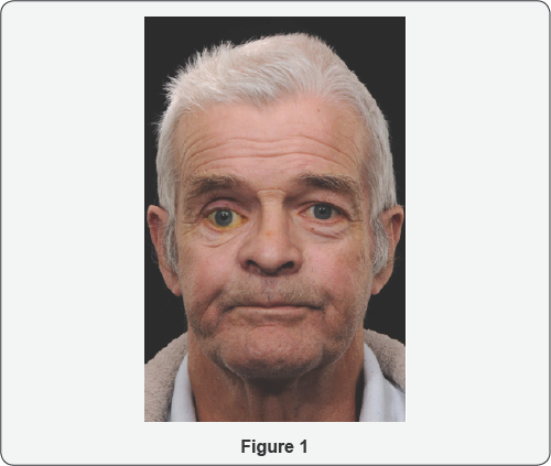
A 65-year-old man was referred to the Orbital clinic from the department of Oral Health, following loss of the orbital floor and significant parts of the medial and lateral walls, as a result of maxillectomy for squamous cell carcinoma of the right middle turbinate. There was no ocular or medical history of note. On examination, his visual acuities (VA) were 6/9 right eye and 6/5 left eye. He had a right hypoglobus of 10mm without a palate implant, and 6mm when in-situ. There was a secondary pseudoupper lid retraction. He had a relative enophthalmos of 8mm with the implant in-situ. Ocular movements were full. A HESS chart was recorded (with implant) indicating a right hypoglobus; diplopia was recorded with red/green glasses, suggesting image suppression.
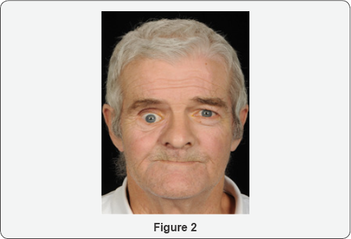
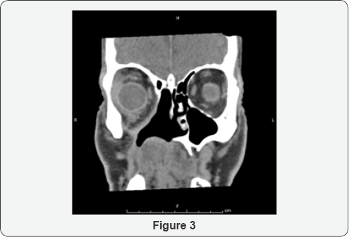
An assessment of his CT scan showed an extensive hemi- maxillectomy with resection of the orbital floor, much of the lamin papyracea, the maxillary antrum and hard palate. Significant enophthalmos was noted. Soft tissue thickening in the region of the surgical margins was noted (Figure 1). As the patient had undergone post-operative radiotherapy, a conservative approach using NASHA for volume augmentation was decided as an initial step. Floor and medial wall reconstruction with implant/dermo fat graft at the level of the equator of orbit was deferred following discussion with the patient. Correction of lid position would be deferred until later.
He underwent an injection of Orbital (non-animal stabilized hyaluronic acid) NASHA. Retrobulbar block was used comprising of Lignocaine 2% with adrenaline 1:200000; 1.5mls was injected, deep, below the globe. The plunger was initially withdrawn to ensure blood vessels were not entered, and a staged augmentation was performed, Three mls of Perlane (Q-Med, Uppsala, Sweden) was injected above the periorbitum of the floor, using an 18G needle. Immediately after the procedure, vision and optic nerve function were monitored and were normal (Compare Figures 2 &3).
At review 1-week later, a 3mm improvement in hyppoglobus and 4mm improvement in enophthalmos was noted. However, he was aware of vertical diplopia, and this was controlled with a base- down 3-dioptre prism. This was thought to be due to loss of right eye suppression with the new globe position.
A further review was performed at 8-months post-injection and a slight increase in enophthalmos and hypoglobus were noted.At 13-months after the initial injection, encouraged by initial results, a further injection of 3mls of Perlane (Q-Med, Uppsala, Sweden) was injected using the same technique as before with tightening of the lower lid by means of a lateral canthal elevation. This resulted in an improvement in hypoglobus to 2-3mm. His diplopia persisted without prisms, although over the next 8-months started coping better without prisms and the diplopia resolved with normal binocular single vision. The long-term plan was to continue with annual orbital NASHA injections


Discussion

From an ophthalmic point of view, mid-face reconstruction aims to provide support to orbital contents, minimize changes in globe position, orbital volume and eyelid function [1]. Symmetrical orbital volume is a requirement for binocular single vision. Complications following reconstructive surgery, and radiotherapy, include resorption of orbital fat and wound contracture, exacerbating enophthalmos and diplopia. Post-maxillectomy radiation therapy increases the risk of orbital sequelae, such as fat atrophy and scar formation, and ocular sequelae, such as optic atrophy, cataract formation, ocular surface dryness, and ectropion [2].
Orbital reconstruction may range from minimal to complex. Minimal bone resections or small orbital floor defects may not require reconstruction. On the other end of the spectrum, subtotal/ total floor defects (>75% surface area) and multi segmental defects will necessitate rigid reconstruction. Primary reconstruction of total maxillectomy defects with preservation of orbital contents remains a complex problem without a perfect solution [3]. In a case series of 66 patients with sinonasal malignancy that had eye-sparing surgery, [2] the most common abnormality following subsequent reconstruction was globe mal position (enophthalmos or hypophthalmos) (34/66 patients, 63%), associated with lack of adequate rigid reconstruction of subtotal or total orbital floor or multisegment orbital defects [2]. The problem can be exacerbated with post-operative radiotherapy [4].
In our case, the patient was keen for correction of his vertical dystopia from a functional and cosmetic point of view. Surgery would have been quite complex due to the absence of the floor, medial and lateral walls. The option of volume augmenatation with a dermo fat graft was considered, but there was a possibility that this graft might not survive due to lack of circulation. There was also a risk of recurrence of the original pathology. A conservative approach was taken and floor augmentation with NASHA was decided as the first stage of the procedure. This addressed both the hypoglobus and enophthalmos to the patient's satisfaction, to the extent that need for further orbital surgery was obviated. Furthermore, binocular single vision returned with the use of prisms.
There are no reports addressing reconstruction of the orbital component of the post-maxillectomy defect using injectable materials for volume augmentation that we are aware of. A minimally invasive approach to orbital volume augmentation using agents such as injectable calcium hydroxylapatite (CaHA), polyacrylamide gel, hydrogel pellet expanders, micro-fat grafts and NASHA have been well described in the anophthalmic socket (post-enucleation socket syndrome). There are only a handful of reports in the context of sighted globes. These include silent sinus syndrome, [5] deep set eyes, [6,7] trauma, [7] and facial hemiatrophy (Parry Romberg syndrome) [7]. We are not aware of any reports of use of filler in the context of carcinoma.
NASHA is produced from non-animal sources by bacterial fermentation. It is well tolerated, has a low immunogenic potential, and there is low risk ofantigenic contamination and hypersensitivity reactions [8]. A major advantage in sighted globes is that its effects can be reversed by dissolving it with hyaluronidase. Disadvantages include a relatively short life span compared to other products and the requirement to inject through an 18G needle in order to maintain particle integrity [8]. However, blunt cannulas can be used. Some studies have reported a 1:1 correlation between the desired augmentation volume (enophthalmos reduction) and each millimetre of product injected, [7] whilst others have not [9]. (We feel the augmentation effect depends on the remaining bony walls and the integrity of the periorbita). Twelve months is the longest duration of action reported [7,9]. Injection of NASHA is recommended in the anterior orbit, and behind the equator of the globe, in order to achieve globe elevation and axial globe advancement respectively [7,9,10]. Injections may be intraconal or extraconal. Proponents of the former cite better globe advancement and reduced anterior migration of filler; [11] proponents of the latter cite risk of vascular compression in sighted globes with intraconal placement [8].
Possible complications include risk of intravascular injection and embolization with loss of vision in sighted eyes. Commonly reported complications include injection site tenderness, ecchymosis and chemosis, [12] stimulation of oculocardiac reflex and associated hypotension, [7,8,12] transient ptosis, [6] anterior migration of filler [13] and inflammatory orbital cellulitis [9].
The benefits of a minimally invasive approach over traditional surgery include reduced operating times, application under local anaesthesia, reduced recovery time and reduced risk of host and donor site morbidity where non-biological materials are used. The benefits need to be weighed against the risks. In the setting of post-neoplasia reconstruction where the prognosis may be poor, use of minimally invasive, albeit temporary, techniques can help to the improve quality and dignity of life for patients, especially where resorting to more complex surgical procedures may be too demanding for the patient in view of their morbidity and reduced life-expectancy [14].

In summary, injectable fillers can provide a temporary solution to volume augmentation of the orbit, for functional restoration and improvement in cosmesis; and may be used in sighted globes with caution. They may be a superior option to traditional surgery in selected cases.

For more Open Access Journals in Juniper Publishers please click on: https://juniperpublishers.com