Wednesday 30 October 2019

Juniper Publishers- Management of Severely Traumatized Eyes Presenting with No-Light-Perception Vision


JUNIPER PUBLISHERS- JOJ Ophthalmology


Mini Review

Presenting visual acuity (VA) has been demonstrated to be the most reliable predictor of the functional outcome in open globe injuries [1-3]. Eyes with no-light-perception (NLP) post-injury are generally related to poor functional outcomes [4]. Traditionally, primary enucleation had been advocated for severely traumatized eyes with NLP in view of the risk of sympathetic ophthalmia [5,6]. Nevertheless, modern immunosuppressives improved the control and treatment of sympathetic ophthalmia and the reported rates of sympathetic ophthalmia is as low as 0.3% in recent studies [7]. In addition, NLP at the acute setting does not necessarily mean that this is irreversible. In the literature, visual recovery rates from NLP to LP or better vary from 4% to 33% [4,8-11].
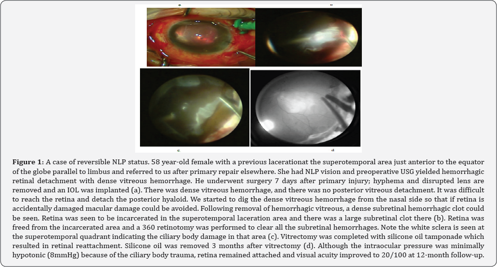
First of all, visual acuity examination in emergency circumstances is not always reliable because the cognitive status of the patient (e.g., unconsciousness, anxiety) may easily influence the result, especially if the examiner is inexperienced. Moreover, most importantly, visual acuity may be profoundly impaired to the extent of NLP due to treatable pathologies (e.g., severe corneal edema, hyphema, dense vitreous and subretinal hemorrhage, particularly in the presence of retinal detachment, extensive choroidal hemorrhage), correction of which may reverse NLP status (Figure 1). Hence, after ascertaining the vision is NLP, clinician must try to identify if the underlying cause is reversible or irreversible. In eyes with severe media opacity, B-scan ultrasonography often aids in diagnosis.
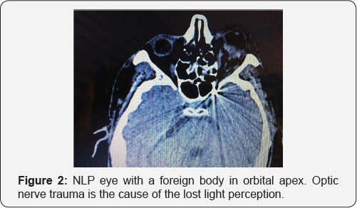
Only in cases of optic nerve transection or complete avulsion, the ophthalmologist may be certain that NLP status is permanent [8,9,12,13] (Figure 2). Relative afferent pupillary defect (RAPD) is an indicator of optic nerve damage; however, it may be falsely positive in the presence of severe hyphema, or subretinal hemorrhage [14]. CT or MRI imaging is successful to demonstrate optic nerve damage (e.g., avulsion, transection) in most of the cases. Even in these cases with no visual potential, the aim should be the anatomic salvage of the globe. Primary enucleation may only be justified if the eye is deformed to an extent that anatomic closure of the wound is not possible.
Secondary vitreoretinal surgery has an unquestionably critical role in preservation of vision and globe. In one study, all the eyes which had recovered from NLP without having secondary vitrectomy were observed to become phthisical with NLP vision within 7 months [10]. Another study showed that eyes that underwent secondary vitrectomy were more likely to recover from NLP vision than those that did not [13]. We advocate early secondary surgery between 3-10 days after open-globe injury. Performing vitrectomy several days after injury allows media clarity, wound stabilization and posterior vitreous detachment to set in. It also has an advantage of decreased uveal congestion which might otherwise result in uncontrollable hemorrhage [15]. However, deferring vitrectomy more than 2 weeks has severe disadvantages (proliferative vitreoretinopathy, ciliary body destruction and consequent phthisis) [16]; hence should be avoided.
Some clinical features may help to predict the visual prognosis of traumatized eyes with NLP after vitreoretinal surgery. Bhagat et al. [13] suggested that NLP eyes that had recovered LP or better vision on the first postoperative day after primary repair might be more likely to recover from NLP at the last follow-up compared to the eyes having NLP on the first postoperative day (71% vs. 8%) [13]. Salehi-Had et al. [10] showed that eyes which had recovered LP or better vision within 5 days of primary repair and/or had vitreoretinal surgery within 5 weeks of the initial injury achieved a more favorable visual outcome [10]. Ciliary body damage, closed funnel retinal detachment, or choroidal damage, on the other hand, has found to be risk factors for developing NLP post trauma [12]. Presence of RAPD and zone 3 wound extending beyond rectus insertion are other clinical features that have been shown to be associated with poor visual prognosis [8]. However, it should be kept in mind that these clinical findings do not necessarily indicate a definite NLP status. Awareness of visual prognosis prior to surgery is important in appropriate management strategy as well as patient counseling.

Given the low risk of sympathetic ophthalmia and advances in vitreoretinal surgical techniques, we recommend globe salvaging procedures for severely traumatized NLP eyes, unless the anatomic reconstruction is impossible. Appropriate primary repair and timely vitreoretinal intervention may give a chance for visual recovery. Even in cases with permanent visual loss, anatomic preservation of the globe should be the primary purpose as the idea of losing an eye is a devastating situation to the patient and the family. Secondary enucleation may be carried out for blind painful cosmetically unacceptable eyes or at the patient's request.

For more Open Access Journals in Juniper Publishers please click on: https://juniperpublishers.com


Juniper Publishers- Visual Functions and Aging


JUNIPER PUBLISHERS- JOJ Ophthalmology


Opinion

Although visual functions impairment are long been well-recognized as an important factor in aging process, their contribution to the problems of older persons on tasks in the natural environment are not well known. The literature has shown age-related deterioration of several visual function including visual acuity, contrast sensitivity, dark adaptation, spatial vision and visual processing speed, orientation and motion perception, stereopsis and rivalry, and functional visual field.
All this age-related visual impairment that occurs during normal (non-pathological) aging can be the result of optical, retinal, and central visual pathways/cortical factors/changes. Although optical factors can contribute to age-related declines, more recent research suggests that the declines are primarily the result of cortical changes (sensory and perceptual processing) and retinal/neuronal changes. There are studies showing an age-related degeneration in intracortical inhibition in V1 and in temporal processing speed in areas 17 and 18 [1]. Others studies have shown the trans-synaptic retrograde degeneration of retinal ganglion cells following retrogeniculate visual pathway lesions[2]. Neurophysiological studies suggest that processing declines may be due in part to changes in the level of neurotransmitters associated with cortical inhibition [3].
Recent findings indicate that visual impairment in the elderly has significant influence on health and functional status, with prognostic value as an independent predictor of mortality[4]. Others studies have found evidence that visual impairment increases the risk of falls and fractures, institutionalization and disability to perform basic activities of daily living. Similarly, elderly individuals with decreased visual acuity are 5 times more likely to show impaired cognitive and affective performance [5]. Research programs on how functional/structural eye changes during the course of adulthood can provide information about risk factors or biomarkers for the development of diseases or common conditions. For example, is important to know the biological (e.g., neural, optical) and environmental (e.g., smoking, dietary) factors that can influence the normal versus the pathological aging (e.g., normal visual field for the age).

This deterioration of visual functions related to age makes important to recognize whether this decline can be recovered or prevented. One promising approach in ameliorating age-related declines in visual function is perceptual learning used to improved visual task performance with practice. The results of our researches on Alzheimer Disease versus Normal-Aging will eventually be useful to develop and evaluate interventions to slow or reverse aging-related declines in vision, thereby improving quality of life.

For more Open Access Journals in Juniper Publishers please click on: https://juniperpublishers.com


Juniper Publishers- A Novel Technique for the Removal of Anteriorly Migrated Dexamethasone Intravitreal Implant


JUNIPER PUBLISHERS- JOJ Ophthalmology


Abstract

The migration of a dexamethasone intravitreal implant (Ozurdex) into the anterior chamber can cause corneal decompensation which may necessitate removal. The implant often fragments when forceps are used and standard vitrectomy instrumentation is suboptimal due to the small opening of the port relative to the size of the implant. We propose the use of a phacoemulsification hand piece for removal of the implant, which has a lumen large enough to assume the implant, infusion to maintain the depth of the anterior chamber, and the ability to emulsify the implant.
Keywords: Phacoemulsification; Dexamethasone; Implant; Migration


Introduction

The Dexamethazone intravitreal implant [1] has been approved by the United States Food and Drug Administration for the treatment of macular edema associated with retinal vein occlusion, noninfectious uveitis involving the posterior segment, and diabetic macular edema. The implant is contraindicated in patients whose posterior lens capsule is torn or ruptured because of the risk of migration into the anterior chamber. However, laser capsulotomy in pseudophakic patients is not a contraindication [1]. The migration of the implant into the anterior chamber, especially if soon after its implantation, can cause corneal decompensation which may necessitate its removal [2,3]. Reported techniques for removal include the use of a vitrector, forceps, viscoelastic, or a Sheets glide [4,5]. However, the implant often fragments when forceps are used and standard vitrectomy instrumentation is often ineffective due to the small opening of the port relative to the size of the implant [3]. Others have reported successful relocation of the implant into the posterior segment with mydriatics and head positioning, external digital force, or manipulation with a needle in the clinic [6-8]. Nevertheless, the implant may still migrate into the anterior chamber yet again [9].


Technique

The experimental surgical video provided demonstrates a beveled, clear corneal incision created with a 2.5 millimeter keratome blade in a porcine eye after inflation of the chamber with viscoelastic. A phacoemulsification hand piece (CENTURION® Vision System, Alcon Laboratories, Inc. Fort Worth, TX) was then introduced into the anterior chamber and used on a high-vacuum setting to engage the implant on its long axis, allowing for efficient emulsification and aspiration.


Discussion

We propose the use of a phacoemulsification handpiece for removal of the implant, which has a lumen large enough to assume the implant, infusion to maintain the depth of the anterior chamber, and the ability to emulsify the implant. The diameter of the Ozurdex implant is 0.46 milimeters, while the diameter of the opening of many phacoemulsifcation probes is 0.5-0.9 milimeters. This technique utilizes the procedures and instrumentation of cataract phacoemulsification that are familiar to all ophthalmologists.


Declarations

Presented at the Sociedad Panamericana de Enfermedades Inflamatorias Oculares, Las Vegas, NV, November 14, 2015.


Competing Interests

TA is a consultant for Allergan. AM has no relevant interests.


Authors' contributions


Conception of project and preparation of manuscript - AM and TA.

For more Open Access Journals in Juniper Publishers please click on: https://juniperpublishers.com


Juniper Publishers- Anterior Chamber Foldable Intraocular Lens for Correction of Aphakia


JUNIPER PUBLISHERS- JOJ Ophthalmology


Abstract

Purpose: To assess the outcomes of anterior chamber foldable intraocular lens (IOL) for aphakia correction.
Materials and methods: Retrospective non-comparative case series study for patients who underwent an operation for aphakia correction by the means of injection of an angular supported foldable IOL performed for 20 eyes of 15 patients. Student’s paired t-test was carried out to compare pre-operative and post-operative visual acuity (VA), IOP & corneal endothelial cell count.
Results: 15 patients were included in the study; 12 males & 3 females. Mean age was 55.38±17.67 years. Mean follow-up duration was 5.95±3.14 months. The mean log MAR visual acuity was 1.26±0.46 pre-operatively and 0.78±0.57 post-operatively (P = 0.003). The change in IOP and corneal endothelial cell count were not statistically significant.
Conclusion: Injection of angular support foldable IOL in the AC is a useful technique for the correction of aphakia.


Introduction

Posterior capsular rupture is a well-known intra-operative complication of cataract surgery [1]. In complicated cases with insufficient capsule and lost zonular support it is not possible to insert a posterior chamber intraocular lens (PC IOL) [2]. Several techniques including posterior chamber sclera fixed lens, anterior chamber sulcus supported lens and anterior chamber iris claw lens have been reported to result in good visual out¬come in the management of aphakic eyes without capsular support [3]. The polymethylmetacrylate (PMMA) ante¬rior chamber (AC) intraocular lens has long been considered as a standard method of aphakia correction. This has been linked to various complications includ¬ing bullous keratopathy, increase of intra ocular pres¬sure (IOP), and endothelial cell loss or even visual loss [4]. Technological advances in lens design (angular support) and in lens mate¬rial (acrylic) have permitted the safe use of lens in the anterior chamber for refractive error correction [4,5].
The aim of the current study was to determine the outcome of using the anterior chamber (AC) foldable lens for unilateral aphakia.


Material and Methods

This is a retrospective non-comparative, consecutive case series study, in which 20 eyes of 15 patients underwent an operation for aphakia correction by the means of injection of angular support foldable intraocular lens in the anterior chamber between January 2014 and December 2014 in Dar El Ouyon Hospital. Written informed consent was obtained after explaining the nature of the procedure from all patients before surgery. Preoperatively, each patient underwent a detailed ocular history (indicating the cause of aphakia) and a standard eye examination including testing of uncorrected and best corrected distance visual acuity (BCVA) using Snellen visual acuity chart (converted into log MAR units for statistical purposes), measurement of the intra ocular pressure (IOP) by Goldmann applanation tonometer, slit lamp examination focusing on the corneal details, and gonioscopy, fundus, and retinal periphery examination. Biometry, anterior chamber depth and corneal White-to-White (WTW) diameter were measured using the IOL Master 500 (Carl Zeiss Meditec AG). Specular microscopy was done to reveal endothelial cell count and abnormal forms using inclusion criteria were unilateral or bilateral aphakia with no capsular support or clinical keratopathy, no evidence of glaucoma, open irido corneal angle, AC depth > 3 mm, WTW diameter > 11.5 mm, and improvement of the visual acuity with corrective trial lenses.
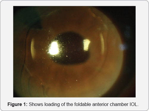
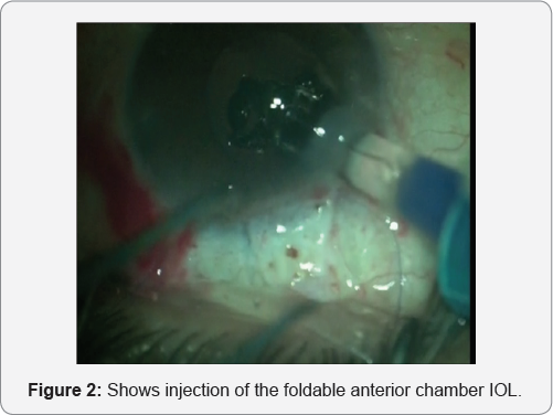
Patients with clinical keratopathy, glaucoma, uveitis, peripheral anterior synechia or clinical maculopathy were excluded. All surgeries were conducted under local peri-bulbar anesthesia using Bupivacaine 0.5% + Xylocaine 2%. One 1-mm paracentesis was made at the upper nasal position and one 2.80 mm clear corneal tunnel incision was made at the upper temporal position with the keratome. A viscoelastic (Viscoat, Alcon) substance was used to reform the AC. The foldable single piece acrylic intraocular lens with the angular support was then injected gently in the anterior chamber, (Figure 1&2). A peripheral iridectomy was done using Vanas scissors. The viscoelastic substance was removed from the anterior chamber with the Simcoe cannula. At the end of the procedure, a subconjonctival depot of dexamethazone and gentamicin was given.
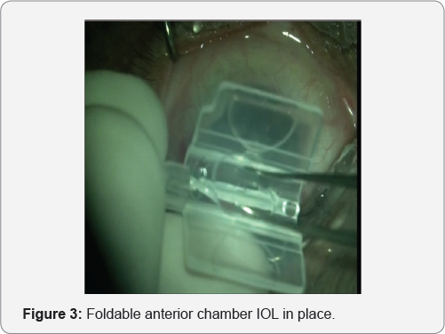
Follow-up visits were scheduled as follows: one day, one week, two months, six months, and one year following surgery. At each follow-up visit, BCVA and IOP were assessed and corneal endothelial cell count and morphology were analyzed. Figure (3) shows foldable anterior chamber IOL in place. Student’s paired t-test was carried out to compare post-operative and pre-operative visual acuity (VA) and intraocular pressure (IOP). The analysis was performed using statistical software Epi-Info 7 and IBM SPSS 19. P-values less than 0.05 were considered statistically significant.


Results

Twenty (20) eyes of fifteen (15) patients were included in this study. The demographics and baseline eye characteristics for all patients are presented in . Eight patients were males (10 eyes, 50%) and seven were females (10 eyes, 50%). Mean age was 50.75 ± 11.275 years (range 30-72 years). Aphakia causes included: 2 planed ICCE (10%), 6 inadvertent posterior capsule rupture with vitreous lost during planed phacoemulsification (30%) and 12 post-traumatic lens subluxation (60%). The mean follow-up duration was (10.8 ± 2.02) months (range 6-12 months). Sixteen (80%) patients showed improvement of the post-operative BCVA of one line or more; two (10%) showed no improvement and two (10%) patients showed deterioration in their post-operative BCVA. The mean visual acuity in log MAR was (1.14 ± 0.46) pre-operative and (0.605 ± 0.489) post-operative (P < 0.05). Post-operative complications are presented in. Two patients (10%) had post-operative intraocular hypertension (over 21 mmHg). Two (20%) patients had pupil distortion, one patient (10%) had retinal detachment and one (10%) patient had macular edema. No corneal problem requiring lens removal was registered.


Discussion

There are many options for management of aphakia with no capsular support [5]. Anterior chamber IOL is one of these options. Foldable lenses with angular support are currently used in the anterior chamber for refractive error correction [6]. In this study, we injected a foldable lens with angular support in the anterior chamber to correct the aphakia. All patients presented with aphakia without capsular support from various etiologies. Post-traumatic lens subluxation was the leading cause of aphakia (60%) in our study. Sixteen (80%) patients showed significant improvement of the BCVA from (1.14 ± 0.46) pre-operative to (0.605 ± 0.489) post-operative (P < 0.05). Previous studies using an iris claw lens to correct aphakia reported significant VA improvement and a low rate of complications [5,7]. However, despite the good visual outcome, implementation of iris claw techniques requires specialized equipment and a long training period. The insertion of a foldable lens in anterior chamber is a very easy procedure through a 2.80 - 3 mm clear corneal tunnel.
In our study, 2 eyes (10%) developed of post operatively a transient IOP elevation, which was managed with topical medications. This change in IOP was not statistically significant (P=0.32). It was also reported in other similar studies [1-3] in which other approaches were used to correct aphakia. Two cases (10%) with pupil distortion were observed postoperative, which was also noted, in another study (2). One case (5%) developed macular edema post-operative, which resolved on topical steroids and non-steroidal anti-inflammatory drops. One case (5%) developed post-operative retinal detachment. A vitrectomy and silicone oil injection was done. These two complications resulted in lower VA in our study.


Conclusion

Our results showed that foldable IOL injection in the anterior chamber improved visual acuity; the complications were similar to those reported in the literature using other methods. Although the visual outcome was good for most patients in this series, more extended follow up and a large series of patients are needed to ascertain the effectiveness and safety of this procedure.


What was Known

Rigid anterior chamber IOLs were commonly used for visual rehabilitation in cases of aphakia. This was associated with many complications such as elevated intraocular pressure, anterior uveitis and corneal decompensation.


What this Paper Adds


Using foldable anterior chamber IOL provides easy, safe and effective method for correction of aphakia.
For more Open Access Journals in Juniper Publishers please click on: https://juniperpublishers.com


Juniper Publishers- Pathogenesis of Duane’s Retraction Syndrome


JUNIPER PUBLISHERS- JOJ Ophthalmology


Aetiology

The most accepted theories until now are: Agenesis or partial development of the 6th cranial nerve nucleus in the brain stem and Branches from the 3rd nerve are redirected to innervate the lateral rectus. So lateral rectus has both Subnormal or no innervations from the 6th cranial nerve and anomalous innervations from the 3rd nerve. According to the amount of anomalous innervations going to the lateral rectus; different presentations and patterns of Duane’s syndrome results (Figure1).


Duane Type 1

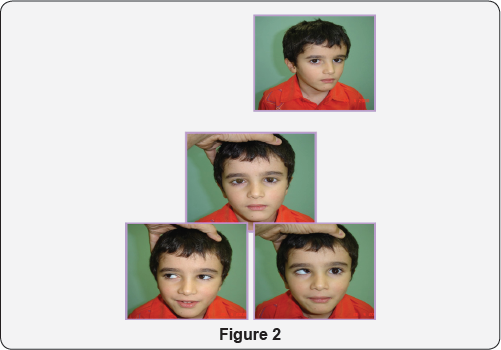
No innervations from the 6th nerve. Mild anomalous innervations from the 3rd nerve so the clinical picture is that limited abduction and mild narrowing of the palpebral fissure in adduction (Figure 2).


Duane Type 2

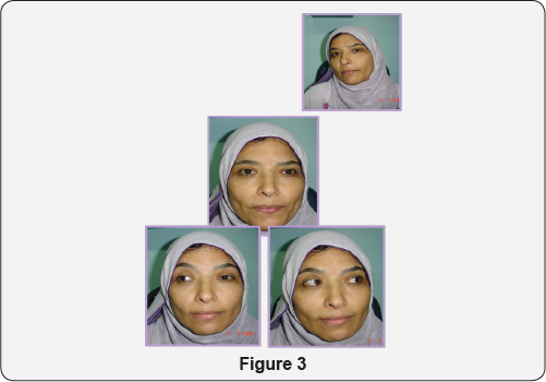
Normal innervations from the 6th nerve. Moderate anomalous innervations from the 3rd nerve so the clinical picture includes normal abduction and limited adduction together with narrowing of the palpebral fissure in adduction (Figure 3).


Duane’s Type 3

No innervations from the 6th nerve. Nerve fibers to the medial rectus is divided 50/50 between medial rectus and lateral rectus so; the clinical picture is that limited abduction (no innervations from the 6th), limited adduction due to equal contraction of both medial and lateral recti on adduction (Figure 4).


Duane’s Type 4

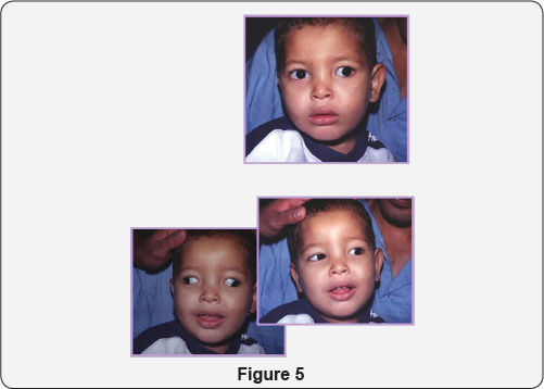
No innervations from the 6th nerve. Most of the fibers of the medial rectus go to the lateral rectus so that with attempted adduction, abduction occur (simultaneous divergence) (Figure 5).

For more Open Access Journals in Juniper Publishers please click on: https://juniperpublishers.coFor more articles in  JOJ Ophthalmology (JOJO) please click on: https://juniperpublishers.com/jojo/index.ph
For more Open Access Journals please click on: https://juniperpublishers.com

Saturday 12 October 2019

Juniper Publishers: Diplopia and Torticollis in Adult Strabismus


JUNIPER PUBLISHERS- JOJ Ophthalmology


Abstract

Objective: To expose the diagnosis of an adult patient with vertical and unilateral divergent strabismus and to approach the surgical treatment in order to remove the symptoms of diplopia and signs of torticollis.
Methods: The measurement of the angle of deviation, the Lancaster test, the Bielchowsky test and the study of ocular motility, both in examination room (versions) and in the surgery room (test of ductions), demonstrated the diagnosis: right eye hipertropia (HTR) and right eye exotropia (XTR). Surgical intervention was performed on the right upper rectus muscle and on the right inferior oblique muscle, with the aim of resolving the deviation of both directions in only one surgical intervention.
Results: The intervention was satisfactory for both the patient and the medical team, because we were minimized the signs and symptoms. After 4 months the residual deviation angle was minimal and allowed the ability of fusion.
Conclusion: The diagnostic and treatment protocol performed in this case show the optimal resolution of the diplopia and torticollis that the patient suffered.
Keywords: Strabismus; Torticollis; Diplopia; Lancaster test


Introduction

We believe that, in some cases, in strabismus surgery there may be more than one option in the choice of surgical protocol, and each professional and his experience sets a more personalized pattern of action. Our objective is showing through this case, for ophthalmologists and other professionals who visit adult patients with strabismus, our experience in the surgical treatment of vertical and divergent deviation with torticollis and an associated diplopia [1-5].


History, Data and Method of Diagnostic Exploration

Patient 41 years old. The patient refers diplopia in increase for a year and torticollis for years. Tilt the head to the left shoulder to try to compensate the diplopia. Ocular antecedents: Lasik refractive surgery (myopia -5.00 both eyes).

Family history: Glaucoma.

Pre-surgery exploration

  1. Visual acuity without correction right eye = 1
  2. Visual acuity without correction left eye = 1
  3. Refraction (cycloplegic) right eye = +1.50-0.50x170°
  4. Refraction (cycloplegic) left eye = +0.50-0.75x145°
  5. Corneal topography: Central ablation in both eyes.
  6. Normal ophthalmic examination.
  7. . Synoptophore without correction in superaversion = 22Δ XTR / 28 Δ HTR. Primary position of gaze = 17Δ XTR / 28Δ HTR. In Infraversion = 9 Δ XTR / 30Δ HTR.
  8. Left eye dominance
  9. Stereopsis test (Titmus / Fly): There is no stereopsis.


Method Surgical

The day of the surgery is performed a tests of duction: right upper oblique muscle (- - -)

Surgical protocol

Backward movement of right upper rectus muscle 3mm. Backward movement of right inferior oblique muscle 6mm.

Post-surgery exploration

The synoptophore after the intervention in the primary position was 3Δ HTR. There is no existence of stereopsis, but it can even fusion the images. The patient is subjectively satisfied. It is proposed, 4 months after the surgery, to begin orthoptic treatment to strengthen the ability of fusion images [5-10].


Discussion

The deviation of an adult’s strabismus is diagnosed and resolved the vertical and divergent strabismus in only one surgical intervention. It was discarded the differential diagnosis of nonocular torticollis by the examination tests. It was considered, for the good maintenance of the obtained result and maintenance of the ability of fusion, to perform an orthoptic treatment after surgery. We consider that the patient will have more visual quality in their daily life when resolving their symptoms of diplopia, signs of torticollis and ocular asymmetry. We emphasize the exploration by the Lancaster test in patients with diplopia for its ease of use, graphical representation and its diagnostic ability and the following of the case over time.


Conclusion


The diagnostic and treatment protocol performed in this case show the optimal resolution of the diplopia and torticollis that the patient suffered.

For more Open Access Journals in Juniper Publishers please click on: https://juniperpublishers.com


Juniper Publishers: Comparative Study between Synoptophore and Cover Test with Prisms at Far Vision


JUNIPER PUBLISHERS- JOJ Ophthalmology


Abstract

Objective: Based on the measurement of strabismus angle with prisms at far vision, which is the most widely used method, we compare it with the measurement performed with synoptophore. We evaluate which factors influence the measurement with synoptophore and with Cover Test with prisms at far vision.
Method: In a sample of 64 patients with a mean age of 27.6 years, we consider two common clinical methods of measuring the angle of ocular deviation: synoptophore and cover test with prisms. Given the differences, we find between the measurement of the angle of ocular deviation and what factors may influence the variability with both methods.
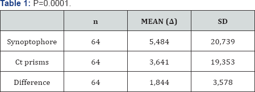
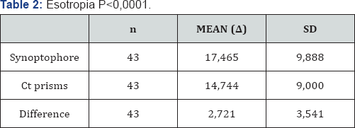
Results: Comparing the measured deviation angle in 64 patients with both methods at far vision, we found a statistically significant difference, with an average of 1,844 prism dioptres. If we compare both methods in esotropia or exotropia, in the 43 patients with esotropia found a statistically significant difference (mean difference 2.721Dp). On the other hand, in the 21 patients with exotropia there was no a statistically significant difference (mean difference 0.048Dp.). Considering gender, 35 men of the sample, we find statistically significant differences (mean difference 2.171Dp), and women, 29 patients of the sample, we also found statistically significant differences (mean difference 1.448Dp).
Conclusion: In esotropia we find that there are statistically significant differences in cover test with prisms at far vision. On the other hand, in exotropia we don’t find statistically significant differences. In both the groups of patients with previous strabismus surgery and non-surgery there are no statistically significant differences. The fact that we find a greater deflection angle with the synoptophore at convergent strabismus confirms the ability of dissociation.
Keywords: Synoptophore; Cover test; Dissociate prism dioptre; Strabismus; Exotropia; Esotropia


Introduction

The stereoscope invented by Charles Wheatstone in 1830 used for oculomotoras and sensory evaluation. Its main use is to assess and treat strabismus, also used in vision therapy for the treatment of amblyopia [1]. It simulates far vision conditions and presents separate images for each eye. It studies oculomotoras and sensory capabilities mainly in strabismus patients [2]. Although the measure of the deviation angle is usually performed with prism bar or loose prisms, it is worth considering the synoptophore, given its characteristics.
Compared with measurement with cover test (CT) and prisms, in synoptophore we obtain:
  1.  Ahigher value of deviation as it is more dissociating.
  2.  More simplicity and comfort in the measurement of the 9 positions of gaze thanks to its mechanical structure.
  3.  The values are more reproducible, making it easier for monitoring the case.
  4.  More accurate values in high-angle strabismus, since in these cases the measure is not performed in leaps.
  5.  More objective measurement


Materials and Methods

In a sample of 64 patients, with a mean age of 27.6 years, we consider two common clinical methods of measuring the angle of ocular deviation: synoptophore and Cover Test with prisms at far vision. We study which differences we find between both methods and what factors may influence the variability of the results. We studied the influence of factors such as the direction of the horizontal deviation (esotropia/ exotropia), if they had vertical deviation or some sort of alphabetical syndrome, if they had undergone strabismus surgery previously and finally the patient’s gender.


Results

Comparing the measurement of the deviation angle in 64 patients with synoptophore and cover test with prisms at far vision we find:

Comparison in factors (Table 1):

Type of deviation

  1.  Esotropia: P<0,0001 (Table 2)
  2.  Exotropia: P=0,9426 (Table 3)

History of strabismus surgery

  1.  With previous surgery: P=0,0176 (Table 4)
  2.  Without previous surgery: P=0,0036 (Table 5)

With previous surgery

  1.  Esotropia: P=0,0104 (Table 6)
  2.  Exotropia: P=0,9001 (Table 7)

Without previous surgery

  1.  Esotropia: P=0,0003 (Table 8)
  2.  Exotropia: P=0,8675 (Table 9)

Gender

  1.  Male: P=0,0017 (Table 10)
  2.  Female: P=0,0280 (Table 11)
We also assessed whether other factors such as vertical deviations or alphabetic syndromes could influence the comparison. Given the low percentage of patients who showed vertical strabismus or alphabetical syndrome, comparative under these factors was dismissed [3-8].


Discussion

Despite significant statistical differences in convergent strabismus, in regular motor examination we always include the measurement with synoptophore as well as the measurement with prism at far vision.
We consider that the greater accommodative capability that convergent strabismus present, both at far and near vision, allows the synoptophore, as being more dissociating, to find greater deviation angles. We believe that although with synoptophore there is a risk of causing greater dissociation in some type of strabismus, for us its use for justified the measure, because it is more accurate, objective and reliable. Anyway we still complete the study with the measurement with prisms. In our protocol for measuring ocular alignment we consider the measurement with synoptophore, objectively, a complementary study to consider.


Conclusion


In esotropia we found that there was statistically significant differences respect to the measurement at distance with prisms at far vision. In exotropia we didn’t find statistically significant differences. Both in patients with previous strabismus surgery and non-surgery there are statistically significant differences. The fact that we find a greater deflection angle with the synoptophore at convergent strabismus confirms the ability of dissociation that we initially suspected occurs with Synoptophore [9-12].

For more Open Access Journals in Juniper Publishers please click on: https://juniperpublishers.com


Juniper Publishers: Eplerenon as Adjunctive Therapy for Choroidal Neovascularisation with Serous Pigment Epithelial Detachment and Subretinal Fluid in Age Related Macular Degeneration

JUNIPER PUBLISHERS- JOJ Ophthalmology

Abstract

Objective: To investigate possible effect of eplerenon 50 mg in serous pigment epithelial detachment (sPED) and subretinal fluid (SF) resolution after initial intravitreal application of 2 mg aflibercept in treatment of neovascular age-related macular degeneration (nAMD).
Method: Case presentation of two females with occult choroidal neovascularisation with sPED due to the nAMD treated with three consecutive intravitreal injections of 2 mg aflibercept. On control check up 6 weeks after last injection, per oral 50 mg Eplerenon (Inspra) once daily is prescribed. For follow up we used visual acuity, intraocular pressure, Amsler grid test, color fundus (CF), optical coherence tomography (SD OCT) baseline, 6 weeks after last intravitreal injections, 6 weeks after initiation of eplerenon as well as potassium serum concentration 6 weeks after initiation of eplerenon therapy is monitored.
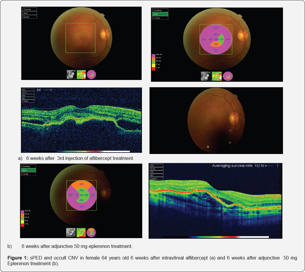
Results: Morphologic improvement evident in reduction of central macular thickness and height of sPED >10% followed with stabile visual acuity was registered in both patients after 6 weeks of eplerenon prescription.
Conclusion: Eplerenon given as adjunctive therapy for treatment of sPED in nAMD could have beneficial effect but further more detailed and more controlled clinical investigations are recommended.
Keywords: Serous pigment epithelial detachment; Eplerenon; Neovascular age related macular degeneration


Introduction

Serous pigment epithelial retinal detachment (sPED) is sharply demarcated, smooth, dome shaped elevation of retinal pigment epithelium (RPE) [1]. SPEDs are manifestation of neovascular age related macular degeneration (nAMD), polypoidal choroidal vasculopathy (PCV) and central serous chorioretinopathy (CSR). Controversy remains about etiology of sPEDs [2-5]. The over activation of the mineralocorticoid receptor (MR) pathway has been shown to cause fluid accumulation in the retina, choroidal vasodilation, and to promote retinal neovascularization in hypoxic conditions [1-4,6-8]. According the role of mineralcorticosteroid receptors in retina, eplerenon has shown beneficial effect in treatment of CSR [5].
A well-known complication of PED is a RPE tear caused by contraction of the elevated PED and retraction of RPE resulting in absence of RPE underneath the neurosensory retina [5,6]. RPE rip could be serious vision threating complication of laser photocoagulation, photodynamic therapy and anti-VEGF therapy. There is an evidence that sPED with height > 350 ?m are more high risk for development of RPE and consecutive macular atrophy [7]. Anti VEGF is nowdays the main therapeutic approach for nAMD and despite chronic presence of sPED and subretinal fluid in regular and continuous anti-VEGF regimes have comparable and benefitial effect in visual and functional outcome [8]. Due to the costs of antiVEGF and patient compliance with intravitreal injections alternative treatments are rational approach to those cases [5].


Case Presentation

Case series of 2 patients with occult choroidal neovascularisation (CNV) due to the nAMD with sPED >350 чm,treated with three initially consecutive intravitreal injections of 2 mg aflibercept (EYLEA®) in private practice. After 6 weeks after last intravitreal aflibercept therapy per oral Eplerenon 50 mg (INSPRA®) once daily was prescribed as persistent sPED exists and relatively large collection of subretinal fluid (>50 чm) presented in macular region. Both patients are females, age of 71 and 64. Visual acuity baseline 30 ETDRS (Early treatement diabetic retinopathy) letters.
After 12 weeks of last treatment with anti VEGF and 6 weeks after Eplerenon prescription visual acuity was 70 and 75 ETDRS letters respectively. Intraocular pressure was with normal limits during all controls (less than 21 mmHg). Both patients had detailed ophthalomologic examination on all consecutive visits. It included complete anterior segment of the eye exam which was within normal limits in both cases. Fundoscopic exam in mydriatic conditions show soft drusen on both eyes, complete resolution of the blood on eplerenon baseline visit and no blood on consecutive visit in treated eye. Both patients had incomplete posterior vitreous detachment, no vitreoretinal changes. Fellow eye has high risk AMD in younger patient (presence of large soft drusen in macula). In other patient fellow eye has soft drusen and occult juxtafoveal CNV. Both patient were treatment naive.
Both patients were previously for 5 years treated with oral beta blockers (metoprolol 25 mg once daily) and ACE inhibitors (enalapril 10 mg twice daily) for arterial hypertension and received statins (atorvastatin 10 mg once daily) for regulation of hyperlipoproteinemia type IIa. Other systemic and ocular health problems were not present. Both patients presented type A personality. Central retinal thickness and central sPED height measured 6 weeks after eplerenon 50 mg treatment and 12 weeks after last intravitreal injection of 2 mg aflibercept seemed to be evidently reduced. Visual acuity during treatment with eplerenon 50 mg was stable through 6 weeks 70 and 75 ETDRS letters respectively. Both patients are educated for self testing Amsler grid, and Amsler grid tests performance was stable and unchanged during all follow up period. Potassium plasma level was monthly monitored in both patients (within normal limits <4,1 mmol/L). Younger patient is now switched to pro re nata (PRN) anti VEGF therapeutic regime, as signs of CNV activity in both cases considered are visual acuity, Amsler grid test, signs of hemorrhage on clinical exam, increase in retinal thickness due to the intraretinal edema on OCT (Figure 1). Other patient refuse further antiVEGF treatment and she is still taking eplerenon combined with antioxidant and vitamin supplements.


Discussion

The purpose of this case presentation is to show effect of eplerenone, a mineralocorticoid receptor antagonist, as a treatment option for reduction of SRF and sPED in cases with nAMD. The role of mineral corticosteroid receptors (MR) in retinal disorders is emerging and the potential association with physiological traits are investigated in CNV. The place of MR antagonists for retinal diseases treatment are discussed [1-4].
Central subfield thickness at follow-up visit after eplerenon initiation significantly decreased more than 10% from baseline which was previously reported in cases with chronic CSR [5]. Following therapy with eplerenon, there was a significant reduction in SF, reduction in CST, and improvement visual acuity in eyes with chronic CSR [5]. A majority of eyes with CSR presented by Singh and al [5] (47.1%) demonstrated stable SRF on treatment, while 35.3% had complete resolution of SRF. Results of this study indicate that eplerenone could be beneficial in the treatment of unresolved CSR. Considering that some cases in nAMD morphologically reminds on CSR could be rational approach to treat those patients with mineralcorticosteroid antagonists with the goal of serous transudation reduction and to reduce number of intravitreal injections which could be serious burden for patients. Also, presence of subretinal and sub RPE fluid is still not mandatory indication for continuous and strict anti VEGF regime. There are evidences that antiVEGF therapy in cases with large serous PED are prone to RPE rip [7,8]. As it is still controversial, there could be some benefit of anatgonists of mineralcorticosteroid receptors in some cases with resistent and non compliant patients with sPED. Further more precise conclusions and role of mineralcorticosteroid receptors in nAMD request more controlled and precise clinical investigations.

For more Open Access Journals in Juniper Publishers please click on: https://juniperpublishers.com


Juniper Publishers- Pathogenesis of Duane’s Retraction Syndrome

JUNIPER PUBLISHERS- JOJ Ophthalmology

Aetiology

The most accepted theories until now are: Agenesis or partial development of the 6th cranial nerve nucleus in the brain stem and Branches from the 3rd nerve are redirected to innervate the lateral rectus. So lateral rectus has both Subnormal or no innervations from the 6th cranial nerve and anomalous innervations from the 3rd nerve. According to the amount of anomalous innervations going to the lateral rectus; different presentations and patterns of Duane’s syndrome results (Figure1).


Duane Type 1

No innervations from the 6th nerve. Mild anomalous innervations from the 3rd nerve so the clinical picture is that limited abduction and mild narrowing of the palpebral fissure in adduction (Figure 2).


Duane Type 2

Normal innervations from the 6th nerve. Moderate anomalous innervations from the 3rd nerve so the clinical picture includes normal abduction and limited adduction together with narrowing of the palpebral fissure in adduction (Figure 3).


Duane’s Type 3

No innervations from the 6th nerve. Nerve fibers to the medial rectus is divided 50/50 between medial rectus and lateral rectus so; the clinical picture is that limited abduction (no innervations from the 6th), limited adduction due to equal contraction of both medial and lateral recti on adduction (Figure 4).


Duane’s Type 4

No innervations from the 6th nerve. Most of the fibers of the medial rectus go to the lateral rectus so that with attempted adduction, abduction occur (simultaneous divergence) (Figure 5).

For more Open Access Journals in Juniper Publishers please click on: https://juniperpublishers.com