Wednesday, 5 February 2020
Juniper Publishers: Virtuous Engineering is the Need of Hour: A Perspe...
Juniper Publishers: Virtuous Engineering is the Need of Hour: A Perspe...: Civil Engineering Research Journal Abstract The best quality of someone or something that creates something valuable can be con...

Tuesday, 4 February 2020
Long Term Follow-Up of Persistent Outer Retinal Defects following Macular Hole Surgery- Juniper Publishers
Juniper
Publishers- JOJ Ophthalmology
Introduction
Pars plana vitrectomy, peeling of internal limiting
membrane and gas tamponade is currently the standard treatment for
macular hole with high success rate and generally favorable visual
outcomes. Previous studies have indicated that a significant number of
surgically repaired macular hole patients may have persistent outer
retinal defects (microholes) detected with OCT. These are associated
with lower best corrected visual acuity (BCVA) following surgery [1-4].
Previous studies have also reported that such outer retinal defects
represent discontinuities in the ellipsoid zone (EZ) and the external
limiting membrane (ELM) and that there was increased normalization of
the EZ and ELM over the first 12 months of post-op follow-up associated
with improved visual acuity [5-6]. The purpose of this study is to report a series of such cases with long (greater than 1 year) follow up.
Methods
Retrospective case series reviewing medical records
of adults undergoing pars plana vitrectomy (PPV) and gas tamponade for
repair of stage 2 to 4 idiopathic macular holes from 2006 through 2009
at Casey Eye Institute and Devers Eye Institute was performed. Peeling
of internal limiting membrane (ILM) was not performed in any of the
cases. Demographic data, visual acuity, clinical course and optical
coherence tomography (OCT) in post-surgical follow-up visits were
reviewed and recorded. Time domain (TD) Stratus OCT was available and
performed in the early postoperative period followed by spectral domain
(SD) OCT in the later follow up visits. The study was approved by the
institutional review board at Oregon Health and Science University and
was conducted in accordance with the Helsinki Declaration.
Results
During the study period, 80 eyes of 73 patients (25
males and 58 females, mean patient age 67.4 years) with idiopathic
macular holes underwent macular hole surgery with standard pars plana
vitrectomy without Internal limiting membrane peeling, gas tamponade
with either short-acting non-expansile concentrations of sulfur
hexafluoride (SF6) or longer-acting non-expansile perfluoropropane
(C3F8) or hexafluoroethane (C2F6). Macular hole closure was achieved in
61 eyes (76.3%). Post-operative OCT data was available in 43 of these
eyes (70%). Of these 43 eyes, 11 cases (25.5%) presented with persistent
outer retinal defects on OCT at least three months after surgery. The
group consisted of nine females and two males with mean age of 66.8±8.2
years at the time of surgery. Analysis of right versus left eye and
short versus long-acting gas tamponade failed to show any statistically
significant difference among eyes who had persistent outer retinal
defect.
Mean follow up time of these 11 cases was 60.5±43.2
months, with a range of 3 to 118 months. Of these eleven cases, three
had less than one year of follow up due to death (one) or decision not
to follow up further (two), during which time the outer retinal defect
persisted in all three. Another patient had a re-opened macular hole at
approximately a year after surgery but did not undergo further
treatment. Seven eyes had longer follow-up with a minimum of 6 years
(mean 89.8±18.6 months, ranging 72 to 118) (Table 1). Of these eyes, three (42.8%) developed spontaneous late closure of the outer retinal defects (Figure 1).
All three had improved visual acuity after the outer defect was closed
(from average 20/50 to 20/25). The final visual acuity in patients
without a persistent outer retinal defect at the last follow-up was
better than those with a defect, but the difference was not
statistically significant due to small sample size.


Discussion
In our study, the overall rate of macular hole
closure after single primary PPV was similar to previously
contemporaneous reported closure rates using the same technique without
ILM peeling [7-14].
Of note, the routine ILM peel and other advances have increased the
anatomical success rate of MH surgery to more than 90% hole closure.
Successful macular hole closure after surgery is determined clinically
by bio microscopy and a negative Watzke-Allen test and is often verified
with OCT. Several previous studies have evaluated ultra structural OCT
imaging of the foveal region in an attempt to correlate this anatomy
with visual outcomes and have suggested that outer retinal features are
more important than inner retinal features in determining visual acuity [1,2,15-18]. Several studies have reported similar rates of outer retinal defects following successful macular hole surgery [1,2,17,18].
Histopathologic examination of eyes after macular hole surgery has
demonstrated that hole closure start by re-approximation of the edges of
the hole to the retinal pigment epithelium followed by growth of Müller
cells and astrocytes into the hole to fill in the photoreceptor cell
layer [19-24].
This is followed by circumferential and radial contraction of the glial
plug pulling the photoreceptor cell layer toward the center of the
hole. During this process, there is a potential for anterior
displacement of tissue and therefore the presence of a space between
migrating glial and photoreceptor cells from the retinal pigment
epithelium in the central area. This can be viewed in OCT as persistent
outer layer defect and can be seen as focal foveal detachment, ellipsoid
zone disruption or both. Other possible explanations for such defects
include outer retina (Müller cells/ photoreceptor or RPE) damage from
long standing macular hole or surgical trauma during vitrectomy. These
defects can also be associated with decreased visual acuity, although
case reports of outer retinal defects with good visual acuity are
present in the literature [15,19].
Further migration of the glial cell (and/ or photoreceptors) and their
adherence to the retinal pigment epithelium centrally is probably
responsible for healing of such defects and disappearance of the central
OCT defect.
To our knowledge, no other study has investigated the
long term outcome (to the extent of average 60 months post operatively)
of such defects after successful macular hole surgery. A variety of
lesions with similar morphology have been described and referred to as
macular microholes [25].
They are small lamellar defects in the outer retina or retinal pigment
epithelium that occur through a variety of mechanisms, including
spontaneous vitre oretinal interface changes, trauma, photo toxicity,
abortive macular hole formation and other unrecognized causes. The
condition is non progressive, occurs in patients of all ages, and is
compatible with good visual acuity. The retinal changes described in our
study can provide a presumptive patho physiological mechanism and
natural course for such lesions as well. In our study, we observed that a
significant percentage of the outer retinal defects may heal after a
long period of follow up. Although we noticed that such healing can be
associated with improved visual acuity, such observations are very few
in number and also may have been confounded by other factors in the long
follow up interval. Therefore, further studies need to be performed to
determine whether such observed changes in the visual acuity is truly
related to healing of the outer retinal defect. Major limitations of our
study include being retrospective and of relatively small size.
Surgeries were performed by several surgeons and methods. The low
resolution of Stratus images should be recognized as a limitation,
particularly since the premise of the study is to identify subtle outer
retinal defects. Also, 25% of closed macular holes were lost to
follow-up in our center potentially causing significant follow-up bias.
There was a long gap between the early follow up and the late follow up
of many subjects which confounded the interpretation of interval changes
in visual acuity. Similar studies with a larger number of patients and
more frequent and regular follow up evaluation are needed to further
investigate these lesions. And their final outcome.
Conclusion
In this study, a quarter of patients recovering from
macular hole surgery had persistent postoperative outer retinal defects
at one year. About42.8%of these defects improved spontaneously over
time. Visual acuity may improve if the defect closes, although further
studies are needed to investigate such changes.
Financial Disclosure
This research is supported by grant P30 EY010572 from
the National Institutes of Health (Bethesda, MD), and by unrestricted
departmental funding to the Casey Eye Institute from Research to Prevent
Blindness (New York, NY).
For more articles in JOJ Ophthalmology (JOJO) please click on: https://juniperpublishers.com/jojo/index.php
Monday, 3 February 2020
Post-Maxillectomy Orbital Volume Augmentation Using Non- Animal Stabilized Hyaluronic Acid (NASHA) Injection: A Temporary Solution to Complex Reconstruction in Selected Cases
Juniper
Publishers- JOJ Ophthalmology
Case Report
A 65-year-old man was referred to the Orbital clinic
from the department of Oral Health, following loss of the orbital floor
and significant parts of the medial and lateral walls, as a result of
maxillectomy for squamous cell carcinoma of the right middle turbinate.
There was no ocular or medical history of note. On examination, his
visual acuities (VA) were 6/9 right eye and 6/5 left eye. He had a right
hypoglobus of 10mm without a palate implant, and 6mm when in-situ.
There was a secondary pseudoupper lid retraction. He had a relative
enophthalmos of 8mm with the implant in-situ. Ocular movements were
full. A HESS chart was recorded (with implant) indicating a right
hypoglobus; diplopia was recorded with red/green glasses, suggesting
image suppression.
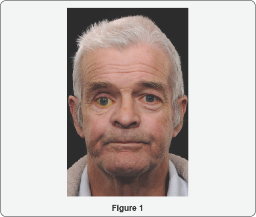
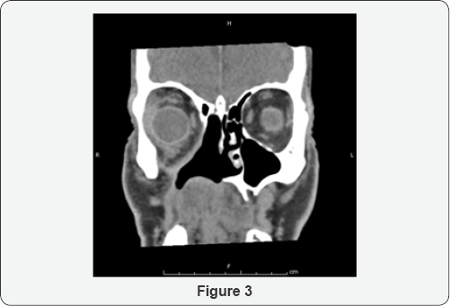
An assessment of his CT scan showed an extensive
hemi- maxillectomy with resection of the orbital floor, much of the
lamin papyracea, the maxillary antrum and hard palate. Significant
enophthalmos was noted. Soft tissue thickening in the region of the
surgical margins was noted (Figure 1).
As the patient had undergone post-operative radiotherapy, a
conservative approach using NASHA for volume augmentation was decided as
an initial step. Floor and medial wall reconstruction with
implant/dermo fat graft at the level of the equator of orbit was
deferred following discussion with the patient. Correction of lid
position would be deferred until later.
He underwent an injection of Orbital (non-animal
stabilized hyaluronic acid) NASHA. Retrobulbar block was used comprising
of Lignocaine 2% with adrenaline 1:200000; 1.5mls was injected, deep,
below the globe. The plunger was initially withdrawn to ensure blood
vessels were not entered, and a staged augmentation was performed, Three
mls of Perlane (Q-Med, Uppsala, Sweden) was injected above the
periorbitum of the floor, using an 18G needle. Immediately after the
procedure, vision and optic nerve function were monitored and were
normal (Compare Figures 2 &3).
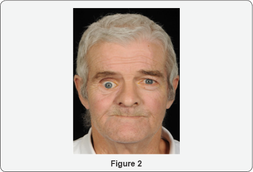
At review 1-week later, a 3mm improvement in
hyppoglobus and 4mm improvement in enophthalmos was noted. However, he
was aware of vertical diplopia, and this was controlled with a base-
down 3-dioptre prism. This was thought to be due to loss of right eye
suppression with the new globe position.
A further review was performed at 8-months
post-injection and a slight increase in enophthalmos and hypoglobus were
noted.At 13-months after the initial injection, encouraged by initial
results, a further injection of 3mls of Perlane (Q-Med, Uppsala, Sweden)
was injected using the same technique as before with tightening of the
lower lid by means of a lateral canthal elevation. This resulted in an
improvement in hypoglobus to 2-3mm. His diplopia persisted without
prisms, although over the next 8-months started coping better without
prisms and the diplopia resolved with normal binocular single vision.
The long-term plan was to continue with annual orbital NASHA injections
Discussion
From an ophthalmic point of view, mid-face
reconstruction aims to provide support to orbital contents, minimize
changes in globe position, orbital volume and eyelid function [1].
Symmetrical orbital volume is a requirement for binocular single
vision. Complications following reconstructive surgery, and
radiotherapy, include resorption of orbital fat and wound contracture,
exacerbating enophthalmos and diplopia. Post-maxillectomy radiation
therapy increases the risk of orbital sequelae, such as fat atrophy and
scar formation, and ocular sequelae, such as optic atrophy, cataract
formation, ocular surface dryness, and ectropion [2].
Orbital reconstruction may range from minimal to
complex. Minimal bone resections or small orbital floor defects may not
require reconstruction. On the other end of the spectrum, subtotal/
total floor defects (>75% surface area) and multi segmental defects
will necessitate rigid reconstruction. Primary reconstruction of total
maxillectomy defects with preservation of orbital contents remains a
complex problem without a perfect solution [3]. In a case series of 66 patients with sinonasal malignancy that had eye-sparing surgery, [2]
the most common abnormality following subsequent reconstruction was
globe mal position (enophthalmos or hypophthalmos) (34/66 patients,
63%), associated with lack of adequate rigid reconstruction of subtotal
or total orbital floor or multisegment orbital defects [2]. The problem can be exacerbated with post-operative radiotherapy [4].
In our case, the patient was keen for correction of
his vertical dystopia from a functional and cosmetic point of view.
Surgery would have been quite complex due to the absence of the floor,
medial and lateral walls. The option of volume augmenatation with a
dermo fat graft was considered, but there was a possibility that this
graft might not survive due to lack of circulation. There was also a
risk of recurrence of the original pathology. A conservative approach
was taken and floor augmentation with NASHA was decided as the first
stage of the procedure. This addressed both the hypoglobus and
enophthalmos to the patient's satisfaction, to the extent that need for
further orbital surgery was obviated. Furthermore, binocular single
vision returned with the use of prisms.
There are no reports addressing reconstruction of the
orbital component of the post-maxillectomy defect using injectable
materials for volume augmentation that we are aware of. A minimally
invasive approach to orbital volume augmentation using agents such as
injectable calcium hydroxylapatite (CaHA), polyacrylamide gel, hydrogel
pellet expanders, micro-fat grafts and NASHA have been well described in
the anophthalmic socket (post-enucleation socket syndrome). There are
only a handful of reports in the context of sighted globes. These
include silent sinus syndrome, [5] deep set eyes, [6,7] trauma, [7] and facial hemiatrophy (Parry Romberg syndrome) [7]. We are not aware of any reports of use of filler in the context of carcinoma.
NASHA is produced from non-animal sources by
bacterial fermentation. It is well tolerated, has a low immunogenic
potential, and there is low risk ofantigenic contamination and
hypersensitivity reactions [8].
A major advantage in sighted globes is that its effects can be reversed
by dissolving it with hyaluronidase. Disadvantages include a relatively
short life span compared to other products and the requirement to
inject through an 18G needle in order to maintain particle integrity [8].
However, blunt cannulas can be used. Some studies have reported a 1:1
correlation between the desired augmentation volume (enophthalmos
reduction) and each millimetre of product injected, [7] whilst others have not [9].
(We feel the augmentation effect depends on the remaining bony walls
and the integrity of the periorbita). Twelve months is the longest
duration of action reported [7,9].
Injection of NASHA is recommended in the anterior orbit, and behind the
equator of the globe, in order to achieve globe elevation and axial
globe advancement respectively [7,9,10].
Injections may be intraconal or extraconal. Proponents of the former
cite better globe advancement and reduced anterior migration of filler; [11] proponents of the latter cite risk of vascular compression in sighted globes with intraconal placement [8].
Possible complications include risk of intravascular
injection and embolization with loss of vision in sighted eyes. Commonly
reported complications include injection site tenderness, ecchymosis
and chemosis, [12] stimulation of oculocardiac reflex and associated hypotension, [7,8,12] transient ptosis, [6] anterior migration of filler [13] and inflammatory orbital cellulitis [9].
The benefits of a minimally invasive approach over
traditional surgery include reduced operating times, application under
local anaesthesia, reduced recovery time and reduced risk of host and
donor site morbidity where non-biological materials are used. The
benefits need to be weighed against the risks. In the setting of
post-neoplasia reconstruction where the prognosis may be poor, use of
minimally invasive, albeit temporary, techniques can help to the improve
quality and dignity of life for patients, especially where resorting to
more complex surgical procedures may be too demanding for the patient
in view of their morbidity and reduced life-expectancy [14].
In summary, injectable fillers can provide a
temporary solution to volume augmentation of the orbit, for functional
restoration and improvement in cosmesis; and may be used in sighted
globes with caution. They may be a superior option to traditional
surgery in selected cases.
For more articles in JOJ Ophthalmology (JOJO) please click on: https://juniperpublishers.com/jojo/index.php
Friday, 31 January 2020
Schwartz Jampel Syndrome- A Case Report- Juniper Publishers
Juniper
Publishers- JOJ Ophthalmology
Itroduction
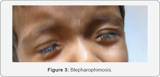
Blepharophimosis is a general diminution of palpebral
fissure in all its dimensions. The lids usually show ptosis, dystopia
canthorum, lateral displacement of the lateral puncti, or abnormalities
of the lashes such as ditichiasis or misdirected and stiff lashes. The
other ocular defects associated with congenital blepharophimosis include
strabismus, nystagmus, amblyopia, microphthalmus, anophthalmus,
epicanthus inversus, microcornea and hypermetropia [1,2]. Schwartz-Jampel syndrome, an autosomal recessively transmitted disease, is a rare presentation of blepharophimosis.
Case History
A 2 year old male child, having dysmorphic features
was referred from the department of Pediatrics for Ophthalmic
assessment. The child was the first born of healthy non- consanguineous
parents after an uneventful pregnancy. His mental and motor development
was normal and he acquired independent walking at 16 months. Fine pincer
grasp developed by 9 months of age. At the age of 2 years the child
could talk only two words with meaning. Social development of the child
was poor because of his abnormal appearance and poor language
development. The parents noted the abnormal facial expression at the age
of 18 months.
On examination the child had a short stature. The
head posture was normal. Forehead did not show excessive wrinkling. The
child had blepharophimosis (Figure 1).
Lid crease was present. The child also had hypertrichosis. The globe
examination was normal. The extraocular movements were normal. There was
no refractive error. Fundus was normal. The child demonstrated pursing
of lips giving him a 'whistling face' appearance and restricting his
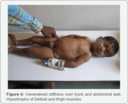
mouth opening (Figure 2). The shape of the chest was abnormal with sternal protrusion and sub-costal retraction (Figure 3).
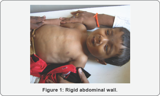
There was stiffness of his abdominal wall. The upper and lower limbs
demonstrated hypertonia. The deep tendon reflexes were exaggerated. He
had a waddling gait. The child had a high pitched voice (Figure 4).
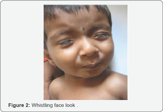
Discussion
Schwartz-Jampel syndrome is a rare autosomal
recessively transmitted disease, characterized by generalized myotonic
myopathy, typical facial features, skeletal dysplasia, contracture of
joints, growth retardation and bone maturation delay [3].
However a few cases showing dominant inheritance have also been
reported. It is classified into 3 types based on age and severity
- Type 1A
- Type 1B
- Type 2
Type 1A
The type 1A disease is diagnosed in mid-childhood
with recognition of myotonic facies with convex profile, short palpebral
fissure, telecanthus, dimpling or quivering of the chin, prominent
eyebrows, low hairline, low-set ears, flat base of the nose,
micrognathia, microstomia, sometimes high-arched palate. The child
exhibits progressive myotonia, muscle wasting and orthopaedic problems
with decreased linear growth myotonia plateus by mid childhood.
Additional findings reported in a few cases are myopia, hypertrichosis,
and strabismus. The continuous myotonia is probably responsible for both
muscular hypertrophy and peculiar facial appearance.
Type 1B
Type 1B is more severe than 1A, Bone dysplasia is present at birth.
Long bones are shortened, femurs are dumbbell shaped. Bone epiphyses are
large and vertebral bodies are flat.
Type 2
Type2 disease is more severe. Onset is neonatal,
there is short limb dysplasia and long bones are bowed. Early death is
frequent [4].
The diagnosis is predominantly on the basis of the typical dysmorphic facies [5].
EMG showing continuous discharges further supports the diagnosis. The
gene defect in SJS type 1 is located in the 1p34-p36 of chromosome 1,
whereas it is different in type 2 [6,7]. Perlecan the major proteoglycan of basement membranes is altered in patients with Schwartz- jampel syndrome disease [8].
However, a significant amount of molecular heterogeneity exists,
genomically and proteomically, within SJS type 1. Currently no known
correlation exists between the specific mutations found and the specific
features of a given case However, the new mutations found by Stum et
al. In 2006 have been discovered so recently that not enough time has
elapsed to explore such possibilities. The new findings should be
important tools to help find correlations among genetic variants,
perlecan forms and levels, and clinical subtypes. Other facts yet
unknown also may influence the severity and the specific characteristics
of the disease [5]. The genetic tests for perlecan gene are not easily available in the commercial laboratories.
The child was diagnosed as having type 1A type of
Schwartz- Jampel syndrome since the typical facial features became
manifest at the age of 18 months. The old pictures of the child taken on
his first birthday showed normal facial features. Medications that have
been found useful in myotonic disorders such as phenytoin and
carbamazipene may help to reduce the abnormal muscle activity. Warm
baths are helpful in reducing stiffness. Botox injections are reportedly
found useful to relieve blepharospsm.
Patients are generally treated with Carbamizipene
2030mg/kg body weight and most of them show improvement. Carbamazipene
probably works by inhibiting neuronal sodium channels and may have
direct effects on neurotransmitter systems. Orbicularis oculi myectomy,
levator aponeurosis resection and lateral canthopexy are some surgical
procedures which may be tried if the response to carbamazipene or botox
is not adequate. The parents of the child were educated regarding the
genetic nature of the disease and were referred to the geneticist. This
particular child has not reported for follow-up as he belongs to a
remote village far from our hospital and is probably reporting for
follow-up at a nearby city.
Conclusion
Schwartz-Jampelsyndromeisararecauseofblepharophimosis. The condition can
be managed with medications in most of the? cases. Surgery may be
required if the condition does not improve with drugs.
For more articles in JOJ Ophthalmology (JOJO) please click on: https://juniperpublishers.com/jojo/index.php
Thursday, 30 January 2020
Eye Loss Due to Disseminated Molluscum Contagiosum Skin Infection Involving the Eyelids in an Immune Competent Child- Juniper Publishers
Juniper
Publishers- JOJ Ophthalmology
Introduction
Molluscum contagiosum is a viral infection of skin
and mucous membranes caused by a double-stranded DNA poxvirus. The virus
causes a characteristic skin lesion consisting of a single or multiple
round pearly white umblicated papules [1]. Molluscum contagiosum is largely if not exclusively a human disease although there are few reported cases in some animals [2]. Distribution is worldwide, but it is more common in areas with hot climate [3].
The virus is transmitted directly through skin to skin contact with
other infected patients or indirectly through contact with contaminated
fomites such as bath sponges and towel. The virus can also be
transmitted to other areas in the same patient by autoinoculation [4].
Although all age groups can be affected, it commonly occurs in two age
peaks: children and adults. Children are usually infected by casual
contact and young adults infected by sexual contact [5].
Clinically molluscum contagiosum lesions are usually asymptomatic;
however, some lesions may become pruritic or tender due to associated
eczema or inflammation. There are no systemic symptoms [6]. In most cases lesions resolve spontaneously without treatment over the course of several months [7].
On examination, the skin lesions are round, dome shaped, pearly, flesh
colored, firm papules with central umblication. They are usually 2-5mm
in diameter (except for giant molluscum which may reach few
centimeters). Beneath the umbilicated center is a white, curd-like core
that contains molluscum bodies. Lesions may be single or multiple
distributed on the skin of the head- including the eye lids, neck,
trunk, the limbs, and around the genital area [8]. Rarely, it may involve the palms, the soles, mucous membranes of the mouth, or conjunctiva [9,10].
Immuno compromised patients - children and adults, such as HIV patients
and patients on immunosuppressive therapy, tend to have atypical and
more wide spread and persistent lesions [5,11].
The diagnosis of molluscum contagiosum is clinically
evident by the characteristic appearance of the skin lesion. In atypical
or giant lesions, a biopsy can be done to reach diagnosis.
Histopathology reveals characteristic intracytoplasmic inclusion bodies
(molluscum or Henderson-Paterson bodies) [12]. Other tests include complement fixation test (CFT) and polymerase chain reaction (PCR) [13].
Treatment in healthy individuals is not always necessary because most
cases are self limiting. Indications include: relieving symptoms and
discomfort, improving cosmetic appearance, persistent lesions, and
reduction of autoinoculation and spread to other contacts [14]. Many modalities exist [15]. Treatments can be divided into three categories: destructive- physical and chemical), immune modulators, and antiviral [16].
Case Report
A four years old female child presented to the
dermatologist with disseminated skin lesions involving the whole body
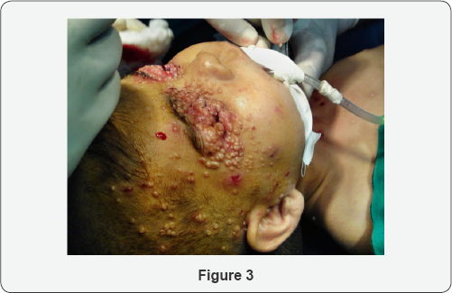
surface area. The lesions were scattered all over the face, neck, trunk
and limbs with larger concentrations around the eyelids- both eyes- and
the genital region. The lesions were round pearly white umblicated
papules typical of molluscum contagiosum. The patient consulted many
dermatologists before she was referred to an ophthalmologist for eye
examination. On examination, the lesions were more confluent and
concentrated around the eyelids skin and eyelid margins making it
difficult to open the balpebral fissure for inspection of the
conjunctiva and corneal surface, and the condition was associated with
secondary pyogenic infection and discharge around the eyelid margin.
Examination under general anesthesia to facilitate eyelids opening and
subsequent surgical removal revealed infective keratitis on the right
side with profuse pus discharge and extensive corneal stromal ulceration
and melting.


The treating dermatologist and ophthalmologist
started surgical excision of as many lesions as possible. The eye
postoperatively was treated with intensive topical antibiotics eye drops
and eye ointment for several days until the infection was resolved and
healing of the corneal surface took place. The patient was referred to a
pediatrician for the investigation of the possible cause of immune
deficiency. The patient did not return subsequently for follow-up (Figure 1-3).
Discussion
Molluscum contagiosum is usually described as a benign and self limiting skin infection that does not always require treatment [17]. However, this may not be the case when the eye is involved [18]. Ocular manifestations may present as a range of complications [1,6,19].
Lesion located on or near the lid margin may give rise to secondary
chronic follicular conjunctivitis. Unless the lid margin is examined
carefully, the causative molluscum lesion may be overlooked therefore it
can be easily misdiagnosed and mistreated. Prolonged follicular
conjunctivitis or secondary bacterial infection can result in keratitis
usually in the form of fine punctate epithelial erosions or sub
epithelial opacities. Corneal vascularisation, scarring and
opacification may result in visual acuity loss. Molluscum contagiosum
infection commonly involves the face and hands. Itching and scratching
facilitate extension of infection to other parts of the same patient;
therefore, the disease usually presents as multiple crops and less
commonly as a solitary lesion which sometimes becomes a confluent
multilobulated giant tumor affecting the eyelid [20,21]. Secondary infection and ulceration can result in permanent scarring.
Molluscum contagiosum is a common pediatric dermatosis in Iraq [22]. Al-Azawi reported a high prevalence of molluscum contagiosum virus (MCV) type I in children age group<10 years [23].
In our clinical practice, molluscum contagiosum infection is wide
spread and eye involvement is very common in Iraq. Predisposing factors
may include low socioeconomic status, crowding, and low personal
hygiene. It affects all age groups especially children in preschool age
and primary school age. This highly contagious infection is usually
acquired from contact with other infected people. They could be family
members or visitors or more commonly other infected children in the
neighborhood or schools.
Molluscum contagiosum infections involving the
eyelids and periocular area are usually managed by ophthalmologists, and
sometimes referred by dermatologists. Although many modalities of
therapy are effective in destruction of the virus, the use of substances
such as liquid nitrogen or chemicals in the vicinity of the eye may be
hazardous [24,25]. Surgical removal by shave excision or curettage is a simple and effective procedure [23].
However, multiplicity of the lesions and young patient age usually
necessitate light general anesthesia given by an anesthesiologist in an
operation theater and therefore cannot be done as an outpatient office
procedure in the minor surgical room. This may result in considerable
suffering to the patient and parents and burden on the health care
providers [3].
Conclusion
Molluscum contagiosum is not always a self-limiting
benign skin infection, but it can cause serious eye complications
especially in the third world. Active treatment is indicated to prevent
secondary complications and limit the spread of the disease to other
people.
Disclosure
The author reports no conflicts of interest in this work.
For more articles in JOJ Ophthalmology (JOJO) please click on: https://juniperpublishers.com/jojo/index.php
Wednesday, 29 January 2020
Eye Loss Due to Disseminated Molluscum Contagiosum Skin Infection Involving the Eyelids in an Immune Competent Child- Juniper Publishers
Juniper
Publishers- JOJ Ophthalmology
Abstract
Background: Molluscum contagiosum is a viral
infection of skin and mucous membranes caused by a DNA poxvirus. It is a
common skin infection in children with numerous ocular manifestations.
Case report: Eye loss due to disseminated molluscum
contagiosum infection of the skin involving the eyelids in a 4 years old
immune competent female child. The clinical history, exanimation,
treatment and follow-up are presented.
Conclusion: molluscum contagiosum is not always a
self limiting benign skin infection and can cause serious eye
complications especially in the third world. Active treatment is
indicated to prevent secondary complications and limit the spread of the
disease to other people.
Keywords: Viral infection; Molluscum contagiosum; Eye lossIntroduction
Molluscum contagiosum is a viral infection of skin
and mucous membranes caused by a double-stranded DNA poxvirus. The virus
causes a characteristic skin lesion consisting of a single or multiple
round pearly white umblicated papules [1]. Molluscum contagiosum is largely if not exclusively a human disease although there are few reported cases in some animals [2]. Distribution is worldwide, but it is more common in areas with hot climate [3].
The virus is transmitted directly through skin to skin contact with
other infected patients or indirectly through contact with contaminated
fomites such as bath sponges and towel. The virus can also be
transmitted to other areas in the same patient by autoinoculation [4].
Although all age groups can be affected, it commonly occurs in two age
peaks: children and adults. Children are usually infected by casual
contact and young adults infected by sexual contact [5].
Clinically molluscum contagiosum lesions are usually asymptomatic;
however, some lesions may become pruritic or tender due to associated
eczema or inflammation. There are no systemic symptoms [6]. In most cases lesions resolve spontaneously without treatment over the course of several months [7].
On examination, the skin lesions are round, dome shaped, pearly, flesh
colored, firm papules with central umblication. They are usually 2-5mm
in diameter (except for giant molluscum which may reach few
centimeters). Beneath the umbilicated center is a white, curd-like core
that contains molluscum bodies. Lesions may be single or multiple
distributed on the skin of the head- including the eye lids, neck,
trunk, the limbs, and around the genital area [8]. Rarely, it may involve the palms, the soles, mucous membranes of the mouth, or conjunctiva [9,10].
Immuno compromised patients - children and adults, such as HIV patients
and patients on immunosuppressive therapy, tend to have atypical and
more wide spread and persistent lesions [5,11].
The diagnosis of molluscum contagiosum is clinically
evident by the characteristic appearance of the skin lesion. In atypical
or giant lesions, a biopsy can be done to reach diagnosis.
Histopathology reveals characteristic intracytoplasmic inclusion bodies
(molluscum or Henderson-Paterson bodies) [12]. Other tests include complement fixation test (CFT) and polymerase chain reaction (PCR) [13].
Treatment in healthy individuals is not always necessary because most
cases are self limiting. Indications include: relieving symptoms and
discomfort, improving cosmetic appearance, persistent lesions, and
reduction of autoinoculation and spread to other contacts [14]. Many modalities exist [15]. Treatments can be divided into three categories: destructive- physical and chemical), immune modulators, and antiviral [16].
Case Report
A four years old female child presented to the
dermatologist with disseminated skin lesions involving the whole body
surface area. The lesions were scattered all over the face, neck, trunk
and limbs with larger concentrations around the eyelids- both eyes- and
the genital region. The lesions were round pearly white umblicated
papules typical of molluscum contagiosum. The patient consulted many
dermatologists before she was referred to an ophthalmologist for eye
examination. On examination, the lesions were more confluent and
concentrated around the eyelids skin and eyelid margins making it
difficult to open the balpebral fissure for inspection of the
conjunctiva and corneal surface, and the condition was associated with
secondary pyogenic infection and discharge around the eyelid margin.
Examination under general anesthesia to facilitate eyelids opening and
subsequent surgical removal revealed infective keratitis on the right
side with profuse pus discharge and extensive corneal stromal ulceration
and melting.
The treating dermatologist and ophthalmologist
started surgical excision of as many lesions as possible. The eye
postoperatively was treated with intensive topical antibiotics eye drops
and eye ointment for several days until the infection was resolved and
healing of the corneal surface took place. The patient was referred to a
pediatrician for the investigation of the possible cause of immune
deficiency. The patient did not return subsequently for follow-up (Figure 1-3).
Discussion
Molluscum contagiosum is usually described as a benign and self limiting skin infection that does not always require treatment [17]. However, this may not be the case when the eye is involved [18]. Ocular manifestations may present as a range of complications [1,6,19].
Lesion located on or near the lid margin may give rise to secondary
chronic follicular conjunctivitis. Unless the lid margin is examined
carefully, the causative molluscum lesion may be overlooked therefore it
can be easily misdiagnosed and mistreated. Prolonged follicular
conjunctivitis or secondary bacterial infection can result in keratitis
usually in the form of fine punctate epithelial erosions or sub
epithelial opacities. Corneal vascularisation, scarring and
opacification may result in visual acuity loss. Molluscum contagiosum
infection commonly involves the face and hands. Itching and scratching
facilitate extension of infection to other parts of the same patient;
therefore, the disease usually presents as multiple crops and less
commonly as a solitary lesion which sometimes becomes a confluent
multilobulated giant tumor affecting the eyelid [20,21]. Secondary infection and ulceration can result in permanent scarring.
Molluscum contagiosum is a common pediatric dermatosis in Iraq [22]. Al-Azawi reported a high prevalence of molluscum contagiosum virus (MCV) type I in children age group<10 years [23].
In our clinical practice, molluscum contagiosum infection is wide
spread and eye involvement is very common in Iraq. Predisposing factors
may include low socioeconomic status, crowding, and low personal
hygiene. It affects all age groups especially children in preschool age
and primary school age. This highly contagious infection is usually
acquired from contact with other infected people. They could be family
members or visitors or more commonly other infected children in the
neighborhood or schools.
Molluscum contagiosum infections involving the
eyelids and periocular area are usually managed by ophthalmologists, and
sometimes referred by dermatologists. Although many modalities of
therapy are effective in destruction of the virus, the use of substances
such as liquid nitrogen or chemicals in the vicinity of the eye may be
hazardous [24,25]. Surgical removal by shave excision or curettage is a simple and effective procedure [23].
However, multiplicity of the lesions and young patient age usually
necessitate light general anesthesia given by an anesthesiologist in an
operation theater and therefore cannot be done as an outpatient office
procedure in the minor surgical room. This may result in considerable
suffering to the patient and parents and burden on the health care
providers [3].
Conclusion
Molluscum contagiosum is not always a self-limiting
benign skin infection, but it can cause serious eye complications
especially in the third world. Active treatment is indicated to prevent
secondary complications and limit the spread of the disease to other
people.
Disclosure
The author reports no conflicts of interest in this work.
For more articles in JOJ Ophthalmology (JOJO) please click on: https://juniperpublishers.com/jojo/index.php
Subscribe to:
Posts (Atom)