Juniper
Publishers- JOJ Ophthalmology
Introduction
Molluscum contagiosum is a viral infection of skin
and mucous membranes caused by a double-stranded DNA poxvirus. The virus
causes a characteristic skin lesion consisting of a single or multiple
round pearly white umblicated papules [1]. Molluscum contagiosum is largely if not exclusively a human disease although there are few reported cases in some animals [2]. Distribution is worldwide, but it is more common in areas with hot climate [3].
The virus is transmitted directly through skin to skin contact with
other infected patients or indirectly through contact with contaminated
fomites such as bath sponges and towel. The virus can also be
transmitted to other areas in the same patient by autoinoculation [4].
Although all age groups can be affected, it commonly occurs in two age
peaks: children and adults. Children are usually infected by casual
contact and young adults infected by sexual contact [5].
Clinically molluscum contagiosum lesions are usually asymptomatic;
however, some lesions may become pruritic or tender due to associated
eczema or inflammation. There are no systemic symptoms [6]. In most cases lesions resolve spontaneously without treatment over the course of several months [7].
On examination, the skin lesions are round, dome shaped, pearly, flesh
colored, firm papules with central umblication. They are usually 2-5mm
in diameter (except for giant molluscum which may reach few
centimeters). Beneath the umbilicated center is a white, curd-like core
that contains molluscum bodies. Lesions may be single or multiple
distributed on the skin of the head- including the eye lids, neck,
trunk, the limbs, and around the genital area [8]. Rarely, it may involve the palms, the soles, mucous membranes of the mouth, or conjunctiva [9,10].
Immuno compromised patients - children and adults, such as HIV patients
and patients on immunosuppressive therapy, tend to have atypical and
more wide spread and persistent lesions [5,11].
The diagnosis of molluscum contagiosum is clinically
evident by the characteristic appearance of the skin lesion. In atypical
or giant lesions, a biopsy can be done to reach diagnosis.
Histopathology reveals characteristic intracytoplasmic inclusion bodies
(molluscum or Henderson-Paterson bodies) [12]. Other tests include complement fixation test (CFT) and polymerase chain reaction (PCR) [13].
Treatment in healthy individuals is not always necessary because most
cases are self limiting. Indications include: relieving symptoms and
discomfort, improving cosmetic appearance, persistent lesions, and
reduction of autoinoculation and spread to other contacts [14]. Many modalities exist [15]. Treatments can be divided into three categories: destructive- physical and chemical), immune modulators, and antiviral [16].
Case Report
A four years old female child presented to the
dermatologist with disseminated skin lesions involving the whole body
surface area. The lesions were scattered all over the face, neck, trunk
and limbs with larger concentrations around the eyelids- both eyes- and
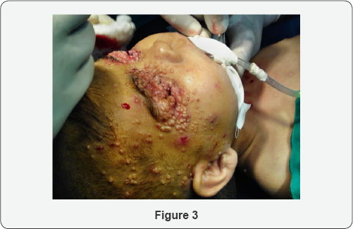
the genital region. The lesions were round pearly white umblicated
papules typical of molluscum contagiosum. The patient consulted many
dermatologists before she was referred to an ophthalmologist for eye
examination. On examination, the lesions were more confluent and
concentrated around the eyelids skin and eyelid margins making it
difficult to open the balpebral fissure for inspection of the
conjunctiva and corneal surface, and the condition was associated with
secondary pyogenic infection and discharge around the eyelid margin.
Examination under general anesthesia to facilitate eyelids opening and
subsequent surgical removal revealed infective keratitis on the right
side with profuse pus discharge and extensive corneal stromal ulceration
and melting.


The treating dermatologist and ophthalmologist
started surgical excision of as many lesions as possible. The eye
postoperatively was treated with intensive topical antibiotics eye drops
and eye ointment for several days until the infection was resolved and
healing of the corneal surface took place. The patient was referred to a
pediatrician for the investigation of the possible cause of immune
deficiency. The patient did not return subsequently for follow-up (Figure 1-3).

Discussion
Molluscum contagiosum is usually described as a benign and self limiting skin infection that does not always require treatment [17]. However, this may not be the case when the eye is involved [18]. Ocular manifestations may present as a range of complications [1,6,19].
Lesion located on or near the lid margin may give rise to secondary
chronic follicular conjunctivitis. Unless the lid margin is examined
carefully, the causative molluscum lesion may be overlooked therefore it
can be easily misdiagnosed and mistreated. Prolonged follicular
conjunctivitis or secondary bacterial infection can result in keratitis
usually in the form of fine punctate epithelial erosions or sub
epithelial opacities. Corneal vascularisation, scarring and
opacification may result in visual acuity loss. Molluscum contagiosum
infection commonly involves the face and hands. Itching and scratching
facilitate extension of infection to other parts of the same patient;
therefore, the disease usually presents as multiple crops and less
commonly as a solitary lesion which sometimes becomes a confluent
multilobulated giant tumor affecting the eyelid [20,21]. Secondary infection and ulceration can result in permanent scarring.
Molluscum contagiosum is a common pediatric dermatosis in Iraq [22]. Al-Azawi reported a high prevalence of molluscum contagiosum virus (MCV) type I in children age group<10 years [23].
In our clinical practice, molluscum contagiosum infection is wide
spread and eye involvement is very common in Iraq. Predisposing factors
may include low socioeconomic status, crowding, and low personal
hygiene. It affects all age groups especially children in preschool age
and primary school age. This highly contagious infection is usually
acquired from contact with other infected people. They could be family
members or visitors or more commonly other infected children in the
neighborhood or schools.
Molluscum contagiosum infections involving the
eyelids and periocular area are usually managed by ophthalmologists, and
sometimes referred by dermatologists. Although many modalities of
therapy are effective in destruction of the virus, the use of substances
such as liquid nitrogen or chemicals in the vicinity of the eye may be
hazardous [24,25]. Surgical removal by shave excision or curettage is a simple and effective procedure [23].
However, multiplicity of the lesions and young patient age usually
necessitate light general anesthesia given by an anesthesiologist in an
operation theater and therefore cannot be done as an outpatient office
procedure in the minor surgical room. This may result in considerable
suffering to the patient and parents and burden on the health care
providers [3].
Conclusion
Molluscum contagiosum is not always a self-limiting
benign skin infection, but it can cause serious eye complications
especially in the third world. Active treatment is indicated to prevent
secondary complications and limit the spread of the disease to other
people.
Disclosure
The author reports no conflicts of interest in this work.
For more articles in JOJ Ophthalmology (JOJO) please click on: https://juniperpublishers.com/jojo/index.php
No comments:
Post a Comment