Juniper
Publishers- JOJ Ophthalmology
Abstract
The OCTA is a novel evolving imaging technology which
utilizes motion contrast to visualize retinal and choroidal vessels. It
showed promises to be used in predicting, grading, guiding and
following treatment of important ocular vascular diseases. The main
advantages of the OCTA are being non-invasive; being blue light and dye
free and providing high quality images in a relatively short time. It
has alleviated important limitations of Fluorescein Angiography (FA),
but still it is not considered as a complete substitute of FA, by
experts. The Combined FA/ICGA and OCTA imaging systems are introduced to
the market. It may have the advantages of the both systems, while
having the least limitations. Resolution of images, field of view, depth
of images and scan speed are important factors when choosing a device.
But one should also take into account that higher image quality needs
more time to be acquisitioned. It could be challenging in busy clinics.
In this article we compared FA and OCTA and also compared widely
available commercial prototype of OCTA devices.
Introduction
Optical Coherence Tomography Angiography (OCTA) is a
novel imaging technology which has considerable advantages over older
angiograms token by Fluorescein Angiography (FA) or Indocyanine Green
Angiogarphy (ICGA). On top of them, one may list advantages like being
non-invasive; no needs for dye; high resolution simultaneous
visualization of the both retinal and choroidal vasculatures;
simultaneous 3 dimensional (3D) visualization of retinal and choroidal
structure; and possibility of segmentation of retinal layers and
capillary plexuses in 3 layers including Superficial (SCP), Middle (MCP)
and Deep (DCP) [1].
One of the promising features of this technology is that beside
qualitative data, it provides quantitative data regarding retinal and
choroidal structural and vascular indices like Vascular Density (VD),
Foveal Avascular Zone (FAZ), etc. Feasible Quantitative data provided by
OCTA; may revolutionize current ophthalmic practice in regards of
predicting [2,3], grading [4,5],
following up treatment in patients with important vascular diseases
like diabetic retinopathy, retinal vein obstruction, choroidal
neovascularization, etc. [6-10].
However, currently the quantitative data are mostly used for
researches, But it seems that in the future when some cut off points are
available by large scale studies. Then, these data could be used for
everyday clinical practices. For an instance, quantitative assessment of
Foveal Avascular Zone (FAZ) could be useful in optimal selection of
therapy in patients with Diabetic Macular Edema (DME) [11] or even grading the severity of Diabetic Retinopathy (DR) [5,12]. It has also shown that FAZ April 19, 2017metrics could change in response to treatment [6] so it could be used in following up patients. But a recent study has challenged these changes [13].
It should be emphasized that FA could also provide data regarding FAZ,
but OCTA is more reliable, precise and also much more feasible. In FA,
frequently dye leakage or DCP and SCP overlaps may influence the FAZ
measurement [14]. Hereby, the OCTA technology, advantages, disadvantages and some commercial prototypes are discussed.
Optical Coherence Tomography Angiography Technology
The principal of this imaging system is detecting
motion contrasts. This device record and compare multiple fast B-Scans
of each vascular layer of retina. It simply presumes that the only
motion inside retina is related to red blood cells (RBC) within
vasculatures. These decorrelation signals are mapped in an OCT
angiogram. Lastly, OCT B-Scan and OCT angiogram join together to
visualize the both histological and vascular structures at the same time
[15].
Optical Coherence Tomography Angiography vs. Fluorescein Angiography
Currently, FA and ICGA are gold standard in
assessment of retinal and choroidal vasculatures. But the FA has
considerable shortages like being invasive; being dye dependent; putting
patients at possible dye mortal side effects (however, rare); clinical
contraindications of dye; putting retina at risk of blue light toxicity;
relative long picture acquisition time (some 15 minutes); disability in
assessing deeper retinal or choroidal layers; disability in providing
3D pictures, disability in providing structural details of retina and
choroid; and not providing quantitative data. It seems that OCTA has
alleviated all above FA's limitations. However, as any other device, it
has inherited some technical limitations [16,17].
One may count: more limited field of view; not providing functional
data regarding vessels like not showing leakages; being more sensitive
to small eye movements; needs for more patients' cooperation and ability
to maintain proper fixation [18].
This later may make the acquisition time in real practice much longer
than official announcements by manufacturers. Different commercial
devices utilize various technologies to improve quality of picture by
reducing motion artifacts [19]; through special algorithms (amplitude decorrelation algorithm, OCT-based or optical microangiography (OMAG) [20], split-spectrum amplitude decorrelation angiography (SSADA), etc.) [21]; and also improving the field and depth of pictures [22].
In this technology, we encounter day to day evolution of imaging system
in terms of eye tracking systems; speed of picture acquisition;
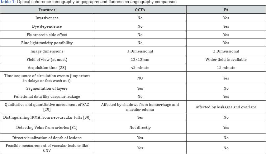
artifact reduction solutions [23]; field and depth of images [22]. Table 1
compares FA and OCTA. The invention of the combined imaging systems
which provide a hybrid FA/ICGA and OCTA images may have the least
limitations.

FA: Fluorescein Angiography; FAZ: Foveal Avascular Zone; OCTA: Optical Coherence.
Tomography Angiography; IRMA: Intraretinal Microvascular Abnormalities
Is it possible to Upgrade SD-OCT Devices to OCTA Device?
The SD-OCT devices can do 26 to 40 thousands scans
per seconds while commercial OCTA's scan speed is some two-fold of this.
The resolution of images (indirectly, as resolution is dependent on
number of scan per section which is limited by fixed scan speed and
acquisition time) and their sensitivity to motion artifacts is
particularly dependent on this. So the SD- OCT can not be utilized to
obtain angiogram as images would be small and clinically useless.
Fortunately, some manufacturers supplied their previous SD-OCT users
with a two-step SD-OCT to OCTA upgrade. Firstly, they upgrade the
hardware of device to higher frequency scan device, then they install
OCTA software module on the device.
Commercial Prototypes Comparison
Most commonly used device in clinical centers, is AngioVue (Opto Vue, Inc., Fremont, Calif., USA) [10].
Recently, Heidelberg Engineering has released its OCTA modules.
AngioPlex (Zeiss . Meditec, Inc., Dublin, Calif., USA) is also an other
widely available device in market Table 2 compare features of these brands, provided by their manufacturer.

All the values are retrived from official websites of manufacturers.
While choosing a device one should consider following
issues. The higher the scan speed is, the lower the effect of motion
artifact would be. And also the resolution of images depends on number
of scans per section. As the scan speed and scan acquisition time are
limited, so it should be taken in to account that the high resolution of
image translate to more acquisition time which is challenging in busy
clinics. It is the reason why some manufacturer has not announced their
device acquisition time, officially. And also some have reduced their
image quality. As the OCTA is not substitute of FA in expert opinions;
and also it is considerably expensive technology; moreover, many clinics
has physical space limitation; so devices that provide hybrid FA/ ICGA
and OCTA, could be an all-in-one reasonable option.
Future of Optical Coherence Tomography Angiography
Currently, the clinical use of OCTA is limited by
its' expensiveness; small field of imaging; slow acquisition time;
quality of images; lack of cut off points for quantitative data; lack of
defined clinical significance of enormous data provided.
While developers are trying to invent faster swept
source devices; smarter eye tracking systems; and also reducing
artifacts that could provide wider higher quality views in matter of
seconds, clinicians should utilize massive data provided by this
technology in their everyday clinical practice by investigating the
clinical relevance of findings. As we are on the edge of new robotic
era, there are promises that images of retina could be efficiently
processed by computers to diagnosis and grade diseases [24]
and also conjunction of surgical or laser devices with imaging system
could revolutionize both the diagnosis and treatment of ocular diseases [25-31].
For more articles in JOJ Ophthalmology (JOJO) please click on: https://juniperpublishers.com/jojo/index.php
No comments:
Post a Comment