Translational Research on BDNF may Lead to New Research Therapy in Glaucoma
Glaucoma is a group of eye disorders, currently recognized to be multi factorial, progressive, leading to reduction in vision and eventual blindness. Glaucoma is characterized by progressive degeneration of the retinal ganglion cells (RGCs) and optic nerve (ON) fibers it is one of the leading causes of vision loss. Usually glaucoma affects the older population. Over 60 million people worldwide were estimated to be affected by glaucoma in 2010, and bilateral blindness from the disease was estimated to be present in 4.5 million people with glaucoma [1] . A generally accepted theory suggests an initial insult to the axons of RGCs in the ON head region, where they exit the eye [2] . Glaucoma is characterized by anomalies such as the RGC degeneration and cell death, loss of RGC axons as well as ON atrophy, impairment of visual function with visual field defects and finally loss of neurons in the lateral geniculate nucleus and visual cortex. Several types of glaucoma are known; these can be divided in primary and secondary. Primary open-glaucoma (POAG) is considered the most common subtype of glaucoma. In POAG, ocular hypertension represents the major risk factor for glaucoma onset and progression. Ocular hypertension is a condition in which intraocular pressure (IOP) is consistently greater than normal. In the presence of ocular hypertension, there is no obvious damage to the ON as detected by an eye examination, ON imaging, or evidence of visual field changes. However, retinal responses to patterned visual stimuli (pattern electro retinogram, P-ERG) together with a transcription factor (Brn3) expressed in RGCs are altered during ocular hypertension in a murine model of glaucoma [3]. It is reasonable to think that ocular hypertension applies some stress to RGCs and their circuitry during a phase preceding the degeneration of RGCs and ON atrophy. In addition, it has been reported that the rate of untreated ocular hypertension patients in developing glaucoma was 9.5 percent in 5 years and 22 percent at 13 years [4].
There are limitations to treating IOP exclusively, including:
Indeed, IOP lowering by means of anti-glaucoma drugs, laser or incisional surgery is unable to arrest the progression of glaucoma till blindness.
These observations suggest that IOP-independent mechanisms contribute to disease progression, and require a new therapeutic approach independent of IOP lowering to prevent vision impairment, RGC death and ON degeneration. Neuro protection by neurotrophic factors was initially investigated for neurodegenerative diseases such as the Alzheimer's disease; evidence suggests that treatments with neurotrophic factors such as the brain-derived neurotrophic factor (BDNF), ciliary neurotrophic factor (CNTF), glial cell line-derived neurotrophic factor (GDNF), neurotrophin-4 (NT-4) increase the survival of neurons in rodent models of injury and disease [5]. BDNF appears to provide the highest level of protection by supporting both protective and regenerative functions. The notion of a neuro protective role for BDNF in retinal degenerations derives from the observation that death of photoreceptors is prevented by intravitreal BDNF administration [6]. BDNF has been shown to protect retinal cells, in particular RGCs, in various models of ON injury and disease [7,8], interestingly, BDNF is effective in a rat glaucoma model as shown by Martin and coworkers [9] using AAV-BDNF transfection. BDNF is a high molecular weight protein locally produced by cells in the ganglion cell and inner nuclear layers [10], its TrkB receptor is expressed in RGCs, amacrine and Müller cells [10,11] that represent the cellular target of BDNF trophic action. RGC take up BDNF and transports it along axons towards target neurons and back to the cell body in the retina [12], BDNF is one of the molecules delivered to the retina by way of retrograde axonal transport [13]. These studies suggest a role for BDNF in retinal injury and diseases. A strong rational supports BDNF treatment in glaucoma. Previous work showed that BDNF delivery to the retina is reduced in glaucoma models [13,14] and BDNF level is reduced in ocular tears [15] and blood [16]. BDNF, but not its receptor TrkB, is reduced in murine models of glaucoma [17,18]. Altogether, these studies suggest that BDNF is expressed in the retina, to help protect neurons maintaining their survival and connections when damaged by injury and diseases. Thus, neuro protection by BDNF in glaucoma can be pursued to protect RGCs. However, the therapeutic approach based on BDNF is promising if the restrictions imposed by complex pharmacokinetic of high molecular weight proteins (for example BDNF low propensity to pass blood- brain barrier following systemic treatment) can be overcome. So far BDNF, as well as other growth factors, has been typically administered to the internal ocular tissues by intravitreous or retrobulbar injection, these methods of treatment are associated with the risk of various complications such as the ocular bulb perforation and infections [19]. Given that glaucoma is a chronic condition, developing over several years, the prospect of chronic, intravitreous administration of BDNF is not realistic. To overcome these obstacles we recently settled a simple method of treatment with BDNF in the form of collyrium. We showed that treatment for a short period with BDNF eye drops was able to increase the retinal level of BDNF in the mouse and rat retina [3]. Remarkably, BDNF topical eye treatment was able to rescue retinal responses to visual stimuli in a murine model of glaucoma during an early phase of degeneration characterized by ocular hypertension, visual impairment and RGC alterations [3] . Thus, the specific anatomical construction of the eye and, possibly, the presence of BDNF carriers offer the possibility for local drug delivery that can avoid the barriers. However, in view of therapeutic approach based on BDNF in glaucoma there are fundamental questions to be answered.
The first question is whether neuro protection by BDNF in glaucoma depends on the stage of retinal degeneration. In our previous work we showed that a short period of treatment with BDNF eye drops was able to restore vision and protect RGCs during an early phase of retinal degeneration in a murine model of glaucoma [3]. However, whether BDNF protects retinal cells at advanced stages of neuro degeneration in glaucoma is still an open question. The second question concerns the durability of BDNF neuro protective effect. Indeed, for glaucoma like other progressive neurodegenerative diseases, it is challenging to identify clinical outcome measures for use in short term proof-of-concept studies. A related question is whether BDNF treatment results in long-term neuro protective effects [20]. Previous results on this issue were contradictory. Interestingly, recent results showed that over expression of BDNF delayed progressive RGC and axon loss in hypertensive eyes [21].
The third question concerns the dose/concentration of BDNF to be used when supplied in the form of eye drops. In other words, does the topical eye application of BDNF represent a safe method of neuroprotection in glaucoma? In a previous work we used high BDNF doses to restore vision in a murine model of glaucoma [3]. This raises concerns over promotion of tumor growth resulting from BDNF taken up from non-retinal tissues; indeed, BDNF, as well as other neurotrophic factors, has been associated with neovascularization and tumor promotion [22]. To reduce the dose/concentration of BDNF we recently formulated BDNF in tamarind seed polysaccharide (TSP) [23], the TSP-BDNF combination appeared to confer a relatively higher bioavailability to BDNF.
Proven neurotrophic factors such as the BDNF should be safe, effective and characterized by long-lasting protection, thus these agents can be taken to clinical trials in glaucoma and other retinal degenerations such as the Retinitis Pigmentosa and age- related macular degeneration (AMD). Drug delivery systems suchas eye drops and, possibly, encapsulated cell technology, AAV-BDNF transfection should be considered for use.
Supported by the Department of Biotechnological and Applied Clinical Sciences (DISCAB), University of L'Aquila, and? the scientific consortium IN-BDNF. We thank Ms. S. Wilson for revising the English style.
Nasolacrimal Duct Obstruction Review-JOJ Ophthalmology
The lacrimal system comprises two components the main and accessory lacrimal glands and their secretions and the lacrimal excretory system [1]. The lacrimal excretory system is divided into the proximal and distal sections. The proximal section includes the punctum, canaliculus, and the common canaliculus [2,3]. The distal lacrimal drainage system consists of the lacrimal sac and the nasolacrimal duct that finally ends under the inferior turbinate and empties into the inferior meatus [2].
The precorneal tear film is composed of aqueous, mucinous and oily components and is necessary for the maintenance of the cornea as well as the maintenance of the ocular surface epithelium. More than 90% of the lacrimal fluid is removed by the excretory system, whereas less than 10% evaporates between blinks. Outflow is mainly regulated by the pumping effect of the orbicularis oculi muscle (Horner muscle) [3,4]. Tears are thus drawn into the lacrimal excretory system after each blink. The passage of tears down the nasolacrimal duct is influenced by gravity, evaporation in the nose, and inspiration and expiration.
A detailed history of any systemic or topical medication, surgery, trauma or scarring, and infection must be obtained. It is valuable to grade the severity of epiphora using a uniform grading system such as the Munk scale [5]. Slit lamp examination starts with recognizing the papilla, presence of a membrane or fibrosis over the punctum, punctum size, tear meniscus height, eyelid margin, conjunctiva around the punctum, eyelid malposition, position of the punctum in the tear lake, and any sign of previous surgery. The Schirmer test [6], tear break up time [7], ocular surface staining, and tear meniscus height will rule out any associated ocular surface abnormalities. Abnormal dye disappearance test is a very maneuver to assess abnormal tear drainage system and is especially helpful in pediatric patients [7].
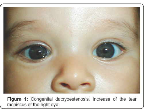
Congenital nasolacrimal duct obstruction is the first cause of pediatric epiphora. Other causes include congenital punctum and canaliculus stenosis and/or atresia, nasal malformations and craneofacial abnormalities. It is frequently seen at birth due to lack of perforation of the valve of Hasner or an inferior and distal nasolacrimal duct opening failure. At birth, half of the nasolacrimal new born pathways are not permeable. A spontaneous apoptosis mechanism takes place between 3rd- 4th weeks after birth. Nevertheless, the obstruction persists in approximately 20% of the patients. Symptoms of congenital nasolacrimal duct obstruction consist of epiphora and dacryocystitis. The diagnosis is easily made in the office by observing epiphora and mattering of the eyelashes (Figure 1). It can be confirmed by compression over the nasolacrimal sac, which results in regurgitation of mucopurulent material in those patients who have developed chronic dacryocystitis. Instillation of 2% fluorescein dye and observation of abnormally delayed passage from de cul-de-sac is helpful in confirming the diagnosis.
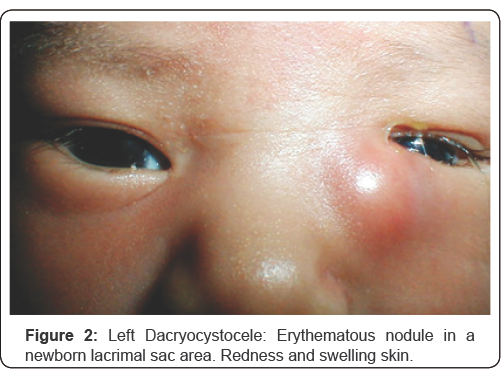
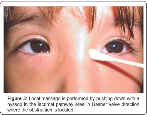
This is a rare entity presented at birth or within the fourth week after birth. The blockage Rosenmüller valve. An edematous, tender and red mass below the medial canthal tendon will be clinically found (Figure 2). Conservative management (medical treatment). If dacryocele is initially sterile, all patients must be treated with warm compresses, local massage and topical antibiotics [8]. Those infected will be treated with broad spectrum intravenous antibiotic therapy [9]. Local massage by pushing down the lacrimal sac is useful and accelerates the lumen duct perforation process (Figure 3). Controversy exists whether conservative management or early probing for decompression are preferable.
Probing consist by introducing a thin metal probe into the lacrimal punctum trough the nasolacrimal pathway, producing a mechanical opening in the obstruction site. It is an operating room procedure under general anesthesia. The ideal time is controversial, most of the time the procedure is performed around the first year of life for those patients that did not show spontaneous improvement or despite conservative treatment [10].
Close dacryointubation is performed by placement of silicone stents through the superior and inferior canaliculus and down to the nasolacrimal duct. This dilates the inferior meatus. The duration of the stent employment ranges from 6 weeks to 6 months [10]. Dacryocystorhinostomy surgical procedure involves the removal of bone adjacent to the lacrimal sac draining directly into the nasal cavity and it is performed when siliconte intubation have failed.
Dermoid cyst, dongenital glaucoma, acute conjunctivitis, corneal abrasion, trichiasis, ocular foreign body sensation and meningoencephalocele must be discarded.
The primary acquired nasolacrimal duct obstruction is caused by inflammation or fibrosis without any precipitating cause. Appears in middle age and elderly females in 3:1 ratio. The obstruction site is located in the lower nasolacrimal fossa and middle nasolacrimal duct. The secondary acquired obstruction is caused by inflammation or fibrosis with precipitating causes as infectious, inflammatory, neoplastic, traumatic or mechanical factors [11,12].
Clinical presentation Patients with primary acquired nasolacrimal duct obstruction most commonly present with a history of epiphora. A chronic dacryocystitis owing to tear stasis can show a mucopurulent discharge at the punctum, or pus can be expressed from the punctum by massage of the lacrimal sac. Two stages can be distinguished [13].
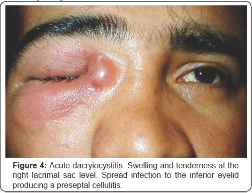
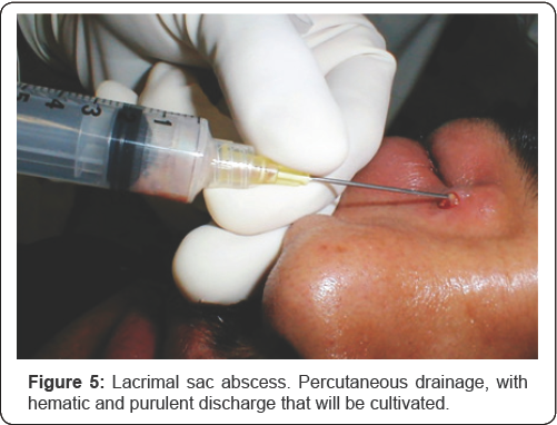
Is an acute inflammation of the lacrimal sac due mostly to the obstruction of nasolacrimal duct. In most cases is a clinical diagnosis. Lacrimal sac bacterial overgrowth and inflammation? occludes the superior and the natural drainage creating a true abscess. Symptoms and signs include a no compressible painful and erythematous mass below the medial canthal tendon. Medical treatment must be initiated because of the risk of extension to the periocular tissues and the orbit (Figure 4 ) including topical and systemic antibiotics, analgesics and antiinflammatory measures. Local heat and massages helps drainage and the opening of the obstruction siteLacrimal sac abscess requires sometimes manual percutaneous drainage, material can be collected and cultivated (Figure 5). Avoid irrigation during the acute phase because the risk of dissemination of the infectious process. Definitive treatment is a dacryocystorhinostomy procedure which can be performed as an external or internal endoscopic. It is preferable to postpone two or three weeks after the acute phase resolution.
Symptoms and signs include recurrent epiphora, swelling and redness at medial canthus and a painless and compressible mass below the medial canthal tendon. The patient usually refers history of previous acute dacryocistitis or chronic unilateral conjunctivitis.
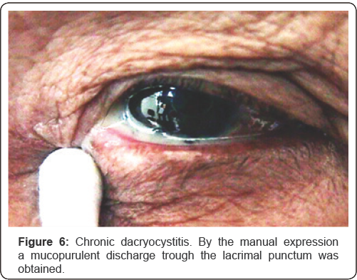
The lacrimal sac is filled with mucoid or purulent discharge that can be expressed frequently with local massage (Figure 6). Medical treatment includes topic and systemic antibiotics and a dacryocystorhinostomy as the surgical first choice. Differential Diagnosis: Preseptal cellulitis, sinusitis, canaliculitis, sebaceous cyst and neoplastic tumours.
For most tearing patients a diagnosis can be arrived at after a thorough history and a few relatively simple office procedures. A small number of cases will require more sophisticated studies to confirm the site of anatomic block. With the various test available, appropriate medical or surgical management can be determined in the vast majority of patient with tear production and drainage imbalance. Nasolacrimal duct obstruction is a common finding and the ophthalmologist must be prepared to recognize signs and symptoms to perform an accurate diagnosis and offer a correct management.
For more Open Access
Journals in Juniper Publishers please
click on: https://juniperpublishers.com
For more articles in Journal
of Physical Fitness, Medicine & Treatment in Sports
please click on: https://juniperpublishers.com/jpfmts/index.php
For more about juniper publishers please click on: https://www.juniperpublishersgroup.com/
Posterior Astigmatism: Improving Refractive Outcomes with Toric IOL Implantation-Juniper Publishers
Cataract surgery is not only a rehabilitative surgery, but also a refractive procedure, largely because of the intraocular lens (IOL) improves in latest years. However, recent studies showed a significant residual astigmatism after phacoemulsification with toric IOL implantation. There are several factors that can cause astigmatism refractive errors, such as IOL misalignment, factors related to the incision, incorrect calculation of toric IOL and corneal measurement errors. We believe that overlooking posterior corneal power is one of the most relevant reasons for refractive errors after cataract surgery with toric IOL.
Cataract is one of the leading causes of blindness worldwide, and its extraction is one of the most performed surgical procedures nowadays. The improvement of phacoemulsification techniques contributes for an increasingly less invasive procedure. Advances in IOL (intraocular lens) calculation, as well as the evolution of IOL technology increase patient's expectations for better results and postoperative spectacle independence [1}. Astigmatism is responsible for 13% of refractive errors [2]. Approximately 20 to 30% of patients submitted to cataract surgery had corneal astigmatism of 1.25 diopters (D) or higher, and around 10% of the patients have 2,00D or higher [3]. Recent studies demonstrate that residual astigmatism after toric IOL implantation is frequent [1,4]. Therefore, the correct astigmatism measurement is crucial for better post-operative results and, consequently, the patient's satisfaction. Furthermore, in present days, the 'gold standard’ in IOL power calculation is optical coherence biometry associated with keratometry. However, the capacity of this technique to determine the true corneal power is limited [5] because it assumes a fixed posterior-anterior curvature ratio, to estimate the posterior corneal curvature influence in the total corneal power [2]. Ignoring the posterior corneal power was recently highlighted as an important factor that leads to errors in toric IOL [5,6]. Posterior corneal refractive power is low when compared to the anterior surface, but when we take the astigmatic power into account, the posterior cornea surface can represent more than 20% of the total astigmatism power of the cornea [5].
Devices for an accurate measurement of posterior
corneal surface have a shorter story when compared to the methods to
evaluate the anterior surface. Nevertheless, this data can currently be
obtained by several methods such as Scheimplug imaging and optical
coherence tomography. This way, total corneal power can be calculated by
using ray tracing or Gaussian optics thick-lens formula [2].
Posterior astigmatism has its own clinical importance demonstrated
since 1890 by Javal, and recent studies show that posterior astigmatism
is usually against the rule and the mean power is around 0.3D (Table 1) [7-10].
When the anterior corneal surface shows with the rule astigmatism, the
posterior astigmatism compensates the anterior surface, and consequently
reduces the total astigmatism. However, if the anterior surface
astigmatism is against the rule, the total astigmatism will increase [9].
Ho et al showed that neglecting posterior astigmatism can cause
absolute errors of 0.2±0.16D in astigmatism magnitude and 7.4±10.3
degrees in astigmatism angle [11].
The surgical prognosis related to the reduction of postoperative
residual refractive cylinder is influenced by the correct calculation of
the total corneal astigmatism and its axis. In conclusion, the efficacy
of toric IOL implantation can be enhanced with the measurement of both
anterior and posterior astigmatism.
The authors declare that they have no conflict of interests regarding the publication of this paper.
For more Open Access Journals in Juniper Publishers please click on: https://juniperpublishers.com
For more articles
in JOJ
Ophthalmology (JOJO) please click on: https://juniperpublishers.com/jojo/index.php
For more about juniper publishers please click on: https://www.juniperpublishersgroup.com
A Review of the Data on the Recently Approved Xen Surgical Gel Stent in the Management of Glaucoma-Juniper Publishers
The cornerstone of glaucoma surgery includes trabeculectomy and tube shunting procedures, which utilize an ab externo approach to divert aqueous humor from the anterior chamber to the sub conjunctival space. The XEN Gel Stent is a 6.0mm tube consisting of porcine-derived collagen that similarly creates a non-physiologic shunt but through an ab interno approach. The XEN gel stent has the potential to effectively lower intraocular pressure and medication use with lower complication rates than traditional glaucoma surgery. This mini-review surveys the data of the XEN implant in current literature.
Glaucoma maintains a significant disease burden worldwide. It is the most common cause of irreversible blindness, affecting over 64 million individuals [1]. Therapy is focused on lowering intraocular pressure (IOP) by a variety of methods, including topical medications, laser, and incisional surgeries. These surgical interventions-traditionally trabeculectomy or tube shunt surgery-rely on creating an additional subconjunctival reservoir for aqueous humor (AH) drainage and subsequent resorption. However, the three-year results of the Tube Versus Trabeculectomy Study found failure rates of 15 and 28 percent, respectively. Serious postoperative complications such as persistent corneal edema, endophthalmitis, and chronic or recurrent iritis were also reported [2].
There has been a recent proliferation in procedures and medical devices that provide similar IOP-lowering effects to trabeculectomy or tube shunt surgery with fewer complications. One such product is the XEN Glaucoma Treatment System (Allergan, Inc., Irvine, CA, USA), which consists of the XEN Gel Stent and XEN Injector. The XEN stent is derived from porcine collagen, measuring 6.0mm long with inner diameters of 140|im, 63|im, or 45|im, although the 45|im stent is currently recommended. Previous studies in animal models have demonstrated no significant inflammatory response to implantation and no signs of degradation of the stent itself [3]. It was recently approved by the United States FDA in November 2016 for use in refractory glaucoma, including those with a history of failed prior surgical treatment, primary open-angle glaucoma (POAG), and pseudoexfoliative glaucoma with open angles that are inadequately controlled on maximal medical therapy.
The XEN stent is inserted into the angle through the scleral spur via an ab interno approach with its disposable injector. Ideally, the device should extend 3.0mm posteriorly from the limbus and into the subconjunctival space and 2.0mm anteriorly into the anterior chamber (AC). Once placed in an aqueous environment, the device hydrates, becoming soft and flexible, helping to maintain its position. By taking advantage of the resistance to flow in a cylinder as determined by the Hagen- Poiseuille equation, the XEN45 produces a pressure gradient 7.56mm Hg at the physiologic flow rate of AH within the AC, at 2.5 microliters/minute [4]. Thus, this stent can theoretically reduce the risk of hypotony seen in IOP-lowering surgeries despite being a valve less device. The fact that it is inserted ab interno obviates the need from conjunctival dissection, theoretically reducing the potential for conjunctival fibrosis and leaving the ophthalmologist with the option to perform ab externo surgeries if necessary in the future.
Several studies were performed evaluating the XEN Gel Stent with multiple inner diameters. Sheybani, Dick, and Ahmed reported on the results of 49 eyes of 49 patients treated with the XEN140 stent (140um internal diameter) without the use of mitomycin C (MMC) [5]. Of the 49 patients, 22 received previous glaucoma surgery, and nine (18%) had a prior laser trabeculoplasty. Complete success was defined as an IOP <18mm Hg and a greater than 20% reduction of IOP at the primary endpoint of 12 months without glaucoma medications. Criteria for treatment failure included visual acuity less than or equal to light perception, need for additional glaucoma surgery, or a less than 20% reduction of IOP at 12 months. This study revealed that the mean IOP reduced from 23.1±4.1mm Hg preoperatively to 14.7±3.7mm Hg at 12 months, a 36.4% decrease. In all, 40% met the criteria for complete success and 88.9% for partial success. Three patients (6%) failed the study criteria and required additional surgery. The most common complication was needling (47%), with nearly half of the cases occurring within the first month.
Another early study by Sheybani and Ahmed used the XEN140 and XEN63 stent (63um internal diameter) without MMC in patients undergoing phacoemulsification [6]. Of the 37 eyes, 47.1% were considered complete successes while 85.3% were qualified successes. Mean IOP significantly decreased from 22.4±4.2mm Hg preoperatively to 15.4±3.0mm Hg at 12 months, and mean medications were significantly reduced from 2.5±1.4 to 0.9±1.0.
One of the first reports involving the exclusive use of the XEN45 stent (45um internal diameter) involved 31 eyes receiving phacoemulsification and MMC treatment at the time of implantation. Mean IOP and medication use were reduced significantly from 20.8±4.6mm Hg to 13.1±3.6mmHg at 12 months and 2.7±1 to 0.9±1.1 at 12 months, respectively, and without significant complications [7].
Pérez-Torregrosa et al. [8] were among the first to describe the efficacy of the XEN45 in patients with mild and moderate glaucoma (defined as a mean deviation between 0 and -12dB on Humphrey 24-2 perimetry) [8]. Additionally, subjects included in this study had pressures<30 while being managed on two or more medications. Twelve months after concomitant phacoemulsification and XEN45 implantation, 27 of 30 (90%) subjects met the successful treatment criteria of IOP≤18mm Hg with no glaucoma medications. The authors reported several intra operative complications, including sub conjunctival hemorrhage with MMC instillation (36.6%), and minor hemorrhage intra camerally (86.6%) and at the scleral exit point (90%). A total of six (20%) stents required relocation, and one eye required re-implantation of the device. One subject was excluded from analysis due to extensive subconjunctival hemorrhage after MMC injection and another due to extrusion of the device into the subconjunctival space intra operatively.
Two studies have addressed the effect of simultaneous implantation and phacoemulsification versus the XEN alone. One such study involved 567 eyes, of which 54% underwent XEN implantation only, and 46% the combined implantation and phacoemulsification [9]. There were no inclusion or exclusion criteria regarding the grade of POAG, and data from all three diameters of XEN stents were not separated. The data revealed a mean preoperative IOP of 21.9±4.2mm Hg that was significantly decreased at 12 months (15.7mm Hg), 24 months (15.0mm Hg), and 36 months (13.2mm Hg) of follow-up. Likewise, a significant decrease in medications by 74, 77, and 74 percent from the mean of 2.7 was observed at 12, 24, and 36 months, respectively. The percentage of patients who were converted to another procedure was 4% by 12 months, 5% by 24 months, and 5% by 36 months. After analysis, it was determined that there was no statistical difference in mean IOP or medications between eyes receiving the implant alone and the combination procedure. Likewise, a 75 patient cohort demonstrated no significant difference in IOP reduction between the standalone and combination procedures at 12 months [10]. The most frequent complications from this study were needling (15.4%) and hypotony defined as an IOP <6mm Hg on postoperative day 1 (12.6%).
The most recent article involving the XEN stent evaluated its use in eyes with suboptimal IOP and medication intolerance, medication noncompliance, or maximum therapy with no history of glaucoma surgery [11]. A total of 13 eyes underwent XEN45 implantation in addition to phacoemulsification if previously phakic. At the 12-month end point. 41.7% were complete successes, with a>20% drop in IOP and discontinuation of all glaucoma medications, while an additional 25% met the IOP goal but remained on at least one medication. Among the reported complications, four eyes required needling, two eyes developed choroidal detachment and hypotony requiring systemic steroids and atropine treatment, one implant extruded, and two eyes required subsequent trabeculectomy.
The current literature available for the XEN Gel Stent demonstrates its effect on IOP and medication reduction in addition to reducing severe intra operative and postoperative complications. The data also suggests a lower early failure rate than those published from trabeculectomy and tube shunt surgery. There remains little data on long-term outcomes of the XEN stent at this time, inherent with a newly approved procedure. Thanks to its earlier implementation outside the USA, data are available for the XEN from mild to refractory glaucoma. However, this data should be evaluated in light of the fact that some studies were performed using the XEN140 and XEN63 stents, which are no longer recommended by the manufacturer.
Of the complications encountered with the XEN stent, subconjunctival hemorrhage and needling were most common, and extrusion of the device was the most common serious complication. It should be noted that the rates of needling varied widely in the literature, from 15.4% to 47%. This could be due in part to varying degrees of experience with the XEN stent and injector as well as different technical approaches such as the use and dose of anti fibrotic used at the time of implantation.
It is well known that cataract extraction provides some improvement in IOP [12]. Thus, the efficacy of the XEN stent may be confounded by those studies in which patients also underwent phacoemulsification. In fact, higher preoperative IOP, older age, and greater anterior chamber depth have all been shown to correlate to the amount of IOP improvement with phacoemulsification in medically managed glaucoma patients [13]. However, two studies with the XEN stent reported no significant difference in IOP reduction and medication use postoperatively between XEN implantation combined with phacoemulsification and XEN implantation alone. Indeed, this is one relationship that will be unlikely to be discerned until more data from more eyes is available for analysis.
Looking forward, several clinical trials in various stages of progress will look to further elucidate the properties of the XEN device and its role in the management of glaucoma. One trial - NCT02036541 - is a Phase 3 trial currently past its primary completion date that will further assess the XEN45 in patients with refractory glaucoma. Another is a parallel assignment, Phase 4 trial for XEN45 in moderate POAG patients (NCT02006693). Yet another study, soon to begin enrollment, will assess the in vivo effects of various interventions on outflow at Schlemm's canal, of which the XEN stent is part of the surgical branch (NCT02807935).
At present, there are multiple surgical options available for effective glaucoma management. The XEN Gel Stent is one such intervention that provides advantages over traditional glaucoma surgery and newer minimally invasive glaucoma surgery (MIGS) procedures while lowering IOP and medication dependence. The device is well tolerated by the ocular tissues and can be placed via an ab interno approach but still creates a non- physiologic subconjunctival shunt or bleb to increase aqueous outflow without the use of a valve system. Since it involves little manipulation of the conjunctiva, implantation of the XEN stent does not preclude future conjunctival surgeries if necessary. Questions remain as to what long-term outcomes of the XEN Gel Stent will demonstrate, as well as which types of glaucoma patients stand to benefit the most from the surgery. Larger-scale studies are needed to resolve these questions and confirm the initial optimistic results.
For more Open Access Journals in Juniper Publishers please click on: https://juniperpublishers.com
For more articles
in JOJ
Ophthalmology (JOJO) please click on: https://juniperpublishers.com/jojo/index.php
For more about juniper publishers please click on:
https://www.juniperpublishersgroup.com/
Keratoconus Progression Diagnosis Evidenced by Corneal Epithelium Mapping-Juniper Publishers
We are reporting a case of a 16y.o. (y.o.b. 1996) young gentleman that we have followed for keratoconus in our center about a year ago. His first visit occurred on February, 2011. At this time he was informed about the different options such as collagen cross-linking, INTACS, the Athens Protocol (the combination of topography-guided partial ablation PRK with CXL), and lamellar keratoplasty. Following a complete clinical investigation, recommendation was given that the 0S be treated with cross-linking with simultaneous partial topography-guided excimer surface ablation (Athens Protocol) [1], which was performed in May, 2011.
The 0D had reasonable good correction (BCVA 20/20) and therefore, due to his age and good corneal thickness (CCT in the vicinity of 500μim), we recommended for him to follow eight months to a year's time in order to evaluate a possible progression and the possibility of intervention. This paper is about our investigation of the 0D keratoconus progression not only by optical anterior segment imaging technologies, but also by the epithelium mapping, which supported our findings relating to correlation of keratectasia and elevated corneal overall epithelium.
Anterior-segment imaging of keratometric, topometric and topographic parameters can be an important tool in the decisionmaking process and prompt action. The optical instruments involved in this report were Biometry by I0L Master (Carl Zeiss Meditec Inc., USA), Anterior-Segment 0ptical Coherence Tomography (AS OCT) by Optovue RTVue (Optovue Inc. Fremont, CA), Placido topography and Scheimpflug topography by Wave light (Erlagen, Germany), specifically the Topolyzerand the Pentacam High Resolution (Oculus Optikgerate GmbH), a.k.a. Oculyzer II, which is a Pentacam HR that has been specially configured to export topometric data to Alcon's refractive suite [2]. The ultrasound bio microscopy (UBM) instrument was the Artemis II + superior (Artemis Medical Technologies Inc. Vancouver, British Columbia, Canada) [3]. To our knowledge this is the first such documented case combining these optical modalities with epithelium mapping by HF ultrasound for keratoconus screening in the peer-reviewed literature.
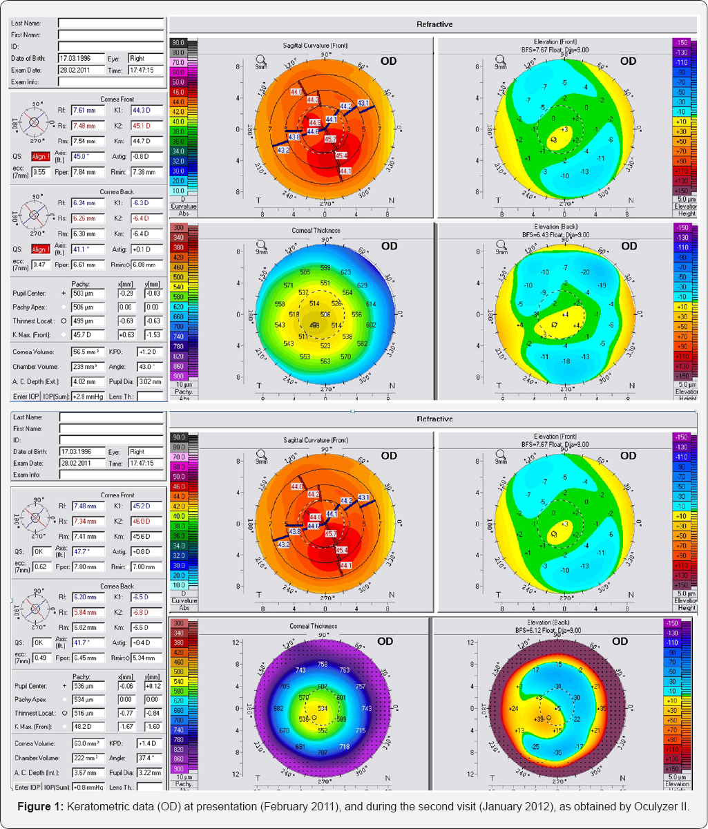
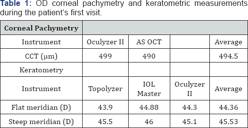
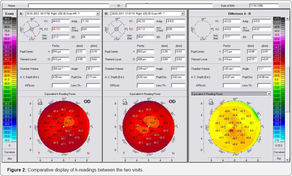
The initial anterior segment parameters recorded during the first visit (late February, 2011) are summarized in (Figure 1 &Table 1). Based on these findings, astigmatism was determined -1.25 D, with the flat axis was determined at 21°. We saw the patient several months later (late January, 2012). The data (Figure 2 & Table 2), indicated keratoconic progression, despite that overall corneal thickness was found increased by an average of 13μim. For example, the difference with the Pentacam (Oculyzer II) preimposed on the old and newer topometric measurement showed a change in the central steepening. For example, sagittal curvature increased, K1 (flat axis) by +0.2 D and K2 (steep axis) by +0.8 D. Likewise, anterior surface elevation was found increased by approximately + 13μim, also there was change between lowest elevation to pupil center (20711, -13 -> +3μiM, 2012 -18 -> +8μim). Based on these findings, astigmatism was determined at 40°
We decided to further evaluate this finding because on topometric terms this finding suggested progression of the keratoconus. We obtained Artemis II+ HF UBM corneal epithelium mapping. Careful evaluation of the UBM findings revealed that the epithelium (Figure 3) was overall thicker compared to a population of 33 patients (50 eyes) [4]. The patient’s epithelium featured mean thickness 56μm (normal population 50.8μim), central thickness 60μm (normal population 52.1μm), and average peripheral thickness 50μm (normal population 49.54μm), while over the cone (located superiorly - temporal) the epithelium was thicker, at 53μm.
We feel that this is a good example where the -at first sight- progression of keratoconus is accompanied by epithelium whose thickness is overall thicker than the control population, particularly at the pupil center by a significant amount (+8μm). This would be impossible to establish with Scheimpflug imaging maps alone. It appears the HF Ultrasound is able, by giving the specific epithelium maps to point to that direction. Obviously, for the definite diagnosis of this theory would be re-evaluation of the cornea with Oculyzer and UBM. In case where the epithelium returned to a thinner thickness we would expect that the Pentacam maps would flatten and that would establish that there is no actual progression of the keratoconus. It is interesting to compare the posterior curvature maps of past and current and see in these maps that there is a change in posterior curvature. One has to take into account that the increase of irregularity by the epithelium thickening may be the factor that creates this bias.
Dr. Kanellopoulos is a Consultant to Alcon Wave light.
For more Open Access Journals in Juniper Publishers please click on: https://juniperpublishers.com
For more articles
in JOJ
Ophthalmology (JOJO) please click on: https://juniperpublishers.com/jojo/index.php
For more about juniper publishers please click on: https://www.juniperpublishersgroup.com/