Thursday 29 August 2019

Evaluation of Aging Disorders by 54 000 Consecutive Ophthalmic Cases-Juniper publishers


Juniper Publishers-Journal of Ophthalmology


Abstract

The Ciliary body is an unusual location of uveal melanomas and usually these kinds of tumors appear with a reduction of vision due to the formation of sectorial cataract or retinal detachment when the tumor spread to a posterior position. We present a 57 years old woman with sudden and severe ocular pain. Ophthalmologic examination showed a hyper mature cataract and superior displacement of the lens with sectorial angular block. The intraocular pressure was 22 mmHg. After papillary dilation aciliary body tumor was observed. The ultrasound study and magnetic resonance imaging confirmed the diagnosis of uveal melanoma.
Background Information:during the years 1991-2015, about 54,000 consecutive cases were examined at the ophthalmic out patient’s clinic. The aim of this study was to find out if any relationship exists between aging disorders, or each is having its own entity, or one disorder could be the driving factor to produce aging and related complications.
Methods:Findings were entered into computerized documentation program that does simultaneous analysis of clinical data. The correlation between aging disorders was analyzed.
Results:Seven disorders were found to be related to aging; hypertension, glaucoma, cataract, diabetes, age related macular degeneration and ischemic neuropathy, in addition to presbyopia. Hypertension was found to be the leading cause for the development of ischemia and therefore may be the stimulating factor for the development of oxidative stress and oxidation. It shows that basic trends of aging are important while investigating the metabolic disorders at molecular level.
Conclusion:Taking both factors; ischemia and oxidative stress into consideration, it is recommended a strategy be adopted, in addition to antioxidant therapy, prophylaxis and prevention of aging includes medication that guarantees healthy relationship between hypertension and cerebral blood flow.
Keywords: Aging disorders; Diabetes; Hypertension; Cataract; Glaucoma; Age related macular degeneration


Introduction

During the years 1991 and 2015, about 54 000 consecutive cases were examined at the Ophthalmic Clinic in Amman - Jordan. Data were entered directly into a computerized documentation program that performs simultaneous analysis of clinical data. Figure 1 shows the age distribution of examined cases. One notices that Jordan has a relatively young population and a significant decrease of incidence starting at the age group 61-70 years.
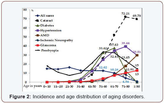
It has been noticed that seven diseases increase with age; Hypertension, Diabetes, Glaucoma, Cataract, Age related Macular Degeneration “AMD” and Ischemic Neuropathy, in addition to Presbyopia. The question that arises, is there any relationship between these six disorders? Or each is having its own entity independent of each other? Or one disorder could be the driving factor to produce aging related complications within the other components?


Materials

The age distribution of examined cases is shown in Table 1. A comparison between aging subjects to all examined cases (Figure 2) gives us the following information;
  • The incidence of aging for all disorders starts to increase at the age of 40 years.
  • Diabetic cases start to decline at the age 61-70 years.
  • Cataract and hypertension starts to decrease at the age group of 71-80 years.
  • Open-angle glaucoma and AMD continue to increase until the age of 80 years and above.
  • The accommodative reservoir decreases from birth up to the age of 30 years and Presbyopia starts to develop in average at age 40 and continues to increase up to the age 61- 70 years.
As the incidence of all disorders start to increase at age 40 years, means that the age of 40 is the starting point of aging. This fact could be confirmed as presbyopia starts in average at the age of 40 years. It is possible that the decline of aging incidence (Figure 2) after the age of 61- 70 years by diabetics and at the age of 71-80 years by cataract and hypertension means a relative early death for these two diseases as compared with the normal distribution of all cases with in the same age group.

Cataract

Cataract increases with age, reaching its maximum 72.21% at the age of 71- ≥ 80 years. Its incidence with age is much higher than all other disorders. “Implications of oxidative stress have been examined in the pathogenesis of cataract in vivo treatment with vitamin E, of the Emory mouse led to a decrease in the rate of cataract progression suggesting that in at least in some entrances an oxidative stress could participate in the formation of cataract [1]. It is known that in addition to heredity, environmental factors play a major role in the development of cataract. In addition, 35.5% of all cataract cases are diabetics, 34% hypertensive, 10.6% have AMD and 8.6% Glaucoma. So cataract is involved in a mixture of additional components that might affect its development and patient’s quality of life. It is therefore expected that cataract patients are also affected from ischemic disorders in the same manner like other aging diseases.

Hypertension

Hypertension increases with age reaching its maximum 39.44% at the age 71-80 years, and then it decreases again. It is known that uncontrolled hypertension causes hypertensive 
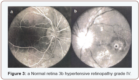
retinopathy. According to its severity, hypertension is classified into four grades (Figure 3) a represents a normal retina.
The ratio of diameter between venules and arterioles is 3:2. (Figure 3) represents hypertensive retinopathy grade IV, showing narrowing of the arterioles, and swelling of the optic nerve head, macular edema, micro hemorrhages and cotton wool spots indicating the presence of severe ischemia. Wayne [2] says “There is increasing evidence that atherosclerosis should be viewed fundamentally as an inflammatory disease. There is evidence that hypertension may also exert oxidative stress on the arterial wall”.

Diabetes

Diabetes increases gradually with age. It reaches the maximum incidence “37.63%” at the age 61-70 years. Its distribution goes parallel to hypertension with the difference that diabetes starts to decline earlier as seen in (Figure 2). There is convincing experimental and clinical evidence that the generation of reactive oxygen species increases in both types of diabetes and the onset of diabetes is closely associated with oxidative stress Some [3] diabetic patient care severely affected with uncontrolled hypertension and the development of hypertensive retinopathy and other vascular disorders in comparison to non-diabetics (Figure 3).
It is expected that diabetic patients with severe hypertension most likely suffer from decreased cerebral blood flow and ischemia of sensitive organs, with the result of getting ischemic neuropathy and renal failure or different vascular obstructions. Similar to hypertension, the prevalence of glaucoma by diabetic patients is 7.7%, much higher than by all examined patients “2.28%”.

Glaucoma

Open angle glaucoma increases with age, starting at the age of 40 years and reaching its maximum “13%” at the age of 80 years. The blood pressure seems to be responsible for aqueous humor formation [4]. The incidence of glaucoma among all patients is 2.28%, and 6.6% among all hypertensive patients. Matt [5] reports of having evidence that antioxidant treatment could help defeat glaucoma. It was found that antioxidant treatment reduces oxidative stress in pressure-treated retinal ganglion cells. This confirms a study done by Weinreb [6] that oxidative damage occurs within hours of elevated hydrostatic pressure or elevated intra ocular pressure.

Age related macular degeneration

Age related Macular Degeneration “AMD” increases with age reaching its maximum 29.81% at the age of ≥ 80 years. The incidence of glaucoma among AMD patients is 6.32% much higher than by all patients included in the study “2.28%” and with hypertension 5.3 %. We experience today exactly what Donders [7] has described Khan HA & Moorhead HB [8] has published, Frank [9] 25 years later that among the four aging blinding diseases, Cataract, Glaucoma, Diabetes and AMD, Agerelated Macular Degeneration is the only one for which there are no effective means of prevention or treatment.
Although it is less common loss of vision in AMD, choroid Neovascularization leading to disc form degeneration produces a more rapid and dramatic and more severe decrease in vision than the atrophic or dry form of AMD. Similar changes were found also among patients in cases of diabetes, AMD, disc form macular degeneration and high myopia, where the clinical appearance of an often round macular scar, that has been recognized to comprise Neovascularization, arising from the chorio-capillaris [10] and proliferative changes at optic nerve head in cases of carotid thrombosis (Figure 4).


Discussion

Aging is a complex of degenerative changes starts at the age of 40 years. Presbyopia is the first measurable sign of aging. The age of 40 years is therefore considered as the starting point for prophylaxis, prevention and rehabilitation in order prolong life expectation and to secure better quality of life for elderly. The presented data show clearly that oxidative stress and oxidative damage initiated much interest by researcher. Oxidative stress is most likely responsible for the ongoing development of oxidative damage and its complications by all mentioned aging diseases. The pathogeneses of these diseases at molecular level gives better insight into a major problem related to aging; the oxidative stress causes a continuous oxidative damage. By the presence of aging diseases and absence of cure, it is expected that prevention with antioxidants might improve or maintain these disorders at acceptable level, but keep fighting to prevent the ongoing oxidative stress is mandatory. Hypertension on the other hand involves all aging diseases as seen in (Table 2).
Ophthalmo-dynamometry is a good method “at least for serial investigations” to measure the blood pressure in both brachialand ophthalmic artery, estimate the vascular resistance and calculate the amount of cerebral blood flow [11] Values that are higher than twice the standard deviation (2xD) of the regression curve are considered as having high cerebrovascular resistance (CVR). The Cerebral Blood Flow (CBF) will be calculated as follows;
In addition to the genetic and environmental factors, uncontrolled hypertension seems to play a leading role in the development of complications related to aging diseases. 47.1% of all hypertensive patients suffer from Glaucoma, and 92.5% of all diabetics are hypertensive. The incidence of glaucoma in hypertension is relatively high “6.6%” in comparison to 2.28% by all examined cases; the incidence of hypertension by AMD patients is 26.7%. As expected hypertension might cause embolism of the central retinal artery CRA the increased vascular resistance measured by ODM on the (Figure 5).Other side not affected, show increased vascular resistance by 59% of all CRA embolism cases, is an alarming sign that such ischemic changes might occur on the other side or elsewhere in the body. Treatment with antihypertensive drugs is important. The positive expected effect of some anti-hypertensive drugs “decrease blood and intra– ocular pressure” could unlikely elevate the cerebral-vascular resistance and decrease the cerebral blood flow as experienced with clonidine [12,13]. It is therefore expected that patients involved within aging diseases suffer from complications related to hypertension and/or a non-wished side-effect of some antihypertensive drugs causing ischemia. Hypertension seems to be the leading and stimulating factor that causes arteriosclerosis, increased vascular resistance and inability to maintain normal blood flow to the whole body, causing severe damage especially to the very sensitive organs, e.g. stroke, myocardial infarction, gangrene, thrombosis and emboli of the central retina or carotid arteries, ischemic neuropathy and renal failure. It might affect the chorio carcinoma and RPE causing ischemia. It is therefore possible that oxidative stress which plays a major role at the molecular level is initiated by ischemia. One comes in agreement with Newell [14] that these macular changes are related to the decreased blood flow of the chorio carcinoma below the fovea. If this theory is correct, increased blood pressure and decreased Blood flow will produce adequate systemic complications within aging diseases. This could be confirmed by the presence of associated disorders as shown in [Table 3] and (Figure 6 a and b).
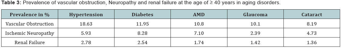
The majority of cases suffering from vascular obstruction are found frequently in hypertension followed by diabetes, AMD, glaucoma and cataract. Ischemic Neuropathy; “ischemic papillitis, retro bulbar neuritis, and other cranial nerves” were found most frequently by diabetics, followed by AMD, in association with hypertension. By calculation the prevalence related to all cases involved in the study, hypertension was found to be the leading disease causing ischemic disorders; Diabetes increases the prevalence of ischemic neuropathy and renal failure. By observing the distribution curve (Figure 2 & 7) one comes to the following life expectation and survival rate of aging diseases. By considering all cases involved in the study independent of age, the survival rate at age 51-60 years was found to be 12.7% and for the age group ≥ 80 years 1.1%. As aging diseases starts to increase by the age of 40 years where the younger population is excluded, it is expected that the survival rate for aging disorders will be different. The prevalence of Cataract, AMD, glaucoma and hypertension continue to increase till the age of 61-70 years. Then the survival rate becomes less. The best survival rate for the age group 71-80 years is for AMD 34.5%, followed by cataract 23.6%, then glaucoma 20.6%, hypertension 16.8% and diabetes 12.3%. The worst survival rate at the age of ≥ 80 years is for diabetics 1.8% and the best was found to be for AMD patients 13.4%. The survival rate of the same aging group for glaucoma 6.1%, cataract 4.1% and hypertension 3% only.


Conclusion

The central regulation between systemic blood pressure and cerebral blood flow is very important. Deregulation of this mechanism through hypertension seems to be the leading factor that initiates oxidant stress, oxidative damage and aging. Antioxidants are therefore very important to prevent further deterioration of aging disorders. For the prophylaxis of aging diseases it is recommended that in addition to antioxidants, special attention be paid to control hypertension and maintain normal cerebral blood flow.


Highlights

Programmed computerized documentation and analysis of aging disorders among 54000 ophthalmic cases.
Correlates between the most common seven aging disorders in ophthalmology.
Demonstrates the importance of paying attention to ischemic systemic disorders associated with ocular complaints.
Modern eye Journals are not paying enough attention to the basic trends of aging.

Suggests new strategy for the prevention of aging disorders.

For more Open Access Journals in Juniper Publishers please click on: https://juniperpublishers.com


Friday 23 August 2019

External Ophthalmomyiasis, Case Report in Campania-Juniper Publishers


Juniper Publishers-Journal of Ophthalmology


Abstract

Objective: To describe a clinical case of external ophthalmomyiasis in a 55 year old man from Umbria, Italy. We are also taking into consideration a differential diagnosis of Onchocerciasis and Cisticercosis.
Design: Case Report Interventional
Participants:1 patient, 2 eyes in an interventional case report
Methods:biomicroscopic examination, surgical removal of the larve, medical treatment, laboratory examination of the specimen and blood screening
Main Outcome Measures:It was not possible to obtain microscopic evidence of the larvae. Blood screening and examination of the specimen was negative but the biomicroscopic evidence of the parasites, together with documented information, made the diagnosis relatively easy
Conclusions:The difference in clinical signs and negative laboratory tests allowed us to exclude all the diseases mentioned above. With case study, we hope to be able to aid in simplifying the diagnosis of this pathology for our colleagues.
Keywords: Ocular Pain; Melanoma; Ciliary Body; Cataract
Abbreviations: BCVA: Best-Corrected Visual Acuity; NMR: Nuclear Magnetic Resonance


Introduction

Ophthalmomyiasis is an infection of the eye by Diptera larvae. Oestrus Ovis is the most frequent cause of ocular myiasis especially in countries with tropical or mild climates [1]. The presence of this pathogen is widely seen in Central and Southern Italy and the islands where sheep farming is common [2]. Ophthalmomyiasis, based on its location, is classified as external, internal and/ or orbital. In its external form the larvae are found on the conjunctiva or on the edge of the eyelid [3]. With accidental contact this can give rise to inflammation of the conjunctiva. The patient may also experience lacrimation, photophobia and foreign body sensation in the eye.


Case History

55 year old male patient, telephoned from Umbria where he was attending an open air public manifestation. He stated that, whilst sitting under a tree the previous evening, he had the sensation that something had “hit” his left eye. He complained of foreign body sensation, pain and photophobia throughout the night. He was advised to come to our private practice. At 14.00 h he arrived. His general condition was good. His left eye was red, painful and intensely photophobic. A biomicroscopic examination revealed numerous tiny worms in rapid movement (Figure 1). The worms were removed by using forceps, cotton buds and continuous irrigation of the inferior and superior conjunctival sac with a iodopovidone 5% solution and netilmycin sulphate 0.455g eye drops equal to 0300g. The patient was then treated with netilmycin sulphate ointment every two hours. At 22.00 h after a second biomicroscopic examination more larvae were removed. Some larvae were preserved in test tubes containing tears and others were placed on slides to be microscopically examined in the laboratory.
The specimens (slides and lacrimal liquid collected in tubes) were examined immediately. One drop of physiological solution at 0, 9% was added to the lacrimal fluid and placed on dry glass slides and observed microscopically (first at 10x field and then 40x). No Diptera larvae were found, because they had dissolved. Giemsa slides at 3% were prepared next. This stain is used to highlight the visibility of other forms of parasites, particularly nematodes and larvae of platelminta cestodes. Thin films of lacrimal liquid were fixed with methanol and then stained with Giemsa at 3% Microscopic examination, at 1000x with an oil immersion lens revealed no parasites.
The following day a blood sample was drawn and biomicroscopic examination revealed the presence of a few slow moving larvae. These were removed with forceps. A complete and thorough examination (vision, IOP, anterior chamber, vitreous and fundus) was normal aside from conjunctival injection. An examination two days later showed the presence of cysts situated in the conjunctiva (Figure 2). The base of the cysts was pearl in color but the upper part was transparent. Haemato chemical tests were negative and a diagnosis of ophthalmomyiasis was made based on physical evidence. An examination six days later revealed a slight reduction in the size of the cysts and an absence of larvae. After eight days the cysts were in a phase of regression and the conjunctiva was no longer inflamed.


Discussions

The term myiasis means the invasion of human tissue by parasites of the type Diptera. The first case of ocular myiasis was described by Keyt in the 1900s [1]. The ocular form can be external, internal or orbital and is commonly associated with Oestrus Ovis [4]. Infestation occurs when the female lays her eggs on the skin or mucous membrane. Stimulated by the warmth these eggs evolve into larvae that are about 1.5 mm in size. The larvae then penetrate the skin or mucous membrane in a few minutes. Penetration inside the eye occurs from perforation of the sclera which allows the larvae to move below the retina. This may leave a hypopigmented trail. Entry into vitreous chamber may occur due to a break in the retina. The larvae can be trapped in the vitreous and gain entry into the anterior or posterior chambers. The risk is partial dislocation or dislocation of the lens and damage to suspensory ligaments (zonulules) [5]. The signs and symptoms of internal ophthalmomyiasis vary accordingly to anterior or posterior positioning of the larvae. The signs of anterior positioning are recurring iritis associated with partial displacement or total displacement of the lens. In posterior positioning one can see vitreous turbidity and possible detachment of the retina [6].
External opthalmomyasis presents symptoms similar to acute catarrhal conjunctivitis as in the case presented. However it is necessary to pay much attention to the treatment, because it could have serious complications such as conjunctival ulceration, endophtalmitis and invasion into other regions of the eye and orbit [4]. Diagnosis is based on anamestetic and clinical data. Identification of the parasite if it is microscopically present. The typical parasitological aspects are, length a little over 1 mm, offwhite color and two hooks in the cephalic segment. Examined under a light the fissures appear tiny and fusiform with cylindrical translucid elements and a black mark at one end. This allows the parasite to be highly mobile and capable of penetrating the conjunctiva [2]. Note, it is extremely difficult to remove and preserve these larvae due to their fragility. Even though some were rapidly placed on slides and others saved in lacrimal liquid in the test tubes, it was not possible for the laboratory to examine them as they dissolved. Treatment requires continual removal of the larvae and antibiotics to prevent further bacterial infection.
We found that irrigation of the conjunctiva with a solution of iodopovidone 5% and nethilmycina, did reduced the number of larvae which helped lower the risk of further infection. It is also necessary to be on the lookout for other conditions that could occur due to larvae having entered the eye. We refer in particular,to cysticercosis and onchocercosis, which , initially, had been considered as possible differential diagnosis.
Cysticercosis is a parasitic disease caused by larvae of platelminta cestode of taenia type A. This cestode has man as its final host and pigs are the intermediary hosts [7]. Clinical symptoms depend on the site of infection and migration. One can see conjunctivitis, iridocyclitis, displacement of the lens, retinal detachment, vitritis, etc. Decreased vision, pain and foreign body sensation are symptoms that the patient may experience [8]. Diagnosis is made by looking for anti cysticercus serum antibodies; enzyme linked immunoelectro transfer blot (EITB).
Onchocercosis is an infectious disease caused by infestation of nematode filariform onchocerca volvulus. The incubation period of 3-12 months shows no clinical signs [9]. The first sign is usually the appearance of subcutaneous nodes. Lesions to the eye, starting at the cornea, begin with opaque white infiltrates and keratitis, followed by corneal opacities. Microfilarie found in the anterior chamber can cause iridocyclitis and glaucoma, whereas, regardless of microfilarie in the posterior chamber, it is possible to find atrophied areas of the retina and choroid. Involvement of the retina can lead to damage of the optic nerve causing reduced vision or eventually blindness [10-11]. Diagnosis is made through anamnestic and laboratory data: an increase in eosinophil, immunoenzymatic tests and the presence of microfilarie. Sample is taken during the night when reproduction of filarie is at its highest.


Conclusion


Although it was not possible to obtain microscopic evidence of larvae, the symptoms, the biomicroscopic aspect of the parasites, together with documented information, made the diagnosis relatively easy. The difference in clinical signs and negative laboratory tests allowed us to exclude any of the diseases mentioned above. With case study, we hope to be able to contribute and aid in simplifying the diagnosis of this pathology for our colleagues.

For more Open Access Journals in Juniper Publishers please click on: https://juniperpublishers.com


Juniper Publishers: Understanding the Relationship of Chronic Toxoplas...

Juniper Publishers: Understanding the Relationship of Chronic Toxoplas...: Advances in Biotechnology & Microbiology Juniper Publishers Authored by Taibur Rahman Abstract Toxoplasma gondii  is...

Juniper Publishers: Preparation of Novel Morphinan Derivative as a Res...

Juniper Publishers: Preparation of Novel Morphinan Derivative as a Res...: Peer Reviewed Chemistry Journals Juniper Publisher Authored by Sureshbabu Jayachandra* Go to Abstract The acid catalyzed...

Wednesday 21 August 2019

Sudden Ocular Pain: An Unusual Debut of a Ciliary Body Melanoma-Juniper Publishers


Juniper Publishers-Journal of Ophthalmology


Abstract

The Ciliary body is an unusual location of uveal melanomas and usually these kinds of tumors appear with a reduction of vision due to the formation of sectorial cataract or retinal detachment when the tumor spread to a posterior position. We present a 57 years old woman with sudden and severe ocular pain. Ophthalmologic examination showed a hyper mature cataract and superior displacement of the lens with sectorial angular block. The intraocular pressure was 22 mmHg. After papillary dilation aciliary body tumor was observed. The ultrasound study and magnetic resonance imaging confirmed the diagnosis of uveal melanoma.
Keywords: Ocular Pain; Melanoma; Ciliary Body; Cataract
Abbreviations: BCVA: Best-Corrected Visual Acuity; NMR: Nuclear Magnetic Resonance


Introduction

Uveal melanomas are the most frequent intraocular primary tumors in adulthood. However, ciliary body location is very uncommon regarding iris or choroidal melanomas. Furthermore, ciliary body location gives symptoms later, which also gives a worse prognosis.


Materials and Methods

Materials and Methods Biomicroscopy, dilated indirect ophthalmoloscopy, Goldmanntonomter, B-Mode echography and ultrasound biomicroscopy were performed. Subsequent RMN imaging was also obtained.


Case report

We report a case of a 57 years old woman who came to the emergency department with a sudden and severe pain in her right eye. The patient also related that she had had a decrease in her visual acuity for last two months. She was a nonsmoker, but would occasionally drink alcohol. Family history was unremarkable for malignancy. Medical history only included controlled hypertension and cholesterolemia. Best-corrected visual acuity (BCVA) at first was 20/200 in the right eye and 20/30 in the left eye.
The examination of the right eye showed a brown pigmented lesion in the lower region of iris, without satellites lesions or papillary deformity. She also presented a hyper mature cataract antero-superiorly displaced endangering the anterior chamber space and the iridocorneal angle (Figure 1). The intraocular pressure was 22 mmHg in the right eye and 16 mmHg in the left eye. After papillary dilation, we found a pigmented tumoral mass behind the iris which was situated between 4 to 6 hours and produced the dislocation of the lens (Figure 2).The retinal examination was of no value because of the presence of the mature cataract. The ophthalmoscopic examination of the left eye was normal.


Results

The study with B-Mode echography and ultrasound biomicroscopy (OTI HF 35-50 Ultrasound System ®) demonstrated a 15 X 10 mm tumor which came from ciliary body and displaced the lens (Figure 3). The nuclear magnetic resonance (NMR) with gadoline confirmed the existence of an intraocular tumor with hyper intense signal in T1 and hypo intense in T2 (Figure 4) with a size of 15 x 10 x 8 mm and heterogeneous catchment of contrast (Figure 5). With these results a ciliary body melanoma was suspiciously diagnosed. The extensive study was negative and a brachytherapy with of iodine 125 (I125) treatments was decided. Actually and after cataract surgery our patient is asymptomatic and without signs of recurrence.


Discussion

Ocular melanoma is the most common intraocular primary tumor in adulthood, which mainly affects white males between fifty and sixty years old. It represents 3.7% of cases of melanoma and it is the second most usual location after cutaneous melanoma [1]. Its incidence is nearby 4.3 cases per million populations [2]. And its most common position is the uveal tract, which is supposed the origin of the 82.5% of intraocular melanomas [1]. Although inside this rate, ciliary body melanoma only represents 12% of uveal melanomas its location is a factor of poor prognosis [3].
The majority of these tumors are asymptomatic until their growth produces a decrease of visual acuity due to the formation of a sectorial cataract or retinal detachment [2]. The presence of dilated episcleral vessels near the tumor (sentinel vessels) is also frequent [4]. Unusually these tumors may also produce an increase in intraocular pressure because of the angular closing by the pigment dispersion produced by myeloma [5] a uveitis due to necrosis of tumor [6] or aleukocoria in children [7]. There is no existing record of a case of ciliary body melanoma that appears with sudden and severe eye pain without a significant increase of intraocular pressure. In the case I am talking about this circumstance happens as well as other manifestations like the presence of a mature cataract, lens subluxation and a sectorial angular closure.
The diagnosis of this pathology is clinical using the slit lamp, accompanied by additional tests such as mode A ultrasound (presence of kappa angle); mode B ultrasound or ultrasonic biomicroscopy (the size, location and extent of tumor is characterized) and orbital NMR (show tumor mass as a hyper intense on T1 and hypo intense on T2) [8]. The confirmation of the diagnosis is obtained by the histological study of the lesion.
Pathological study is also important to establish the prognosis of these melanomas because there are some factors that can add to a worse prognosis for this kind of tumors like epithelioid cell line, high mitotic activity, necrosis of tumor, lymphocytic infiltration or extra sclera extension [9]. Genetic factors are other important prognostic. The mutation in genes 3 and 8 is related to poor prognosis in these tumors [10].
Regarding the treatment of these tumors, there are several alternatives such as radiotherapy, brachytherapy or external beam radiation; local excision (iridocyclectomy) or enucleation. Depending on the size of the tumor and the presence or absence of distant metastases we would choose a treatment or another. Our patient has been treated with brachytherapy due to the characteristics of the tumor and the absence of extraocular disease.
Ciliary body melanomas have a rate of distant metastasis of19% and 33% at 5 and 10 years respectively, being the most frequent liver, lung or bones metastases [1]. This rate is the highest of uveal melanomas, which makes the survival from these tumors, also lower [3].


Conclusion


In conclusion we suggest that sudden ocular pain is a very unusual presentation of already unusual ciliary body melanoma. The objective of this article is describing this way of atypical debut. These tumors are the most metastatic inside the group of intraocular melanomas. However the brachytherapy treatment can be a good choice for this kind of tumors if we detect them before extra ocular dissemination.

For more Open Access Journals in Juniper Publishers please click on: https://juniperpublishers.com


Monday 19 August 2019

The Flash Electroretinogram of Burrowing Owl -Athene cunicularia-Juniper Publishers


Juniper Publishers-Journal of Ophthalmology

Abstract

Background: The burrowing owl (Athene cunicularia) has peculiar visual capacity, presenting conspicuous vision, what can be justified, among other motives, by the presence of cones and rods in the macula and the existence of double cones. The Athene cunicularia presents retinal cell circuit highly organized and complex, with presence of two foveae. Thickening in the ganglion cell layer surrounding the fovea contributes to the change of the refractive index of the retina at this location, thus increasing the sensitivity for detecting motion. The high concentration of photoreceptors in the fovea is the cause of the maximum optical resolution of this region.
Results: We conduct a transversal observational study according to the protocol of the International Society for Clinical Electrophysiology of Vision (ISCEV) that aims at evaluating the retinal response of the species after stimulation with white light, both in photopic and scotopic stimuli. The verification of the p-value and t-value, we obtained for the variables, respectively: a-wave amplitude - 0.02 and 2.94; b-wave amplitude - 0.12 and 1.76; b-wave time culmination - 0.07 and 2.12;b-wave latency - 0.74 and 0.34.
Conclusion:In order to standardize the variables of the electroretinogram of the species, the authors propose to evaluate the electroretinogram components under white light in both stimuli, providing electrophysiological data from the visual stimulation of retinal species, encouraging the Biological Sciences with substrate for future research and clinical evaluations.
Keywords: Owls; Electroretinography; Retina
Abbreviations: CEPAL: Ethics Committee of Faculty of Medicine of Health Sciences Center in Federal University of Rio de Janeiro; ERG: Electro Retino Gram; IBAMA: Brazilian Institute of Environment and Renewable Natural Resources; ISCEV: International Society for Clinical Electrophysiology of Vision


Introduction

The retinal electrical activity corresponds to the initial event processing of visual information. All animal species that have visual apparatus present some retinal response, so that the comparative study of different species allows us to acquire knowledge that can be applied in our daily lives.
It is true that most treatments used in humans result from applications arising from animal research. Knowledge of cytology, histology and physiology of the human race is due to the various animal studies.
The owls are known for their great visual capacity due to their complex retinal structure. As an interesting kind of vertebrates, the investigation of their visual apparatus operation is of most importance. The existence of a previous study on the morphology of photoreceptors and the evaluation of the b-wave of electroretinogram (ERG) in various color spectra in Athene cunicularia (Burrowing Owl) motivated acomplementary electroretinographic study of the specie concerned [1].
The visual capacity of owls presents peculiarities, among them, their big eyes which account for 1-5% of body weight, depending on the species. They have binocular vision, which provides them stereopsis. Their total visual field is 110°, having 70° binocular vision. Their eyes are large to increase their efficiency, especially under low light conditions and are so well developed that they are not rounded, but tubular. They are protected by three structures, the upper eyelids, lower and the nictitating membrane [2].
The retina of Athene cunicularia has mixed fovea, with a predominance of rods, observing the eight rods relationship for two simple cones and a double cone [3-5]. The presence of a double cone only occurs in some species [6-8]. Another important morphological appearance in the retina of these birds (including the Athene cunicularia) is the presence of aster, a structure mainly composed of axons of bipolar cells that have oblique direction and centrifugal to the fovea, allowing for a retinal neuronal rearrangement circuit area resulting from the central area [6,9-11].
The Athene cunicularia presents a retinal cell circuit extremely organized and complex. Thickening in the ganglion cell layer surrounding the fovea contributes to the change of the refractive index of the retina at this location, thus increasing the sensitivity for movement’s detection [12-15]. The high concentration of photoreceptors in the fovea is what accounts for the maximum optical resolution of this region [3]. The Athene cunicularia does not present differentiated retinal electrical response to some light spectra in photopic and scotopic environment, when taking into account the latency and amplitude of b-wave [1].
In order to standardize the variables of the electroretinogram of the species, the authors, following their line of research, have proposed to evaluate the electroretinogram components under white light in both environments (Figure 1 and 2).
The Athene cunicularia has twilight hunting habits and previous histological descriptions identified outer segments of the long rods, which can justify their twilight habit [3, 16-19].
Discrimination of colors in owls was reported, as well as the light wavelength, which is not important for the behavior of owls [20]. Although the vision of the owls is not as sensitive as previously thought, it still has advantages over the diurnal species at low levels of luminosity [21]. The hearing of Owls is essential for their predatory activity, serving to detect and hunt their prey [22, 23] their vision is intended to determine the spatial orientation, distance and speed fly [24].
The cross-sectional observational study of the species in question was chosen due to the existence of previous morphological and electrophysiological documentation [1], which will be supplemented by the electrophysiological data obtained by applying the diffuse Flash ERG in vivo.


Objective

Presenting the electroretinographic record of Athene cunicularia, identifying their components when the retina is stimulated by the white light spectrum in photopic and scotopic conditions, as well as the relationship between the answers found in both responses and their influence on living species. Providing electrophysiological visual stimulation data of retinal species, encouraging the Biological Sciences with substrate for future research and clinical evaluations.


Materials and Methods

Animals

We studied seven species of owls Athene cunicularia, of both genders, provided by the Brazilian Institute of Environment and Renewable Natural Resources (IBAMA). The animals weighed between 180 - 205 g, and height between 21.8 - 26 cm.
The research protocol complies with the requirements of the Ethics Committee of Faculty of Medicine of Health Sciences Center in Federal University of Rio de Janeiro (protocol number 14/2004) and IBAMA (protocol number 02022.000518/2005- 33).

Methodology of Electroretinography Exam

In order to accomplish the study, we considered as inclusion criteria:
  1. Animals of both genders;
  2. Absence of general and ophthalmic anatomical changes;
  3. Weight within the required standards in the literature; and
  4. No vitreoretinal pathologies.

The exclusion factors

  1. Failure to comply with, at least, one of the items of the inclusion criteria.
Following the study protocol, the animals were sedated with ketamine sulfate (20 mg / kg) intramuscular; mydriasis was obtained with topical 1% tropicamide, in a total of three drops per eye. After mydriasis, direct ophthalmoscope was performed on both eyes to exclude vitreoretinal opacities and then topical corneal anesthesia with 0.4% oxybuprocaine hydrochloride at a dose of three drops per eye with thirty second intervals between each drop.
Following the procedures of sedation and anesthesia, three electrodes needle-shaped 13 x 0.45 mm were used for examination. One of the electrodes was introduced into the corneal stroma, causing a self-sealing injury, without the need for sutures or bandages; the other two were inserted into the subcutaneous, one superiorly and between the animal eyes (ground electrode) and the other (reference electrode) in the supraorbital region of the eye to be examined. The electrodes were placed one minute prior to the light stimulus.
Among the fourteen eyes stimulated, seven were destined to photopic response and seven to scotopic response, the choice being made randomly, so that the photopic documentation was obtained of the right eyes and the scotopic documentation of the left eyes.
The electroretinography device used was Neuropack II®, of the company Nihon Koden®. The flash lamp used in all tests, either scotopic or photopic stimuli, had intensity of 20 Joules being performed with stimulator located 30 cm from the animal.
The parameters used for electroretinogram record followed the protocol established by the International Society for Clinical Electrophysiology of Vision (ISCEV) 2004, except for the use of summit Ganzfield (full field), unavailable in the Ophthalmology Department of the Federal University of Rio de Janeiro. The electrodes used were the same and had the same impedance. The duration of the light stimulus was <5 ms. The time to adapt to light to stimulate in photopic environment was 10 minutes and the dark adaptation to stimulus in scotopic environment was 20 minutes [25]. Registered stimuli contemplated white electromagnetic spectrum, visible to man.

Analysis of results

The type of study used was cross-sectional observational analysis, demonstrating the electroretinogram of Athene cunicularia in photopic and scotopic response.
For the evaluation of the data obtained were considered the following conventions:
  • -a-wave amplitude: distance between the most negative point of the a-wave and the level of the beginning of the electroretinogram. Unit in microVolts;
  • -b-wave amplitude: distance between the most positive point of b-wave and the most negative level of the electroretinogram. Unit in microVolts;
  • -b-wave time culmination: distance between the beginning of the stroke and the most positive point of the b- wave. Unit in milliseconds;
  • -b-wave latency: distance between the beginning of the stroke and the most negative point of b-wave. Unit in milliseconds.
With reference to the light stimulus studied, we obtained the mean and standard deviation of the above parameters. Due to the small sample used, the statistical evaluation was performed by Student’s t-test two-tailed paired, compared to the same variable in both environments. We determined the error rate (α) of the applied test in order to validate the comparison made. The values of “p” were evaluated for each comparison (photopic or scotopic) and its specific variable.


Results

The electroretinography results of Athene cunicularia were obtained in both photopic and scotopic responses, by being observed all variables proposed for the analysis. They were registered as uneventful strokes in both environments such as under the protocol. They were identified and recorded in the electroretinogram components: a-wave amplitude, b-wave amplitude, the completion time and latency of b-wave.
The t-Student two-tailed paired test was carried out from the same spectrum of light (white), and the same variable in both stimuli, respecting null hypothesis the equality between stimulated conditions, checking that, with an error margin of 1%, there was no rejection of the null hypothesis for all evaluated variables.
The verification of the p-value and t-value, we obtained for the variables, respectively:
  1. - a-wave amplitude - 0.02 and 2.94;
  2. -b-wave amplitude - 0.12 and 1.76;
  3. - b-wave time culmination - 0.07 and 2.12;
  4. - b-wave latency - 0.74 and 0.34

Discussion

Under the histological appearance of the Athene cunicularia retina, although not object of this study, there are reports of a quite similar structure of vertebrates in general, except for a few peculiarities, such as the presence of rod cells in the macular region and the presence of two foveae [5]. Different histological structures have been described among species of different owls, and probably related to the habit of life of each species [3,26].
The full field ERG expresses the complex electrical response of all retinal cells when stimulated with light [27]. The layout generated by the electrical response is recorded and generates components that can be evaluated separately. Recorded by electrodes, each component represents the activity of various cells of a certain group, and they respond synchronously to the light stimulus. Thus, the ERG record reveals the electrical activity of different cell classes, allowing us to evaluate the retinal functionality.
A comparative study of the b-wave electroretinogram performed under different color spectra has shown, in the studied species, that there is no distinction between the orange color spectrum, yellow and white when comparing the retinal electrical responses in photopic or scotopic stimuli [1]. However, for a better understanding of the electrical activity of the retina it is essential to know the details of the a- and b-waves, which is why the authors chose to study with more details the a-wave and b-wave components of the ERG of the species concerned.
From a clinical point of view, the evaluation of electroretinogram takes place under white light stimulation, which is why the study of the white light was chosen as a reference. The anesthetic ketamine sulfate was used due to its wide application in the veterinary environment and for its inert action on the waves of the electroretinogram, not interfering with the generation of retinal potentials.
It was decided to hold the stimulus under photopic and scotopic stimuli in different eyes, in order to avoid retinal stress in the stimulated eye and a possible corneal edema called out by placing the active electrode on the cornea, which did not occur. The protocol of ISCEV was followed uneventfully, leaving, however, increasingly evident the need for modern electrophysiological equipment, as the summit Ganzfield, which undoubtedly would reduce the loss of light stimulus and significantly increase the fidelity data obtained.
As for the morphological traces, we observed that they are similar to those of the humans, with a- and b-waves, as well as oscillatory potentials. The evaluation of the amplitude of a-and b-waves, as well as the completion time and latency of b-wave were studied because they are relevant measures in the literature and intended to document those components to future standardization of the design of the studied species.
The most clinically important components are the b-wave amplitude and b-wave completion time (Figure 3-6), since the measures relating to a-wave and the b-wave latency are hard to measure and therefore at higher risk of measurement error. In the sample assessed we can confirm the difficulties encountered in the daily clinic actually being the measures of lesser value and most difficult assessment. The latency of the a-wave, for having extremely small amount, about 5 ms, has not been evaluated.
We used the t-Student two-tailed paired test due to the small sample. The data evaluated in the species concerned are unedited from a statistical point, and there are no reports in the literature that refute or ratify why the determination of the “p” value should not exclude data with significance under 95%.
Under the classical evaluation of Granit [28] we could classify the Athene cunicularia retina as a type E, that is, an excitatory type, with a clear PII component, or better, a b-wave. This classification attributed to the retina of the excitatory type a greater activity of the rods; however, now we know that the b-wave is more related to bipolar, horizontal and Müller cells [29-34]. Oscillatory potentials were observed, but were not the object of this study.
The data from the study reflect the reality of the studied species group, and documented all identified values.
The idea that we had in relation to the owls that they have the ability to see either with light or in the dark, may not be confirmed in other species and should continue to be investigated. Their predatory activity and their life habit are due, no doubt, among other characteristics, to the capacity of perception of the light spectrum similarly in both photopic and scotopic stimuli.
The idea that we had in relation to the owls that have ability to see the light and the dark, cannot be confirmed in other species and should continue to be investigated. Their predatory activity and its life habit are due, no doubt, among other characteristics, to the capacity of perception of the light spectrum similarly in both photopic and scotopic stimuli.


Conclusion

The comparative study of the retina in various vertebrate classes has revealed important evolutionary aspects. The Structural differences of the retina as a whole, as well as of the cell types composing it allow us to try to correlate their morphology with the way of life of each animal [3].
The electroretinographic trace of Athene cunicularia under white light stimulus of the white spectrum showed the a- and b-waves, registering all variables proposed for study. From the evaluated data, we can conclude that the retina of the species studied should be classified as of an excitatory type, due to the predominance of the positive component in the ERG.
When comparing the same variable (amplitude of the a- and b-waves and completion time and latency of the b-wave) in both photopic and scotopic stimuli, we observed that retinal electrical response found is identical, confirming the hypothesis that the retina of the studied species responds similarly to white light in both stimuli. This peculiarity is responsible, among other features, for the excellent twilight visual ability of the species, and therefore, directly involved in their life habit. In the biological sciences research the electroretinographic registration of Athene cunicularia may then be a parameter for detailed studies of the physiology of birds and for a better understanding of visual perception as well as the study of eye diseases of the species.


Authors’ Contributions

MP initiated, planned and wrote the manuscript. LR participates in drafting the article and revising it critically for important intellectual content, as well as the analysis and interpretation of data. EF participated in its design and coordination. AM performed the statistical analysis and helped to draft the manuscript, as well as give final approval of the version to be submitted and any revised version. All authors read and approved the final manuscript.


Acknowledgements

The authors thank Carlos Bastos for assistance in all electrophysiological tests, being fundamental in this study; and Fernanda Ramos, for her invaluable contribution in the statistical support. This material is based on work supported by the CEPAL under grant 14/2004 and IBAMA under grant 02022.000518/2005-33. Any opinions, findings and conclusions or recommendations expressed in this article are those of the authors and do not necessarily reflect those of the CEPAL or IBAMA.

For more Open Access Journals in Juniper Publishers please click on: https://juniperpublishers.com


Friday 16 August 2019

Orbital Miasis: Complicating Squamous Cell Carcinoma Conjunctiva A Rare Case Report-Juniper Publishers


Juniper Publishers-Journal of Ophthalmology


Abstract
Miasis refers to invasion into the living tissues of humans and other mammals by the eggs or larvae of flies from the order of Dipteria. The invasive parasitic larvae almost invariably cause massive destruction of orbital tissue especially in neglected patients accompanied by marked inflammatory reactions and secondary bacterial infections. Infestation can be caused by multiple or single larvae. We report a case of 50 yr post menopausal women who was having squamous cell carcinoma of conjunctiva and was a neglected and belonged to low socioeconomic status. The patient ultimately ended with massive orbital miasis.
Keywords:Orbital Miasis; Carcinoma

Introduction

Orbital miasis is very rare worldwide. Miasis is a term first introduced by F. W. Hope. Miai is a Greek word meaning ‘fly’. Miasis refers to invasion into the living tissues of humans and other mammals by the eggs or larvae of flies from the order of Dipteria [1,2]. The invasive parasitic larvae almost invariably cause massive destruction of orbital tissue especially in neglected patients accompanied by marked inflammatory reactions and secondary bacterial infections. Infestation can be caused by multiple or single larvae [3]. Nursing staff, medical and paramedic staff should be aware of this condition, particularly in high risk cases like immobile, open wound and skin malignancy patients.

Case Report

50 year old postmenopausal, normotensive, non diabetic, chronic hukka smoker patient comes to the accident and emergency department of SMHS Hospital with a complaint of blood stained discharge from the left eye and worms seen in the same eye (Figure 1). On examination the upper and the lower lids of the same eye were swollen, thickened and hard and could not be closed. The eye ball and the ocular tissue could not be seen. There was blood stained discharge and numerous live active worms in the left orbit. There was active oozing of blood on the left side. Orbital miasis was diagnosed. After going through the records of the patient, we came to know that patient was already diagnosed as a case of left poorly differentiated squamous cell carcinoma of the conjunctiva. CECT head showed soft tissue density mass with no bone invasion. MRI shows 5x4cm extracranial intraorbital mass with intraorbital component adherent to left lateral rectus muscle with left preauricular lymphadenopathy. The staging of tumor was T4 N1 M0. After cleaning and debridement of left orbital cavity the patient was referred back to the parent institution for emergency exentration and radiotherapy.
The maggots were identified as chyrosoma bezziana.

Discussion

Ophthalmomiasis is a rare form of eye disease and massive orbital or ocular miasis is even rarer [1,3-6]. Flies are small vingid orthopods of the class insecta. Some are biological or mechanical vectors of protozoal viral, bacterial for helminthic disease. Miasis in humans may be benign to asymptomatic or may result in mild to violent disturbances even death. Orbital miasis is the least common form of ophthalmomiasis.
By going through the literature we found very less cases of orbital miasis. Children and elderly persons or dementia patients who could not adequately take care of themselves are usually the victims. Most reported cases have belonged to the lower class of society being farmers, labours or beggars. In children, gonococcal conjunctivitis and in adult’s periocular ulcerated skin cancers may predispose to larval infestation3. Orbital miasis occurs when large number of diptherous larvae known as flies invades and destroy orbital contents. Ophthalmo-miasis may result in complications ranged from minor ocular ulceration to complete blindness and even death [7]. Common ophthalmomiasis inducing agents are cochhomyia hominivorax, hypoderma bovis, caliphora vomitoria, wohlfartia magnifia and chyrosoma. Bezziana the maggets identified in our patients belonged to C. Bezziana. Diagnosis is based on microscopic examination of the sliced caudal ends of the larvae (preserved in 70% alcohol or formalin). A dichomatous key is used to identify the number of interior spiracet opening and shape of body part [8]. Discharge associated with chyrosoma Bezziana foul smells and bloody.
The main predisposing factor for infestation in our patient was bed of necrotic tissue, provided by squamous cell carcinoma. Other important factors were lack of self care, general ignorance, low socioeconomic status and poor hygiene.
Any skin ulceration like diabetic, neurotrophic and malignant ulcers in the exposed areas in these patients are a risk factor and should be treated [9].
Miasis is not a common disorder in humans and it affects primarily wounds of skin, infection with purulent secretion and blood and body secretions are most common factors that attract flies.

Conclusion

Orbital miasis is a rare disease, which is marked by inflammatory reactions and secondary bacterial infections. Life threatening consequences like intracranial invasion may also occur. Old, debilitated and neglected patients are more prone to such disease. Malignant growths often form scaffolding for such infections to occur.

#
Figure 1: The maggots were identified as chyrosoma bezziana.

For more Open Access Journals in Juniper Publishers please click on: https://juniperpublishers.com


Wednesday 14 August 2019

Does Intravitreal Bevacizumab Injection for Retinopathy of Prematurity Treatment Arrest Anterior Segment Development?-Juniper Publishers


Juniper Publishers-Journal of Ophthalmology


Abstract
Purpose: To determine intravitreal bevacizumab (IVB) effect on ocular development by comparing refractive and biometric outcomes of intravitreal bevacizumab (IVB) and laser photocoagulation for treatment of retinopathy of prematurity (ROP).
Methods: A prospective nonrandomized interventional comparative study was conducted in a referral hospital for ROP management. All patients who received either single IVB or diode laser photocoagulation were enrolled. Cycloplegic refraction and biometry was performed before treatment and at the corrected age of 9 months.
Results: The IVB group included 17 patients (28 eyes; gestational age (GA): 28.54 ±2.2 weeks) and the laser group included 17 patients (34 eyes; GA: 28.53 ± 1.6 w). GA, BW and corrected age at the end of follow-up was statistically similar between the two groups. Eyes in IVB group had significantly longer axial lengths and thinner lenses at final visit (p=.037 and p=.002).
Conclusions: Following IVB treatment of ROP, eye development in general and crystalline lens in particular are less affected compared to laser treatment. This supports the idea that anterior segment arrest which was first described for laser therapy of ROP occurs minimally with IVB if at all.
Keywords: Retinopathy of Prematurity; Intravitreal Bevacizumab; Diode Laser photocoagulation; Refraction; Biometry Abbreviations: IVB: Intravitreal Bevacizumab; ROP: Retinopathy of Prematurity; GA: Gestational Age; ETROP: Early Treatment of ROP study; VEGF: Vascular Endothelial Growth Factor; AL= Axial Length; ACD= Anterior Camber Depth; LT= Lens Thickness; V= Vitreous Cavity

Introduction

Retinopathy of prematurity (ROP) is a vasoproliferative disease of preterm neonates which may result in severe complications if left untreated in high risk patients. In 2001 Early Treatment of ROP study (ETROP) showed significant benefit of laser photocoagulation in eyes with type 1 prethreshold ROP [1]. Since then, laser photocoagulation of the avascular retina using either transpupillary or transscleral approach is the standard of care for type 1 ROP [2-6]. In recent years, the use of anti-vascular endothelial growth factor (VEGF) agents mainly bevacizumab, has been increasingly popularized for the treatment of various ocular neovascular diseases including ROP [7-11] Promising results have been reported for IVB injection in ROP especially in patients with severe or aggressive posterior ROP [12].
Previous studies have shown that ROP patients show significant myopia (55.2 to 80.04% in age group under 3 years old) after laser photocoagulation [13-15]. It is well established that myopia associated with prematurity and conventionally treated (cryo- or laser therapy) ROP is not fully explainable by axial length changes. In fact, it may be a result of a disruption of emmetropization called anterior segment arrest consisting of corneal steepening, anterior chamber depth reduction, and lens thickening [16-19]. Recently, a few studies have reported less myopia after intravitreal bevacizumab (IVB) injection in ROP patients in comparison to laser photocoagulation or combination treatments [20-25]. Geloneck et al. [25] speculated that IVB minimally disrupts anterior segment development, hence less myopia. However biometric effects of anti-VEGF agents on ocular growth have not been fully evaluated in a pre- and post-treatment model. Current study was conducted to compare the refractive errors and biometric indices before and after single IVB injection and conventional laser therapy for ROP.

Methods

In this prospective comparative study, from March to September 2013, all premature infants who were scheduled to undergo either diode laser photocoagulation or IVB injection for the treatment of type 1 ROP in Rassoul Akram Hospital, Iran, Tehran were eligible for this study. Informed consent was obtained from the parents of all infants enrolled in the study, fully describing the treatment modalities and ultrasonography technique. Iran University Eye Research Center Ethics Committee approved the study. Screening and management of all patients were performed by retinal specialists (MMP and AS) in accordance to the guidelines of the American Association for Pediatric Ophthalmology and Strabismus [26] and the revised guidelines of the International Committee for the Classification of Retinopathy of Prematurity [27]. For prethreshold disease in zone I or posterior zone II, an intravitreous injection of 0.625 mg bevacizumab (Avastin; Genentech Inc, San Francisco, California, USA) was performed [10]. Infants with prethreshold disease in anterior zone II, received transscleral diode laser photocoagulation of avascular retina [28]. Patients who did not respond to primary monotherapy and needed further intervention were excluded. Also, eyes with media opacity including cataract, corneal opacity and vitreous hemorrhage, and those with other ocular diseases including glaucoma, and congenital vitreoretinal diseases were excluded.
Refractive errors and biometry indices were obtained under cycloplegic condition approximately 30 minutes after instillation of topical Tropicamide (Mydrax; Sina Darou, Tehran, Iran), 3 times with an interval of 5 minutes. Measurements were performed immediately before treatment, and at the age of 9 month.
Handheld retinoscopy was performed by two of the three expert examiners (RA, JK and MSS), masked to the planed treatment. If their results disagreed by more than 0.5 Diopters (D), refractions were repeated and the discrepancy was resolved. Spherical equivalent (SE) ≤ − 0.5 and ≤ − 5.00 D was considered as myopia and high myopia, respectively [29, 30].
Biometry was performed in supine position with the lid speculum in situ via A-scan contact mode ultrasonography (OcuScanRxP; Alcon Lab, Dallas, TX, USA). Measured indices included axial length (AL), anterior chamber depth (ACD), lens thickness (LT), and vitreous cavity length (V). All scans were performed by a single investigator (JK). After instillation of topical Tetracaine 0.5% (Anestocaine; Sina Darou, Tehran, Iran), 10 subsequent scans were recorded in Auto-save mode. Scans were repeated until standard deviation of less than 0.1 was achieved. Care was taken to apply minimum pressure on the cornea during ultrasonography.
Data analysis was done using SPSS software (version 16, SPSS, Inc., Chicago, IL, USA). T tests (paired t test when applicable) and Chi square test were used for analysis of continuous and categorical variables, respectively. P values less than 0.05 were considered statistically significant.

Results

A total of 34 neonates including 17 patients (28 eyes) in the IVB group and 17 patients (34 eyes) in the laser groups were studied. Table 1 shows demographics of the patients. Birth age, birth weight, follow up duration and corrected age at the end of follow-up were similar between the two groups; however, patients in IVB group received therapy significantly earlier (p< 0.001). All patients in the study responded to treatment in terms of resolution of ROP, no recurrence of ROP and no detachment/hemorrhage after treatment.
Results of refractive error measurements are summarized in Table 2. At baseline examination, a marginally significant difference was found in the mean SE between the 2 groups (-3.37 ± 4.68 Diopters [D] in the IVB group and -1.5± 3.97 D in the laser group, p: 0.08) and prevalence of myopia was significantly higher in IVB group (71.04% vs. 35.55%; P= 0.004). At final exam, refractive error in the IVB group and laser therapy group was -1.02 ± 2.96 D vs. -0.12 ± 2.28 D (P = 0. 18) and the rate of myopia in IVB group decreased to 50%, while no significant change was observed in the laser group (38.24%, P=0.36). Finally the absolute change in SE was not significantly different between the 2 groups (P=0.3).
Table 3 shows the biometric measurements. At baseline, no significant difference was found between the two groups in any of the biometric measurements. At final exam, eyes in the IVB group had significantly higher AL and V measurements and shallower ACD and shorter LT measurements compared to the laser group (p=.037, p=.017, p=.002 and p=.002). The biometric changes after treatment were significantly different between the two groups in AL, LT and V measurements (P=0.002, P=0.007 and P< 0.000).
In bivariate correlation analysis, SE change in the laser group correlated significantly to axial and vitreous cavity length changes (p=0.005 and p=0.006). No significant correlation between SE and biometric changes were found in IVB group.
In multivariate analysis, no significant association was found between SE changes and the treatment modality (p=0.46), AL changes (p=0.56), ACD changes (p=0.49), LT changes (p=0.08), V changes (p=0.49), GA (p=0.56) and BW (p=0.74).

Discussion

Although there are few reports of more hyperopic changes following laser treatment of ROP [31] most recent studies comparing IVB and laser monotherapy or combination therapies show a myopic preponderance in laser therapy (Table 4).
Whether the observed myopic shift is attributable to the allocated treatment or the severity of the disease has been a matter of controversy, however, the follow up of the BEAT- ROP clinical trial [10,25] the only large randomized prospective study in the field, demonstrated that the higher degree and frequency of myopia in laser treated eyes (compared to the eyes who received IVB) did occur in spite of no significant difference in myopia severity.
In a process called emmetropization a relatively wide distribution of refractive error in full term newborns, gets narrower toward hyperopia in the first few years of life [30,32]. In this process, vitreous cavity length elongation is balanced by reduction of corneal curvature (from 51 to 44 D), crystalline lens power (by getting thinner) [33]. Myopia associated with prematurity and conventionally (cryo- or laser therapy) treated ROP is not fully explained by axial length, but it is a result of an emmetropization disruption called anterior segment arrest consisting of corneal steepening, anterior chamber depth reduction, and lens thickening [16-19]. Although Geloneck et al. [25] speculated that IVB minimally disrupts anterior segment development; effect of anti-VEGF agents on ocular growth is not fully evaluated.
In the present study despite the initially higher prevalence of myopia among IVB group (71%) in comparison to laser therapy group (35.55%) before treatment, the frequency decreased to 50% at the age of 9 month in IVB group while no significant change was observed in laser therapy group (38.2%). On the other side, biometry results demonstrated that although the eyes in IVB group were initially marginally smaller than those in the laser group, they finally had significantly larger size. At a concordant trend lens thickness in IVB group significantly decreased leading to less frequency of myopia in this group. Such a significant reduction in lens thickness was not observed in the laser group. These observations support the idea that the crystalline lens development (the expected lens thinning) continues following IVB treatment of ROP while it is arrested by laser therapy. This is in consistence with a previous study which suggests that high myopia associated with ROP is primarily a reflection of inappropriately higher lens thickness and power [34]. To the best of our knowledge, it is for the first time that a study reports the refractive and biometric indices of eyes before and after undergoing treatment for ROP.
It has been proposed that anterior segment growth may be slowed by decreased levels of local growth factors as a result of delayed migration of vessels to oraserrata (in premature neonates) alongside photoreceptors maturation arrest [35,36]. It is also known that laser therapy stops retinal vessel development, while vessels continue to develop beyond neovascular ridges upto the oraserrata after IVB injection [10].This may partly explain the pathophysiology of the so-called anterior segment arrest following the laser therapy; however, further experimental investigation is needed.
The present study has several limitations. Although randomization would avoid analytical concerns inherent to the non-randomized design, investigators believed it would be unethical to randomize ROP patients, regardless of their stage of the disease, to the two treatment group. In the current ROP protocol applied in this reference hospital, ROPs in zone I and posterior zone II are treated with IVB and ROPs in anterior zone II are offered the laser treatment. Additionally enrolled infants in the present study have notably higher birth weight and gestational age compared to some studies which may also contribute to the smaller degree of myopia observed in the pretreatment examination in this study compared to other reports. Finally for the investigators a relatively short follow-up was considered an acceptable trade-off for the prospective design. Despite these limitations, current study is the first to report pre- and post-treatment biometric and refractive indices of eyes treated for ROP and its results further support the theory that IVB does not halt anterior segment development in ROP patients as laser therapy does.

For more Open Access Journals in Juniper Publishers please click on: https://juniperpublishers.com