Wednesday, 22 January 2020

Comparing the Sensitivity of Visual Evoked Potential and Standard Achromatic Perimetry in Diagnosis of Optic Neuritis- Juniper Publishers

Comparing the Sensitivity of Visual Evoked Potential and Standard Achromatic Perimetry in Diagnosis of Optic Neuritis- Juniper Publishers

Juniper Publishers- JOJ Ophthalmology

Abstract

Purpose: To evaluate the diagnostic power of Visual Evoked Potential (VEP) and Standard Achromatic Perimetry (SAP) in a group of patients with Multiple Sclerosis (MS) and a history of optic neuritis (ON).
Methods: 136 eyes of 68 patients (11 male and 57 female, average age 27.18 years) with a confirmed diagnosis of MS underwent SAP and pattern VEPs. 67 eyes (49.2%) had no history of ocular involvement and a negative ophthalmologic examination, while 69 had proven ON according to physical examination. The mean deviation (MD) and the pattern standard deviation (PSD) of perimetry and the amplitude & latency of VEP in both normal and abnormal eyes were recorded. MD & PSD<0.5% and pl00 latency more than 118 millisecond were considered abnormal.
Results: MD was abnormal in 93.9% of the eyes with ON, PSD was abnormal in 69.4% and latency was abnormal in 55% of the affected eyes with ON. In overall, VEP can diagnose 55% of the eyes affected with ON and SAP can diagnose 96%. In statistical analysis of MD, PSD, latency and amplitude values with Mann-Whitney Test MD and PSD were statistically significant (MD has p<0.001, PSD has P<0.05, latency has p=0.83 and amplitude has p=0.38).
Conclusion: The results of this study demonstrate that SAP is more sensitive than VEP in the diagnosis of ON. SAP is a less operator and patient dependent technique and the patient test reliability can be assessed with parameters such as False-positive response rate, False- negative response rate, Fixation loss & Short-term fluctuation measurement. Because of these advantages, SAP has better sensitivity than VEP in the diagnosis of ON. Our results suggest that clinical and subclinical visual involvement of ON can be better diagnosed using both SAP and VEP tests, together.
Keywords: Keywords: Diagnostic power of SAP & VEP; Multiple sclerosis


Introduction

Multiple sclerosis (MS) is an autoimmune disease that involves the Optic Nerve, brain and spinal cord by damaging the myelin sheath [1]. The disease can affect patients' visual pathway and therefore lead to Optic Neuritis (ON) which is an eye discomfort accompanied by decreased visual acuity, changes in the visual field, uncontrollable rapid eye movements and double vision (clinical and frequent subclinical evident manifestations) [1-3]. De-myelinating disease leads to less responses of the brain and the optic nerve which is detected by the visual evoked.
potential [3-5]. Alterations in the visual evoked potential (VEP) and standard achromatic perimetry (SAP) have been reported to be useful in patients with MS. Many studies have shown the presence of abnormality in the results of VEP and SAP in optic neuritis [6], but there is little agreement about the prevalence of these abnormalities or about which examination is more sensitive [7-10]. In this article, our aim is to evaluate the diagnostic power of SAP & VEP in a group of patients with Multiple Sclerosis (MS) and a history of optic neuritis.


Material and Methods

Sixty eight patients with ON and MS were recruited from the neurology and neuro-ophthalmology clinics of Rassoul Akram hospital in 2010-2011. The diagnosis of ON/MS was made based on standard clinical symptoms, examination findings, and radiologic abnormalities. In all patients, the disease was in remission and no patient was affected by any general disease apart from MS [11].
The diagnosis of ON was based on clinical signs and symptoms; such as: history of a progressive decrease of vision, color vision deficit, painful eye movements, fundus examination and relative afferent pupillary defect (RAPD) which is diagnosed by a neurologist or neuro-ophthalmologist. Patients voluntarily participated in this study and all participants were enrolled after an informed consent was obtained. All patients underwent full ophthalmic examination, including best-corrected visual acuity (BCVA) measurement, slit lamp biomicroscopy, applantation tonometry, and fundus examination after pupillary dilation. VEP pattern reversal was performed (Sirius Galileo; Esaote Biomedica, Florence, Italy). Latency and amplitude were recorded and AP100 latency>118ms (i.e., 2SD above the mean) was considered abnormal.
Standard achromatic perimetry (SAP) was performed by means ofthe Humphrey Field Analyzer 750 (model 750; Carl Zeiss Meditec, Dublin, CA) using the 30-2 program with the Swedish interactive threshold algorithm (SITA) standard strategy. Two consecutive visual field examinations were performed for each patient, but only the second was evaluated for the purpose of the study and only if reliability indices were normal. The criteria for abnormality were mean deviation (MD) or pattern standard deviation (PSD) and were labeled abnormal by the instrument's software. VEP and SAP were done for all patients with one expert optometrist.
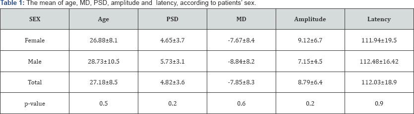
Patient data were collected through questionnaires, respectively, were entered into the SPSS software version 18.0 and were then analyzed. Maximum, minimum, and SD±Mean were reported for the quantitative data and for the qualitative data, the number (percentage) was stated. The Chi-Square test, with the aid of SPPS software was used to evaluate the relationship between qualitative variables and Mann-Whitney test was used for evaluation of normality between data and a P-value of<0.05 was considered valuable. VEP and SAP were done in all 68 patients (136 eyes). The age, sex, MD, PSD, amplitude and latency recorded are shown in Table 1.

Results

From the 68 patients that were included in this study, 11 were male (16.2%) and 57 were female (83.8%). The mean±SD age of the patients was 27.18±8.5 (range:14-61 years old). 49.6% of the eyes that were included in the study were normal and 50.4% had ON. The mean MD value was -7.86db (with SD: 8.4) with a range of -34.49db to7.57db. Also, the mean±SD MD value in normal eyes was -4.89db±7.12 with a range of -34.49db and 7.57db, while the mean value±SD of MD in the affected eyes was-10.63db±8.98 with a minimum of -31.28db and a 5.18db maximum. MD was negative in 19.1 % of the normal eyes and positive in 80.9%, while being negative in 6.1% and positive in 93.9% of the eyes affected with ON. The mean±SD, PSD value was 4.82d3.6 with a minimum of 1.06db and a maximum of 14.46db. PSD value in normal eyes had a mean±SD of 3.55db±3.09 with a minimum and maximum of 1.06db and 14.13db; and the mean value of PSD in affected eyes was 5.78db (SD: ±3.75) with a minimum of 1.34db and a 14.13db maximum. PSD was negative in 59.6% and positive in 40.4% of the normal eyes and 30.6% negative and 69.4% positive in the affected eyes with ON.
The mean±SD latency value was 112.03ms±18.98 with a range of 58ms to 157ms. The mean±SD latency value in normal eyes was 109.98ms±17.03 with a range of 58ms to 157ms while in the affected eyes, the mean±SD value of latency was 114.07ms±19.85 with a 58ms minimum and a maximum of 150ms. Also, latency was negative in 70.6% and positive in 29.4% of the normal eyes, while being 45% negative and 55% positive in the affected eyes with ON.
The mean±SD amplitude value was 8.79μv±6.41 with the minimum of 0.5μv and a maximum of 34.9μv. The mean±SD amplitude value in normal eyes was 10.04μv±6.76 with a range of 1.13μv to 27.90μv and in the affected eyes, the mean±SD value of amplitude was 7.71μv±5.86 with a 0.50μv minimum and a maximum of 27.90μv.
In statistical analysis of MD, PSD, latency and amplitude P<0.05, that statistically significant but latency has p=0.83 and values with Mann-Whitney Test, MD has p<0.001, PSD has amplitude has p=0.38 (Table2).


Discussion

In many studies, subclinical alterations can be helpful in diagnosing some cases of the visual system in both normal and Multiple Sclerosis patients, but we found that there was little agreement about the prevalence of these abnormalities [12-15] and the fact that which abnormality has the most sensitivity(6,14). In our study there was no single examination detected in all cases of visual involvement. So, the VEP P100 latency is the most diffuse and also the parameter of it had been used for detection the optic nerve involvement, but it is not very sensitive for the diagnosis of post chiasmal localizations. The results of our study had been impacted by other related studies too.
On other hand, the result of other studies shows that the VEP P100 latency cannot detect all cases of optic nerve involvement [10]. Also, some studies demonstrate that automated perimetery can too be an excellent tool in evaluating neuro-ophthalmologic disorders. Our studies show that SAP is more sensitive than VEP in the diagnosis of ON. In overall, VEP can diagnose 55% of the affected eyes with ON and SAP can diagnose 96%. In statistical analysis of MD, PSD, latency and amplitude values with Mann- Whitney Test, MD has p<0.001 and PSD has P<0.05 and latency has p=0.83 and amplitude has p=0.38. SAP and is a less operator and patient-dependent technique and the patient test reliability can be assessed with parameters such as False-positive response rate, False-negative response rate, Fixation loss & Short-term fluctuation measurement.
In an article by Ruseckaite R et al. [16], the results on Frequency doubling illusion VEPs and automated perimetry in Multiple Sclerosis is shown. The recordings in the study were obtained from 27 Normal subjects, 26MS patients who had experienced Optic Neuritis (MSON) and 24MS patients without a history of ON (MSNON). Ruseckaite R et al. [16] study also demonstrated that discriminant models based on the Frequency Doubling Technology (FDT) thresholds and multi focal VEP (mfVEPs) were able to diagnose more that 90% of MSON patients, but performed poorly for MSNON patients.
Because of these advantages, SAP can have better sensitivity than VEP in the diagnosis of ON. (96%vs55%). Other advantages of SAP are as follows:
  1. Standardized testing conditions, which allow better serial and inter- institutional comparisons of fields
  2. Less technician dependence which improves sensitivity.
  3. Producing numerical data that are amenable to statistical analysis for comparisons and clinical studies.
In VEP, if a patient loses focus on the target, the test results may be false negative or false positive. For Most clinical situations, the VEP is of limited use. It is subject to numerous factors that may produce abnormal waveforms in the absence of visual pathway damage, including uncorrected refractive error, media opacity, amblyopia, fatigue, and inattention (either intentional or unintentional). In Most cases, the VEP is unnecessary for the diagnosis of optic neuropathy and is less accurate to quantify it than perimetry [8,9]
The two scenarios in which VEPs remain clinically useful are: First, the evaluation of the integrity of the visual pathway in infants or inarticulate adults. In this case, a preserved flash or pattern response confirms intact pathways and an abnormal flash response consistently reflects gross impairment. An abnormal pattern response is less useful, as it may indicate damage or this may be a false- negative result. Second, confirming intact visual pathways in patients with markedly abnormal subjective visual responses of a suspected nonorganic origin [10,11].
Corallo G et al. [17], in a study about conventional perimetry and visual evoked potentials in the assessment of patients with Multiple Sclerosis, demonstrated that the group of asymptomatic subjects had abnormal Conventional Automated Perimetry (CAP) in 1 eye (6.25%), abnormal Short-wavelength automated perimetry (SWAP) in 9 (56.2%), abnormal FDT in 11 (68.7%), and abnormal VEPs in 7 (43.7%). Also, Corallo G et al. [17] show that the combined use of all techniques allowed the researchers to identify silent optic nerve impairment in 15 (93.7%) eyes of MS patients. In another study by Della et al. [18], on the nerve fiber layer analysis with GDx with a variable corneal compensator in patients with Multiple Sclerosis, the result is shown that GDx VCC is less able to detect early defects in MS patients compared to the currently used standard techniques of SAP and VEPs.
At last in our cases, intact pattern had been developed a measure of expected acuity when stimuli of various sizes and confirms an intact visual pathway. Because voluntary inattention or de focusing may markedly reduce the pattern waveform, again, an abnormal or absent pattern response does not confirm organic disease.


Conclusion

According to the results of this study and what was discussed above, we suggest using both VEP and SAP tests for the diagnosis and follow up of Optic Neuritis in patients with Multiple Sclerosis.


Declaration

For more Open Access Journals in Juniper Publishers please click on: https://juniperpublishers.com

Tuesday, 21 January 2020

Retinal Blood Vessels Extraction: Introduction and Future Trends- Juniper Publishers

Juniper Publishers- JOJ Ophthalmology

Introduction

Segmentation play a key part in therapeutic imaging. Segmentation is utilized as a part of a many application, for example, investigation of physical structure, medical screening during evaluation of tortuosity, stenosis and angiogenesis [1]. In clinical analysis, segmentation assists the patients' to detect the level of the severity of the ailments. But, the aforesaid applications demand an adequate segmentation procedures that can isolate diverse sizes of the vessels as well as recognize irregularities in the vessels for better assessment. A portion of the accessible procedures are manual based. Manual isolation of vessel and non-vessel pixels is irksome, complex and time consuming, particularly during the investigation of enormous and composite databases when contrasted with computerized/ automatic segmentation [2]. In spite of the fact that the computerized procedures are deliberated to be precise and quick, despite everything they confront difficulties, for example, trouble in recognizing vessels from the non-vessels because of impediment created by blockage tissues, trouble in segmenting diverse widths of vessels particularly unhealthy vessels because of existence of artifacts in medical images, which leads to misclassification.
The vascular network of retina photograph contain the significant details which are utilized for the identification and exploration of different retinal disorders, for example, hypertension [3], glaucoma [4], and diabetes [5]. The eye's expert utilized fundus camera for capturing retinal photograph of the patients. These retinal photographs are used by the ophthalmologist for inspections, screening and analysis of various retinal disorders. The segmentation of blood vessels in retina images display significant vascular variations which are used for recognition and diagnoses of various ophthalmic abnormalities. The structure of vessel and non-vessel pixels is very homogenous in retinal images, which make vessels hard to isolate from the background pixels. Consequently, it is compulsory to utilize an appropriate image segmentation framework for precise extraction of retinal vasculature. These procedures depend on the image structures, for example, the cross-sectional profiles, identical intensity sections and boundaries [6].
Reviews and studies on the methodologies for extraction of vascular tree in medicinal images are present in the literature. Fraz et al. [7] categorize the retinal vessels extraction methods into seven groups based on the image processing techniques, namely, pattern recognition methods, mathematical morphology approaches, vessel tracking schemes, model based methods, parallel hardware based systems, multi-scale based procedures and matched filter based methodologies. Supervised and unsupervised approaches are in the sub-group of pattern recognition methods. Supervised schemes utilized already learned and trained data to choose whether a pixel belongs to a vessel or not, while unsupervised procedures achieve the vessel extraction with no earlier marked information. The word mathematical morphology is utilized as a tool for extracting image segments that are valuable in the demonstration and explanation of region shapes such as features, edges, skeletons and curved structures. Vessel tracking systems fragment a vessel between two points utilizing neighbourhood data and work at the level of a solitary vessel rather than the whole vascular network. The concept behind multi-scale frameworks for vasculature detection is to isolate facts associated with the blood vessel having variable size at multi scales. The computation time complexity of retinal vessel detection frameworks and requirements for real-time execution is resolved by parallel hardware based implementation of procedures. The matched filter based methodologies analyze the dissimilarities of the intensity level of the cross-section profile of the retinal image with the pre-set template or kernel.


Discussion and Future Trends

The target of this article is to discuss the open issues related to retinal blood vessels segmentation and to guide the scholars towards the interesting research directions. The recent vasculature segmentation methodologies still face trouble in isolating vessels due to image artifacts (such as intensity variations, noise, motion artifacts). A little number of available approaches are competent to detect vessels in medical images of different modalities. Researchers can further investigate to reduce the computation time, especially in supervised methods. A robust technique is required to segment blood vessels in healthy, unhealthy (disease infected) and noisy retinal images, to handle a large datasets containing images of different resolutions, to detect vessels of different widths, to locate vessels at their correct positions and to accurately compute vessels width. Another open area is to compute arteriolar-to-venular ratio (AVR) to isolate artery and veins. Complex preprocessing and postprocessing issue need to be addressed to decrease the computation time. Adaptive capabilities is required to control over-segmentation and under-segmentation under varying image conditions. The human intervention need to be eliminated for selection of region of interest, threshold selection and initial seed point selection.


Conclusion

The extraction of the retinal blood vessels has been a vigorously investigated zone in present age. The perfect localization of the retinal vasculature develops the foundation of numerous automated computer aided systems for analysis and detection of cardiovascular and ophthalmologic disorders. Even though many promising methods and strategies have been developed, there is still opportunity to get better in blood vessel extraction approaches.

For more Open Access Journals in Juniper Publishers please click on: https://juniperpublishers.com

Saturday, 11 January 2020

Optical Coherence Tomography Angiography (OCTA) in Ophthalmology; Technology, Pros, Cons and Commercial Prototypes-


Juniper Publishers- JOJ Ophthalmology


Abstract

The OCTA is a novel evolving imaging technology which utilizes motion contrast to visualize retinal and choroidal vessels. It showed promises to be used in predicting, grading, guiding and following treatment of important ocular vascular diseases. The main advantages of the OCTA are being non-invasive; being blue light and dye free and providing high quality images in a relatively short time. It has alleviated important limitations of Fluorescein Angiography (FA), but still it is not considered as a complete substitute of FA, by experts. The Combined FA/ICGA and OCTA imaging systems are introduced to the market. It may have the advantages of the both systems, while having the least limitations. Resolution of images, field of view, depth of images and scan speed are important factors when choosing a device. But one should also take into account that higher image quality needs more time to be acquisitioned. It could be challenging in busy clinics. In this article we compared FA and OCTA and also compared widely available commercial prototype of OCTA devices.


Introduction

Optical Coherence Tomography Angiography (OCTA) is a novel imaging technology which has considerable advantages over older angiograms token by Fluorescein Angiography (FA) or Indocyanine Green Angiogarphy (ICGA). On top of them, one may list advantages like being non-invasive; no needs for dye; high resolution simultaneous visualization of the both retinal and choroidal vasculatures; simultaneous 3 dimensional (3D) visualization of retinal and choroidal structure; and possibility of segmentation of retinal layers and capillary plexuses in 3 layers including Superficial (SCP), Middle (MCP) and Deep (DCP) [1]. One of the promising features of this technology is that beside qualitative data, it provides quantitative data regarding retinal and choroidal structural and vascular indices like Vascular Density (VD), Foveal Avascular Zone (FAZ), etc. Feasible Quantitative data provided by OCTA; may revolutionize current ophthalmic practice in regards of predicting [2,3], grading [4,5], following up treatment in patients with important vascular diseases like diabetic retinopathy, retinal vein obstruction, choroidal neovascularization, etc. [6-10]. However, currently the quantitative data are mostly used for researches, But it seems that in the future when some cut off points are available by large scale studies. Then, these data could be used for everyday clinical practices. For an instance, quantitative assessment of Foveal Avascular Zone (FAZ) could be useful in optimal selection of therapy in patients with Diabetic Macular Edema (DME) [11] or even grading the severity of Diabetic Retinopathy (DR) [5,12]. It has also shown that FAZ April 19, 2017metrics could change in response to treatment [6] so it could be used in following up patients. But a recent study has challenged these changes [13]. It should be emphasized that FA could also provide data regarding FAZ, but OCTA is more reliable, precise and also much more feasible. In FA, frequently dye leakage or DCP and SCP overlaps may influence the FAZ measurement [14]. Hereby, the OCTA technology, advantages, disadvantages and some commercial prototypes are discussed.


Optical Coherence Tomography Angiography Technology

The principal of this imaging system is detecting motion contrasts. This device record and compare multiple fast B-Scans of each vascular layer of retina. It simply presumes that the only motion inside retina is related to red blood cells (RBC) within vasculatures. These decorrelation signals are mapped in an OCT angiogram. Lastly, OCT B-Scan and OCT angiogram join together to visualize the both histological and vascular structures at the same time [15].


Optical Coherence Tomography Angiography vs. Fluorescein Angiography

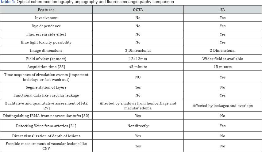
Currently, FA and ICGA are gold standard in assessment of retinal and choroidal vasculatures. But the FA has considerable shortages like being invasive; being dye dependent; putting patients at possible dye mortal side effects (however, rare); clinical contraindications of dye; putting retina at risk of blue light toxicity; relative long picture acquisition time (some 15 minutes); disability in assessing deeper retinal or choroidal layers; disability in providing 3D pictures, disability in providing structural details of retina and choroid; and not providing quantitative data. It seems that OCTA has alleviated all above FA's limitations. However, as any other device, it has inherited some technical limitations [16,17]. One may count: more limited field of view; not providing functional data regarding vessels like not showing leakages; being more sensitive to small eye movements; needs for more patients' cooperation and ability to maintain proper fixation [18]. This later may make the acquisition time in real practice much longer than official announcements by manufacturers. Different commercial devices utilize various technologies to improve quality of picture by reducing motion artifacts [19]; through special algorithms (amplitude decorrelation algorithm, OCT-based or optical microangiography (OMAG) [20], split-spectrum amplitude decorrelation angiography (SSADA), etc.) [21]; and also improving the field and depth of pictures [22]. In this technology, we encounter day to day evolution of imaging system in terms of eye tracking systems; speed of picture acquisition; artifact reduction solutions [23]; field and depth of images [22]. Table 1 compares FA and OCTA. The invention of the combined imaging systems which provide a hybrid FA/ICGA and OCTA images may have the least limitations.
FA: Fluorescein Angiography; FAZ: Foveal Avascular Zone; OCTA: Optical Coherence.
Tomography Angiography; IRMA: Intraretinal Microvascular Abnormalities


Is it possible to Upgrade SD-OCT Devices to OCTA Device?

The SD-OCT devices can do 26 to 40 thousands scans per seconds while commercial OCTA's scan speed is some two-fold of this. The resolution of images (indirectly, as resolution is dependent on number of scan per section which is limited by fixed scan speed and acquisition time) and their sensitivity to motion artifacts is particularly dependent on this. So the SD- OCT can not be utilized to obtain angiogram as images would be small and clinically useless. Fortunately, some manufacturers supplied their previous SD-OCT users with a two-step SD-OCT to OCTA upgrade. Firstly, they upgrade the hardware of device to higher frequency scan device, then they install OCTA software module on the device.


Commercial Prototypes Comparison

Most commonly used device in clinical centers, is AngioVue (Opto Vue, Inc., Fremont, Calif., USA) [10]. Recently, Heidelberg Engineering has released its OCTA modules. AngioPlex (Zeiss . Meditec, Inc., Dublin, Calif., USA) is also an other widely available device in market Table 2 compare features of these brands, provided by their manufacturer.
All the values are retrived from official websites of manufacturers.
While choosing a device one should consider following issues. The higher the scan speed is, the lower the effect of motion artifact would be. And also the resolution of images depends on number of scans per section. As the scan speed and scan acquisition time are limited, so it should be taken in to account that the high resolution of image translate to more acquisition time which is challenging in busy clinics. It is the reason why some manufacturer has not announced their device acquisition time, officially. And also some have reduced their image quality. As the OCTA is not substitute of FA in expert opinions; and also it is considerably expensive technology; moreover, many clinics has physical space limitation; so devices that provide hybrid FA/ ICGA and OCTA, could be an all-in-one reasonable option.


Future of Optical Coherence Tomography Angiography

Currently, the clinical use of OCTA is limited by its' expensiveness; small field of imaging; slow acquisition time; quality of images; lack of cut off points for quantitative data; lack of defined clinical significance of enormous data provided.

While developers are trying to invent faster swept source devices; smarter eye tracking systems; and also reducing artifacts that could provide wider higher quality views in matter of seconds, clinicians should utilize massive data provided by this technology in their everyday clinical practice by investigating the clinical relevance of findings. As we are on the edge of new robotic era, there are promises that images of retina could be efficiently processed by computers to diagnosis and grade diseases [24] and also conjunction of surgical or laser devices with imaging system could revolutionize both the diagnosis and treatment of ocular diseases [25-31].
For more Open Access Journals in Juniper Publishers please click on: https://juniperpublishers.com


Friday, 10 January 2020

Results of 50 Phacoemulsification Surgeries with Injectable Lens Implantation at Indus Medical College Hospital Tando Mohammad Khan- Juniper Publishers


Juniper Publishers- JOJ Ophthalmology

Abstract

Purpose: To evaluate the results of phacoemulsification surgeries at department of ophthalmology at Indus Medical College Hospital Tando Mohammad Khan.
Material and Methods: This study in which only 50 eyes of 43 patients were included to treat by phaco emulsification was conducted from 1st January to 30th June 2016. 31 patients were male whereas 12 were female. 20 were right eyes, 16 were left while 7 patients were undergone bilateral phaco surgeries within 7 to 16 days.1 patient was in age group C, another 1 was in group D, 8 were in group E, 12 were in group F and remaining 21 were in group G. 21 were suffering from diabetes, 12 were hypertensive, 2 were with cardiac problems using pace maker and 2 was involved with HCV infection. Patients suffering from ocular diseases: open angle glaucoma 3, pseudoexfoliation 4, pigment dispersion syndrome 1, chroniciritis 2, cholestrosisbulbi 1, asteroid hyalosis 1, age related macular degeneration 2 Eyes were dilated with mydriacil/phenylephrine eye drops, local anesthesia as retrobulbar as well as facial block (von lint technique) were given using 2% xylocaine inj without adrenaline. 2.8mm incision, capsulorexhsis with bent 27 gauge needle, followed by hydrodissection and in some hydrodelienation with small caliber irrigation cannula, copious 2% methylcellulose used to save endothelial cells as well as to maintain anterior chamber, all 4 steps of phaco followed with divide and conquer method and finally injectable intraocular lens implanted. Every operation ended with subconjunctival injection of dexamethasone 2mg plus gentamicin 20mg.
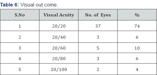
Results: 37 eyes gained 20/20 visual acquity on first post-operative day, 3 eyes gained 20/40, 5 gained 20/60 which over a period of five days improved to 20/20 after using topical prednisolone 1mg along with moxifloxacin eye drops, 3 gained 20/80 corrected with glasses, 2 were having 20/100 because of macular diseases.
Conclusion: In our experience phacoemulsification is an excellent technique which saves time, gives early rehabilitation depending upon the patience, experience and skill of surgeons.
Keywords: Cataract; Phacoemulsification results


Introduction

The term cataract is defined as an opacification of crystalline lens, having different morphological types; subcapsular; anterior and posterioir, cortical, nuclear opacification [1]. Cataract further divided according to density: grade 1 to grade 4. Cataract leads to decreased or blurring of vision.
Mostly senile [2] with other important causes like trauma, dibetes, myotonic dystrophy, atopic dermatitis, neurofibromatosis type 2, steroid induced, chronic iritis, high myopia, retinitis pigmentosa, gyrate atrophy, Stickler syndrome etc. [3].
With the passage of time lens increases in weight and thickness as new layers of cortical fibers are formed concentrically, the lens nucleus undergoes compression and hardening (nuclear sclerosis). Crystalline (lense proteins) are changed by chemical modifications and aggregation into high- molecular-weight proteins [4].
The only treatment for cataract is surgery either large incision ECCE or phacoemulsification, small incision early rehabilitation and with good visual outcome. The technique and results of cataract surgery have changed dramatically during the past three decades. In all over the world we have moved from intracapsular cataract extraction as the preferred technique to almost exclusively extracapsular techniques. Smaller incisions have become thestandard, with phacoemulsification now being the method of choice for most of surgeons [5].


Material and Methods

This study in which only 50 eyes of 43 patients were included to treat by phaco emulsification with injectable intraocular lens implantations at Indus Medical College Hospital Tando Mohammad Khan from 1st January to 30th June, 2016. All eyes were dilated prior surgery with mydriacil, phenylephrine eye drops, local anesthesia given using retrobalbur and facial (von lint) with 2% lidocaine (xylocain injections). Phaco done with infinity (Alcon) machine under Taggaki microscope.
Out of 43 patients 12 were females, 31 were males (Table 1), patients were divided into different age groups as: there was no patients in group A and B, group C and D have only one patient respectively, group E includes 8, group F includes 12 and group G includes 21 (Table 2).
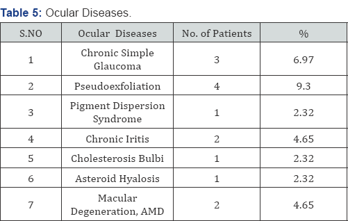
Out of 50 eyes 20 were right, 16 left and 7 were both eyes (Table 3). 37 (86.04%) Patients Were suffering with systemic diseases like diabetes (48.83%), hypertention (27.90%), cardiac problem, using pacemaker (4.65%), HCV (4.65 %) underwent surgery after having fitness from their physicians (Table 4) 14 (32.55%). Patients were suffering from ocular diseases like Glaucoma (6.97%), pseudoexfoliation (9.30 %), Pigment dispersion syndrome (2.32%), old healed iritis with peripheral ant Synaechae (4.65%), Cholesterosisbulbi (2.32%), asteroid hylosis (2.32%), Age related macular degeneration (4.65%) (Table 5).
After asceptic techniques, drapping and using 2 drops of 10% povidine solution instilled into eye, after 1 minute copious irrigation done, incision started with 2.8mm phaco knife, capsulorexhsis done with 27 gauge bent needle, hydrodissection and in some hydrd ilea nation using small caliber irrigation cannula, copious use of 2% methylcellulose to save endothelium as well as to maintain anterior chamber. All 4 steps of phaco followed and finally injectable intraocular lens implanted. Wound closed with stromal hydration. Every operation finished with sub conjunctival injection of Dexamethasone 2mg plus gentamicin 20mg, and eye kept pached for 24 hours.


Results

50 eyes of 43 patients were undergone surgery by phacoemulsification with injectable intraocular lens implantation, 37 eyes (74%) improved visual acuity to 20/20 at first postoperative day, 3 eyes (6%) improved upto 20/40, 5 eyes (10%) upto 20/60, 3 eyes (6%) upto 20/80, and 2 eyes (4%) improved upto 20/100 (Table 6). Figure 1 a, b, c shows injectable lens implantation during one of our surgery.


Discussion

Phacoemulsification is a very safe and less time taking technique depending upon good dilation of pupil pre- operative and during surgery as well as the patience, experience and skill of surgeons. It is established that the smaller phacoemulsification wound gives less induced astigmatism, faster visual rehabilitation and improved wound security than ECCE [6-11]. Smaller wound heals more rapidly with less risk of leakage, viscoelastic do not leave the eye through small incision [12]. 37 (74 %) of my patient improved visual acuity upto 20/20 on first and secondday, 3 (6%) developed striate keratitis and treated with topical steroid and regained 20/20 on 5th post operative day. In 5 (10%) visual acuity corrected.
With glasses with in -1.50 D sphere and 0.75 cylinder at 90degrees, 2 (4%) who were suffering with age related macular degeneration remained after BCVA at 20/100. In our study not a single case suffered with post operative endophthalmitis same as in a study done by Cooper et al. [13].
Out of these 50 eyes only 3 developed striate keratitis, reason was hard nucleus more than grade 3 density needed high phaco power and time by the technique divide and conquer same as described by Gimbel [14]. Topical steroids were being prescribed and on 5th postop day vision become 20/20. Though it was fairly high 20% in one study by Popiela G et al. [15] but in our experience it was only 6 %, a grade 3 nucleus (severely dense) and long absolute phaco time as independent predictors for endothelial cell loss [16]. Phacoemulsification in the capsular bag by directing probe away from the corneal endothelium and keeping the lens fragments at deeper plane are the measures which would be helpful in minimizing the chances of corneal edema and striate after phacoemulsification same as suggested by Zetterstrin C [17] and Pirazzoli G et al. [18].


Conclusion

Small incision surgery such as phacoemulsification with injectable intraocular lens implantation is a very safe and less time taking technique which depends upon the experience and skill of surgeon who strictly follows selection and exclusion criteria and with a good knowledge when to abandon or convert the technique and always keeps the lens fragments in the capsular bag with the phaco tip directed away from endothelium, do not follow the lens fragments near the posterior capsule, allow fragments' to follow the tip.


Acknowle dgement


I am thankful to Aftab Ahmed Khan assistant professor for his assistance, Aisha Khan (4th year MBBS) and Atiqa Khan (final year BDS) students for their help in compiling data. Special thanks to my assistant Moin Shaikh who assisted all cases. Jawaid Iqbal assistant professor who is always helpful.
For more Open Access Journals in Juniper Publishers please click on: https://juniperpublishers.com


Thursday, 9 January 2020

Patient-Centered Care: Where Policy Meets Practice- Juniper Publishers


Juniper Publishers- JOJ Ophthalmology


Commentary

A 59-year-old woman is referred to you by her primary care provider (PCP) for a routine eye examination. She had never undergone a comprehensive eye exam previously and was looking forward to the opportunity to have her eyes examined by an ophthalmologist. She purchased reading glasses for the first time 5 years ago, but now even those spectacles were not addressing her visual needs. Her complaints to her PCP prompted the referral. Additional history revealed that this patient was recently laid off and was now insured on Medicaid, which had been expanded in her state following the passage of the Affordable Care Act.[1]
On examination, her visual acuity was 20/40 OD and 20/60 OS. Angles were open OU 360 degrees and IOP measured 25mmHg OD and 32 OS mmHg. SLE revealed clear corneae, deep and quiet anterior chambers, and 1+nuclear sclerosis. Central corneal thickness measurements were 550um OD and 555um OS. Optic nerves evidenced moderate cupping, 0.7OD and 0.8OS.
Patients such as this 59-year-old woman enter our offices every day. Her visual fields and optic nerve imaging confirmed mild glaucoma OD and moderate glaucoma OS. Her physician set a target pressure of 16-18mmHg OD and 14-16mmHg OS. Now that her diagnosis has been established and a plan discussed with the patient, it is clear based on her history shared at the beginning of her examination that the reliance only on topical medication would not be realistic. Although generic medications may be are affordable options, given the list of medications that she manages for her diabetes and hypertension, it is clear that additional options will need to be considered.
This case reminds us of the importance of considering the whole patient rather than solely the patient's ocular presentation. Being realistic in the beginning of a patient's relationship with the provider is critical for establishing the basis for an effective partnership to slow or halt the progression of disease. It is also important to understand the healthcare landscape that shapes patients' choices for coverage of their care. For example, in the United States, the Affordable Care Act (ACA) that was launched in 2010, expanded Medicaid coverage to previously uninsured patients in more than half of the country [1]. This patient had purchased insurance under the ACA, Now that her insurance may be discontinued, she is concerned that she will be uninsured again.
Given her circumstance, the physician decided to begin treatment with generic latanoprost once daily as a monocular trial in the left eye; the option of selective laser trabeculoplasty (SLT) is also discussed. The patient returned Measuring and her intraocular pressures are essentially unchanged, measuring 22 OD mm Hg and 29 OS mm Hg. The patient admitted not being able to afford the medication and agrees to proceed with SLT SLT is performed in both eyes and six weeks later IOP measured 18mHg OD and 20mmHg. Additional SLT treatment is considered to get her pressures closer to her target.
This case brings up three discussion points the author wishes to highlight:
  1. The benefits of treatment with SLT
  2. The importance of understanding the context of care
  3. Why eye health needs to be a population health priority
The benefits of treatment with SLT in early and advanced glaucoma has been reported by others [2,3]. Notably, in a series of 26 eyes with early glaucoma, investigators reported greater than a 20% reduction in intraocular pressure and a reduction in intraocular pressure less than 21mmHg in 62.9% of treated eyes, and with retreatment an additional 7.4% were noted to evidence a reduction in intraocular pressure. Among 44 eyes with advanced glaucoma, an IOP reduction greater than 30% reduction in IOP and an IOP less than 18 mm Hg was noted in 50% of eyes treated. These eyes were followed for 1 year [3] When compared to either medications or surgery over a 5-year period, laser trabeculoplasty has been noted to be the most cost effective[4]. Thus, for our patient, given her challenges with the affordability of medications, SLT is a reasonable option.
In the United States, the landscape for patients who struggle to afford healthcare is currently uncertain, given current efforts to repeal the Affordable Care Act [1]. Thus, understanding the context in which this patient must consider her care is important. Personalizing the treatment plan for patients should consider the realities of the impact of care, in addition to the best, evidence-based options. At 59 years of age, this patient is not yet eligible for Medicare and thus, the cost effectiveness of the laser trabeculoplasty [4] is an important consideration. In addition, the outcome of the proposed treatment is critically important. If the patient can be managed on fewer medications or on no medications, then her quality of life will benefit.
Consider the evidence of the effectiveness of surgical intervention if the patient had presented with angle closure glaucoma. If this patient had presented with angle closure glaucoma, clear lens extraction may have been an option for her, particularly given the results of the EAGLE study. The EAGLE study assessed the effectiveness of clear lens extraction in patients with angle closure glaucoma. In a randomized controlled study design, randomizing patients to either laser iridotomy or surgery, the investigators reported clear lens extraction to be more effective and cost effective than laser iridotomy [5]. Regardless of the outcome of the debate related to the Affordable Care Act, [1] it is likely that as a discipline, it will be important to assemble additional data such as the EAGLE5 and the Cantor evaluation related to laser trabeculoplasty [3] to support the reimbursement of specific clinical decisions, as metrics for reimbursement move from volume to value [6]. Timing for our patient's procedure is important, given the imminent changes in her healthcare coverage.

The last point highlights a recent report from the Institute of Medicine (now named the National Academy of Medicine) released in the fall of 2016, entitled, "Making Eye Health a Population Health Imperative: Vision for Tomorrow" underscores importance of preventing irreversible blindness and suggests strategies for minimizing vision impairment. Nine recommendations are offered addressing public awareness, the need for greater evidence to guide policy, importance of expanding access, enhance the capacity of public health resources, and the need for promoting community action [7]. 0ur patient represents a missed opportunity for earlier diagnosis and intervention. Encouragement by other providers with whom she may have interfaced even five years prior to her presentation would have given her a greater likelihood of avoiding irreversible loss of vision related to glaucoma. 0nly by elevating the eye health in the minds of nonophthalmic clinicians, the public, and insurers will the needle significantly move in preventing blindness. Our patient has benefitted from work of many innovators and researchers, her ability to maintain her visual lifeline for the rest of her life rests largely with her as she navigates the healthcare landscape. The provider is her partner in this journey, effectively guiding her with a focus on the context of her care.

For more Open Access Journals in Juniper Publishers please click on: https://juniperpublishers.com


Wednesday, 8 January 2020

Juniper Publishers- JOJ Ophthalmology- Juniper Publishers

UFO (Unidentified Full Objects) Sighted in The Cornea: Can We Make The Diagnosis By Means of in vivo Confocal Microscopy?

Introduction

In vivo confocal microscopy (IVCM) is a powerful diagnostic technique that provides minimally invasive, high resolution, steady-state assessment of the corneal cellular structure [1]. Rapid scanning is used to recreate a full field of view and to get a “real time” viewing [2] Because of its ability to analyze living tissue at cellular levels, IVCM represents a valid tool for clinical diagnosis and management of corneal diseases [3]. It may be useful in the areas of infective keratitis, corneal dystrophies, refractive surgery, and contact lens wear, where it allows for differential diagnosis and detection of subtle short and longterm changes [2]. In our study, we evaluate the efficacy of IVCM in the diagnosis of corneal disease.


Materials and Methods

Thirty eyes of 30 patients with corneal diseases were included in the study. All patients underwent IVCM and color picture of the anterior segment. A color photograph of the entire ocular surface of each eye was obtained using a slit lamp and Ekta chrome, 16x magnification.
Heidelberg Retinal Tomograph with Rostock Corneal Module (HRT-RCM) (Heidelberg Engineering, GmBH, Dossenheim, Germany) was used to evaluate the corneal structure. The system design and use of this confocal microscope have been described in detail [4]. Before the examination, a drop of a topicalanesthetic, proparacaine hydro chloride ophthalmic solutionof 0.5%, was administered to the cornea, and a drop of 2.5% hydroxyl propyl methyl cellulose was placed at the tip of the objective to serve as an immersion fluid. The patient was asked to focus on a fixation device to allow for the alignment of the objective to the region of interest. Sections of the peripheral and central cornea were imaged. Real-time images of all layers of the cornea were detected through the use of a low-light camera and recorded. The images were digitized and stored in computer memory.
Round hyper-reflective bodies seen with in vivo confocal microscopy were defined as UFOs (Unidentified Full Objects). Frames containing UFOs were selected and analyzed by a masked observer (MP), to ascertain whether confocal images alone were sufficient to formulate a correct diagnosis. The masked observer was then provided with the color picture of the cornea and asked to re-assess his/her previous diagnosis if needed.


Results

Fifteen out of the 30 patients presented Acanthamoeba keratitis (AK); 4 conjunctival pigmented lesion; 3 Map-Dot- Fingerprint keratopathy; 3 post-LASIK Diffuse Lamellar Keratitis (DLK); 2 fungal keratitis; 2 epithelial in-growth and 1 corneal pigmented lesion. In vivo confocal microscopy allowed for correct diagnoses in 22 cases (73%), whereas in 8 the diagnosis was incorrect. Patients with AK and fungal keratitis were correctly diagnosed. DLK patients were generically diagnosed as having "corneal scarring" and subsequently correctly diagnosed through examining the color picture. Out of the 8 misdiagnosed cases, 7 were correctly diagnosed once the color picture of? the cornea was provided. One patient affected by Map-Dot-fingerprint was misdiagnosed as suffering from AK even ofter Examining the colour picture (Table 1)(Figure 1&2).


Conclusion

In vivo confocal microscopy is a non-invasive examination that provides relevant information on corneal anatomy [5]Its role in the clinical setting has been the most described in the management of infectious keratitis [6]. Even if corneal scraping and biopsy remain the gold standard in the micro biology diagnosis, IVCM may facilitate early diagnosis and the initiation of targeted antimicrobial therapy. It is particularly valuable in challenging cases such as contactlens- related AK [7]. Acanthamoebacysts, trophozoites and fungal hyphae can be identified by using IVCM directly [8]. In a prospective, doublemasked, observational study [9], the sensitivity of IVCM in recognition of Acanthamoebacysts and fungal elements was 88.3%, and specificity was 91.1%. As previously stated by the American Academy of Ophthalmology which reported level II evidence for the adjunctive role of IVCM in the diagnosis of AK [6,10]. Nevertheless, clinical pictures are instrumental in getting the correct diagnosis. In our study, UFOs were mostly mis interpreted as Acanthamoeba cysts, probably because that they are easily identified by this tool thereby yielding a high rate of false positive findings. One single image of IVCMis deemed insufficient if correct diagnoses are to be made, as findings may well overlap in different diseases. Nevertheless, when integrated with bio-microscopic findings, this tool is essential if prompt, accurate and non-invasive diagnoses of corneal disease are to be formulated.

For more Open Access Journals in Juniper Publishers please click on: https://juniperpublishers.com

Tuesday, 7 January 2020

Juniper Publishers- JOJ Ophthalmology- Juniper Publishers

A Comparison of Automated Refractions Using Plusoptix S04 Photoscreener, Nidek AR-20 Handheld Autorefractor and Nidek ARK-510A Auto Ref-Keratometer- Juniper Publishers

Abstract

Purpose: To evaluate automated refraction measurements using the Plusopti X S04 photo screener (OP), Nidek AR-20 hand held auto refractor (NAR) and compare them to the more traditional Nidek ARK-510A (ARK).
Methods: 102 patients aged 3 to 81 years were included. All patients underwent on-cycloplegic automated refraction using the PlusoptiX S04 photo screener, Nidek AR-20 handheld auto refractor and the Nidek ARK-510A. This was followed by measuring Cyclo plegic auto refraction using the Nidek AR-20 handheld auto refractor and Nidek ARK-510A.
Results: Without Cyclo plegia, the mean sphere values were significantly different for both the OP (-0.41 D, SD±2.39D) versus ARK (-0.60D, SD±2.24) groups with a p=0. 008, and for NAR (-0.48, SD±2.01) Versus ARK groups with a p=0.029. With cycloplegia, there was no significant difference in the mean sphere values between the OP versus ARK groups as compared to the OP versus NAR and NAR versus ARK groups (p=0.049 and p=0.001 respectively).
Conclusion: Our study indicates similarities between the OP photo screener and the traditional table-mounted ARK results after cycle plegia. These results were not reflected when comparing the NAR handheld auto refractor to the ARK. Although efficient as screening tools, we recommend caution when using handheld instruments.
Keywords: Refractometer; Refractive errors; Myopia; Hyperopia; Astigmatism; Cycloplegia; Middle east; Lebanon


Introduction

Refraction is a clinical test used to determine the refractive state of the eye and to assess the individual's need for visual aid. Although hretinoscopy is still considered the gold standard for determining the refractive state of the eye, it is subject to interobserver variability [1,2]. Over the past few decades, new advancements have introduced the auto refractors which have gained a popular routine use among ophthalmologists [3,4]. Auto refractors are easy to use, time saving and relatively accurate in measuring the amount and type of refractive error. Today, many auto refractors are available in the market (Canon, Nikon, Topcon, Carl Zeiss Meditec, Nidek etc.). A disadvantage of most auto refractors is they need to be mounted on a table due to their relatively large size. As such, ametropia measurements may therefore be difficult in bed ridden patients, disabled persons and very young or handicapped children. Handheld auto refractors or photo screener scans are useful in such scenarios.
Plusopti XS04 (OP) (Plusoptix GmbH; Nuremberg, Germany), is a third generation vision screener designed to screen for refractive errors, anisocoria, and strabismus in children starting at the age of 6 months [5]. Screening is quick with both eyes screened simultaneously and can be performed by non-medical personnel, with a screening distance of 1 meter [5]. The device measures refraction, pupil size, and corneal reflexes. Results with abnormal limits are displayed as "refer". Referral criteria include anisometropia, astigmatism, hyperopia, myopia, anisocoria and corneal reflexes [5].
To date, multiple studies show that the OP vision screener is useful when used in screening for amblyopia oramblyogenic risk factors [6-10] small-angle strabismus [11] is considered effective when compared to cycloplegic pediatric ophthalmic evaluation [9,10] and can be used to screen patients with intellectual disability foramblyogenic risk factors with 95% sensitivity and 50% specificity [12].
In our manuscript, we explore the accuracy of both the OP vision screener when used as suggested in noncyclopleged patients, the Nidek AR-20 (NAR) (NidekCo. Ltd, Gammagori; Aichi, Japan) handheld auto refractor on non cyclopleged and cyclopleged patients and compare the results with those obtained before and after cycloplegia using a more traditional table- mounted NidekARK-510A (ARK) autorefractor/keratometer (NidekCo. Ltd, Gammagori, Aichi, Japan).


Materials and Methods

We prospectively evaluated 102 consecutive patients presenting to the outpatient clinic during August 2013, without any bias to their age, gender, diagnosis or previous medical history. Patients were excluded if they could not be refracted because of poor cooperation or any ocular pathology. After the initial history was taken, a trained ophthalmology fellow (H.B) and professional optometrist (J.H) used all 3 instruments to obtain non Cyclo plegic automated refraction. This was followed bycycloplegia using 1 drop of Mydriacyl (tropicamide 1%, Alcon, Puurs, Belgium) in each eye twice (10 minute interval). Twenty to 30 minutes later, Cyclo plegic auto refraction was obtained using the ARK, and NAR auto refractors.
Data entry and statistical analysis were performed using SPSS 22.0 for Windows (SPSS Inc, Chicago, IL, USA). Analysis variables included:gender, age (years), auto refraction (sphere, cylinder, axis) and spherical equivalent for both eyes. For each of the 3instruments, the data were divided into 4 main categories: sphere, cylinder, axis and spherical equivalent. Spherical equivalent (sphere power+(0.5 x cylinder power)) measured in diopters was calculated from the auto refraction measurements. For the ARK and NAR handheld auto refractor, data were further divided into non cycloplegic and Cyclo plegic auto refractiCases with missing variables were excluded from the analysis. Two types of statistical testing were used to determine differences or similarities between the OP, NAR handheld auto refractor, and the ARK. Analysis of variance (ANOVA) testing was performed to compare the 3 groups. A variable ratio (F) was calculated to determine overall statistical differences. Paired-samples t-tests were performed to compare means between 2 groups. Significance was defined as p values less than or equal to 0.05.


Results

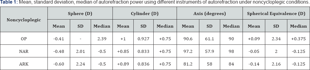
One hundred two patients (204 eyes) were initially recruited. Nineteen eyes (9.3%) could not be refracted by OP (one patient could not be refracted for both eyes by neither OP nor the NAR) and were excluded from the analysis. A total of 185 eyes completed the study on all 3 instruments and were included in the analysis (45.7% males, 54.3% females). The patients ranged in age from 3 years to 81 years (mean 41.02±19.7years, median 43 years).Using OP, the mean recorded sphere on 185 eyes was -0.41 diopters, mean cylinder was +1 diopters, and mean axis were measured at 90.6° (Table 1). The median spherical value was Plano (range -6.75 to +10). The mean spherical equivalent determination of the OP group was +0.09 diopters (range -6.125 to +10.5). A myopic spherical value was found in 47.6% of the eyes 42.7%were hyperopic and 9.7% were plano. Astigmatism was identified in 96.8%

OP:PlusoptiX S04 (Plusoptix GmbH; Nuremberg, Germany); NAR: Nidek AR-20 (Nidek Co.Ltd, Gammagori; Aichi, Japan); ARK: Nidek ARK-510A (NidekCo.Ltd, Gammagori; Aichi, Japan).

Non cycloplegic auto refraction results for nidek AR-20 handheld autorefractor and nidek ARK-510A

The mean sphere value recorded by the NAR for 185 eyes was -0.48 diopters, mean cylinder was+0.85 diopters, and mean axis were 97.16°. The median spherical value was -0.5 diopters (range -6.75 to+8.5). The mean spherical equivalence for the NAR group -0.05 diopters (range -6.125 to+9.12). A myopic spherical value was identified in 60.5%; 33.5% were hyperopic, and 5.9% were Plano. Astigmatism was identified in 91.9% (Table 3). The mean spherical value recorded using the ARK on 185 eyes was -0.604 diopters, mean cylinder was +0.89 diopters, and mean axis measurement was 81.2° (Table 1). The median spherical value was -0.5 diopters (range -6.25 to+10.5). The mean spherical equivalence measured in this group was -0.136 diopters (range -4.875 to+1G.875). A myopic spherical value was diagnosed in 61.1%, 3G.3% were hyperopic, and 8.6% were Plano. Astigmatism was identified in 95.7% of 185 eyes (Table 3).

Cycloplegicautorefraction results for nidek AR-20 handheld autorefractor and nidek ARK-510A

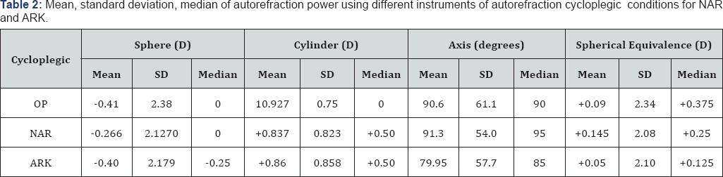
The mean sphere value recorded by the NAR for 185 eyes was -0.266 diopters, mean cylinder was +0.837 diopters, and mean axis were 91.3° (Table 2). The median spherical value was Plano (range -5.75 to+10.5). The mean spherical equivalence for the NAR group +0.145 diopters (range -4.75 to+10.875). A myopic spherical value was diagnosed in 48.6%, 41.6% were hyperopic, and 9.7% were Plano. Astigmatism was identified in 97.3%. The mean spherical value recorded using the ARK on 185eyes was -0.40 diopters, mean cylinder was +0.86 diopters, and mean axis measurement was 79.95° (Table 2). The median spherical value was -0.25 diopters (range -6.25 to+10.5). The mean spherical equivalence measured in this group was +0.05 diopters (range -4.875 to+10.875). A myopic spherical measurement was diagnosed in 52.4% of eyes, 35.1% were hyperopic, and 12.4% were Plano. Astigmatism was identified in 95.7% of eyes (Table 4).
OP: PlusoptiX S04 (Plusoptix GmbH; Nuremberg, Germany); NAR: Nidek AR-20 (NidekCo.Ltd, Gammagori; Aichi, Japan); ARK: Nidek ARK-510A (NidekCo.Ltd, Gammagori; Aichi, Japan).
Optix: PlusoptiX S04 (Plusoptix GmbH; Nuremberg, Germany); C-NAR: cycloplegic-Nidek AR-20 (NidekCo.Ltd, Gammagori; Aichi, Japan); C-ARK: cycloplegic- Nidek ARK-510A (NidekCo.Ltd, Gammagori; Aichi, Japan).
Optix: PlusoptiX S04 (Plusoptix GmbH; Nuremberg, Germany); C-NAR: Cycloplegic- Nidek AR-20 (NidekCo.Ltd, Gammagori; Aichi, Japan); C-ARK: Cycloplegic- Nidek ARK-510A (NidekCo.Ltd, Gammagori; Aichi, Japan).
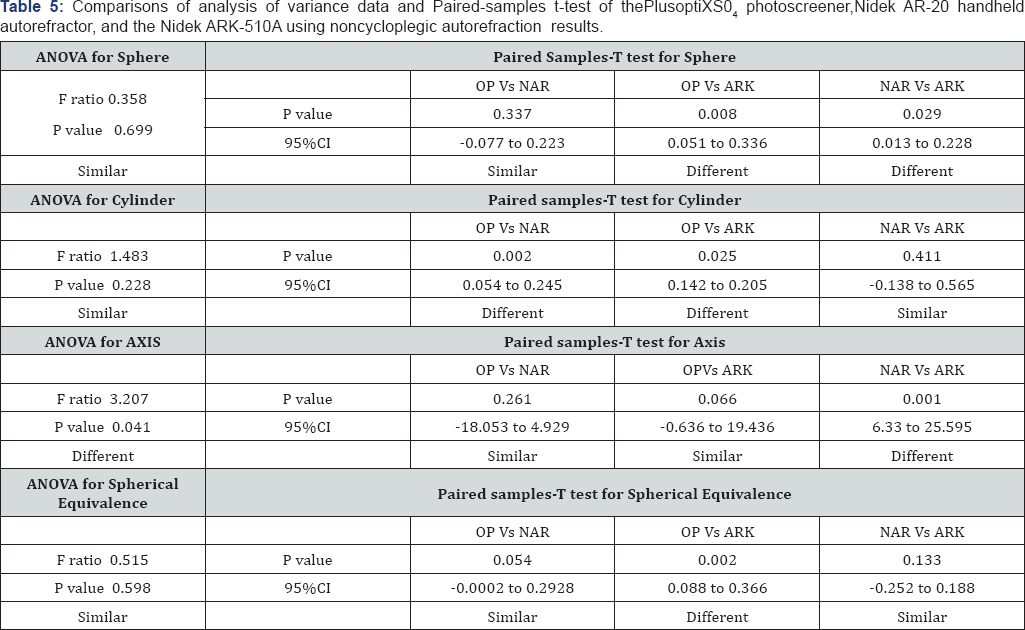
Except, of the axis value, ANOVA testing for auto refraction parameters of all instruments before cycle plegia revealed no significant difference among the 3 groups (Table 5). While paired-samples t-test comparisons of OP and NAR sphere values showed no statistical significance (p=0.34). Comparisons of sphere values for OPversus ARK and NAR versus ARKwere statistically significant (p=0.008 and 0.03 respectively) (Table 5) . Paired-samples t-test comparisons of cylinder values of NAR versus ARK showed similar results. However, the OP versus NAR and OP versus ARK groups showed a significant difference (p= 0.002 and p=0.025 respectively) (Table 5). The axis value analysis by ANOVA revealed a significant p-value (p= 0.034), indicating an overall significant difference between the 3 groups. Conversely, paired-samples t-test results were only significant between the NAR and ARK groups (p=0.001) (Table 5).
Calculated spherical equivalence measurements did correlate with our initial sphere measurements in the NAR versus ARK groups. Paired-samples t-test comparisons between the OP versus ARK demonstrated a difference with a p= 0.002. When comparing the OP versus NAR and the NAR versus ARK groups, no statistical difference was observed (Table 5).
ANOVA: analysis of variance test; CI: confidence interval; F: Variance ratio; OP: PlusoptiX S04 (Plusoptix GmbH; Nuremberg, Germany); NAR: Nidek AR-20 (NidekCo.Ltd, Gammagori; Aichi, Japan); ARK: Nidek ARK-510A (NidekCo.Ltd, Gammagori; Aichi, Japan).
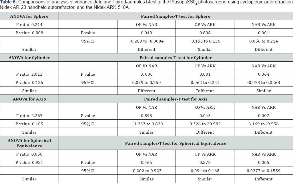
Data from the NAR and ARK instruments were reassessed using Cyclo pelagic autorefractions (C-NAR and C-ARK); ANOVA testing for all refraction parameters did not indicate any statistical differences (Table 6). Similarly, paired-samples t-test comparisons showed comparable results among the OP versus C-ARK groups. However, there was a statistical significance in OP versus C-NAR and C-NAR versus C-ARK groups (p= 0.049 and 0.001 respectively) (Table 6). Paired-samples t-test for the cylinder values of the OP versus the C - NAR group and OP versus C-ARK indicated statistical differences (p=0. 000 and p =0. 001) (Table 6).
For Axis values, paired-samples t-test comparisons of OPversus C-NAR values were statistically similar, whereas those of OP versus C-ARK showed a significant difference (p= 0.04). The C-NAR axis value compared to that of the C-ARK also showed a significant difference (p= 0.07) (Table 6). Finally, when analyzing spherical equivalence, paired-samples t-test comparisons showed similar results except for the C-NAR versus C-ARK group which showed a significant difference with a p=0. 005 (Table 6).


Discussion

Although considered as the gold standard for measuring refractive status, Cyclo pelagic refractions can be time consuming, cause patients discomfort and adds additional costs. As amblyopia is one of the leading causes of visual impairments in adults [13] developing accurate, easy to use, friendly autorefractors that do not require the use of cytoplegic agents could help in identifying significant refractive errors more rapidly and efficiently while avoiding the drawbacks of cycloplegia.As new instruments for refractive error measurements become available, it is of critical importance to evaluate and compare their validity to that of existing reliable technology. The OP photo screener has been marketed toward health care providers as a tool to assess refractive errors in young children without cycloplegia [5,9,10]. Although many studies showed that the OP vision screener is useful as a screening tool for amblyopia or amblyogenic risk factors [6-10] Dahlmann-Noor et al argue that the use of OP as a single screening test in children may miss a significant number of children with amblyopia or amblyogenic risk factor [14].
As in other studies, we found that both the OP vision screener and NAR handheld auto refractor were simple and user friendly, as well as quick in reporting refraction result without the need for direct contact or patient compliance. However, limitations that we encountered while using the OP included the need for a dimly lit room to obtain proper measurements, and its inability to obtain measurements in patients with a number of ophthalmologic conditions. Of the 19 eyes that failed autorefraction by the OP, 6 had a high refractive errors (more than -5), 4 had a history of cataract surgery, 2 had a history of cataract surgery and corneal rings for keratoconus (also failed autorefraction by NAR), 2 had a history of diabetic retinopathy, 1 had a history of a corneal ring for keratoconus, while the remaining 4 had no known ocular problems but were read as "pupil undetected". Disadvantages similar to those claimed by Rajavi et al which may have limited our use included: pupil size, fixation problems and posterior segment pathologies [15]. Limitations with the NAR were mostly a failure of patient cooperation resulting in no measurements being taken especially in children. A child's ability to hold the head still limited our ability to produce repeatable measurements.
The ARK table-mounted auto refractor readings were more frequently myopic compared to the OP and NAR (Table 3 & 4). This may be explained by the fact that it is well known that auto refractive instruments tend to overestimate myopia and underestimate hyperopia [10]. Except for the axis value prior to cycloplegia, when comparing all 3 instruments using ANOVA, our data indicated that there was no significant difference in auto refraction results before and after cyloplegia (Table 5 & 6). Comparing the OP spherical values with values obtained before cycloplegia using the NAR and the ARK indicated that there was a significant difference between the OP and the ARK groups as well as the NAR and ARK groups. This may lead us to conclude that the two handheld instruments may not be as reliable as the well established ARK for diagnosing refractive errors. In contrast, however, the results from both handheld instruments were similar (Table 5). Although the above mentioned results were reflected in the calculated spherical values for both the OP versus ARK and OP versus NAR groups, the NAR versus the ARK group showed similar results.
After cycloplegia on NAR and ARK, our spherical values indicated a difference between OP versus C-NAR and the C-NAR versus C-ARK groups (Tables 6). However, there was no difference between the OP and C-ARK values. Our study results are consistent with data from other studies that claim the OP to be a screening tool to assess refractive without the need cycloplegia [5,9,10]. While in both the cyclopleged and non cyclopleged groups, the OP recorded myopia less often than those recorded by the NAR and the ARK, astigmatism recordings were more comparable in all 3 groups (Table 3 & 4). The astigmatic (cylinder) readings were statistically similar between all 3 instruments by ANOVA however, paired samples-T test detected a difference between the OP and both the NAR and ARK groups before and after cycloplegia (Table 5 & 6). These results could mean that with regards to diagnosing astigmatism (measuring cylinder values) the OP may not be as reliable as the ARK as compared to the NAR.
Although handheld refractors are proving to be more practical, time efficient and provide a value as screening tools, we suggest that they should be used with caution when determining automated refractions especially in children. In the era of refractive surgery, it is important to assess the reliability and reproducibility of newer instruments when compared to existing technology and should not compromise the accuracy of the refractive state of a patient at the expense of practicality and time saving. We do not recommend the use of the hand held instruments for diagnostic purposes, with the exception of remote or inaccessible areas where portable instruments are essential and cycloplegia is difficult.
A positive aspect of our study setup is that our study population was non bias to age during recruitment. To our knowledge the Optixphotoscreener has only been extensively studied in screening younger population groups. This study may give us a better analysis of the use of the open adult patients. Several limitations are inherent in our study. In view of the time constraints and the difficulty of getting young children and elderly patients to cooperate and concentrate for long durations, repeated measurements on all 3 instruments were not documented. Furthermore, and for the same reason of time constraint, two separate individuals obtaining measurements on the handheld instruments may have introduced inter-observer bias. Finally, the same observer documenting measurements from both the handheld and table-mounted auto refractor was not masked to subjective refraction readings, introducing possible observer bias.

For more Open Access Journals in Juniper Publishers please click on: https://juniperpublishers.com