Tuesday, 25 May 2021

Juniper Publishers- JOJ Ophthalmology

A Review of the Data on the Recently Approved Xen Surgical Gel Stent in the Management of Glaucoma-Juniper Publishers

Abstract

The cornerstone of glaucoma surgery includes trabeculectomy and tube shunting procedures, which utilize an ab externo approach to divert aqueous humor from the anterior chamber to the sub conjunctival space. The XEN Gel Stent is a 6.0mm tube consisting of porcine-derived collagen that similarly creates a non-physiologic shunt but through an ab interno approach. The XEN gel stent has the potential to effectively lower intraocular pressure and medication use with lower complication rates than traditional glaucoma surgery. This mini-review surveys the data of the XEN implant in current literature.

Introduction

Glaucoma maintains a significant disease burden worldwide. It is the most common cause of irreversible blindness, affecting over 64 million individuals [1]. Therapy is focused on lowering intraocular pressure (IOP) by a variety of methods, including topical medications, laser, and incisional surgeries. These surgical interventions-traditionally trabeculectomy or tube shunt surgery-rely on creating an additional subconjunctival reservoir for aqueous humor (AH) drainage and subsequent resorption. However, the three-year results of the Tube Versus Trabeculectomy Study found failure rates of 15 and 28 percent, respectively. Serious postoperative complications such as persistent corneal edema, endophthalmitis, and chronic or recurrent iritis were also reported [2].

There has been a recent proliferation in procedures and medical devices that provide similar IOP-lowering effects to trabeculectomy or tube shunt surgery with fewer complications. One such product is the XEN Glaucoma Treatment System (Allergan, Inc., Irvine, CA, USA), which consists of the XEN Gel Stent and XEN Injector. The XEN stent is derived from porcine collagen, measuring 6.0mm long with inner diameters of 140|im, 63|im, or 45|im, although the 45|im stent is currently recommended. Previous studies in animal models have demonstrated no significant inflammatory response to implantation and no signs of degradation of the stent itself [3]. It was recently approved by the United States FDA in November 2016 for use in refractory glaucoma, including those with a history of failed prior surgical treatment, primary open-angle glaucoma (POAG), and pseudoexfoliative glaucoma with open angles that are inadequately controlled on maximal medical therapy.

The XEN stent is inserted into the angle through the scleral spur via an ab interno approach with its disposable injector. Ideally, the device should extend 3.0mm posteriorly from the limbus and into the subconjunctival space and 2.0mm anteriorly into the anterior chamber (AC). Once placed in an aqueous environment, the device hydrates, becoming soft and flexible, helping to maintain its position. By taking advantage of the resistance to flow in a cylinder as determined by the Hagen- Poiseuille equation, the XEN45 produces a pressure gradient 7.56mm Hg at the physiologic flow rate of AH within the AC, at 2.5 microliters/minute [4]. Thus, this stent can theoretically reduce the risk of hypotony seen in IOP-lowering surgeries despite being a valve less device. The fact that it is inserted ab interno obviates the need from conjunctival dissection, theoretically reducing the potential for conjunctival fibrosis and leaving the ophthalmologist with the option to perform ab externo surgeries if necessary in the future.

XEN Gel Stent in the Literature

Several studies were performed evaluating the XEN Gel Stent with multiple inner diameters. Sheybani, Dick, and Ahmed reported on the results of 49 eyes of 49 patients treated with the XEN140 stent (140um internal diameter) without the use of mitomycin C (MMC) [5]. Of the 49 patients, 22 received previous glaucoma surgery, and nine (18%) had a prior laser trabeculoplasty. Complete success was defined as an IOP <18mm Hg and a greater than 20% reduction of IOP at the primary endpoint of 12 months without glaucoma medications. Criteria for treatment failure included visual acuity less than or equal to light perception, need for additional glaucoma surgery, or a less than 20% reduction of IOP at 12 months. This study revealed that the mean IOP reduced from 23.1±4.1mm Hg preoperatively to 14.7±3.7mm Hg at 12 months, a 36.4% decrease. In all, 40% met the criteria for complete success and 88.9% for partial success. Three patients (6%) failed the study criteria and required additional surgery. The most common complication was needling (47%), with nearly half of the cases occurring within the first month.

Another early study by Sheybani and Ahmed used the XEN140 and XEN63 stent (63um internal diameter) without MMC in patients undergoing phacoemulsification [6]. Of the 37 eyes, 47.1% were considered complete successes while 85.3% were qualified successes. Mean IOP significantly decreased from 22.4±4.2mm Hg preoperatively to 15.4±3.0mm Hg at 12 months, and mean medications were significantly reduced from 2.5±1.4 to 0.9±1.0.

One of the first reports involving the exclusive use of the XEN45 stent (45um internal diameter) involved 31 eyes receiving phacoemulsification and MMC treatment at the time of implantation. Mean IOP and medication use were reduced significantly from 20.8±4.6mm Hg to 13.1±3.6mmHg at 12 months and 2.7±1 to 0.9±1.1 at 12 months, respectively, and without significant complications [7].

Pérez-Torregrosa et al. [8] were among the first to describe the efficacy of the XEN45 in patients with mild and moderate glaucoma (defined as a mean deviation between 0 and -12dB on Humphrey 24-2 perimetry) [8]. Additionally, subjects included in this study had pressures<30 while being managed on two or more medications. Twelve months after concomitant phacoemulsification and XEN45 implantation, 27 of 30 (90%) subjects met the successful treatment criteria of IOP≤18mm Hg with no glaucoma medications. The authors reported several intra operative complications, including sub conjunctival hemorrhage with MMC instillation (36.6%), and minor hemorrhage intra camerally (86.6%) and at the scleral exit point (90%). A total of six (20%) stents required relocation, and one eye required re-implantation of the device. One subject was excluded from analysis due to extensive subconjunctival hemorrhage after MMC injection and another due to extrusion of the device into the subconjunctival space intra operatively.

Two studies have addressed the effect of simultaneous implantation and phacoemulsification versus the XEN alone. One such study involved 567 eyes, of which 54% underwent XEN implantation only, and 46% the combined implantation and phacoemulsification [9]. There were no inclusion or exclusion criteria regarding the grade of POAG, and data from all three diameters of XEN stents were not separated. The data revealed a mean preoperative IOP of 21.9±4.2mm Hg that was significantly decreased at 12 months (15.7mm Hg), 24 months (15.0mm Hg), and 36 months (13.2mm Hg) of follow-up. Likewise, a significant decrease in medications by 74, 77, and 74 percent from the mean of 2.7 was observed at 12, 24, and 36 months, respectively. The percentage of patients who were converted to another procedure was 4% by 12 months, 5% by 24 months, and 5% by 36 months. After analysis, it was determined that there was no statistical difference in mean IOP or medications between eyes receiving the implant alone and the combination procedure. Likewise, a 75 patient cohort demonstrated no significant difference in IOP reduction between the standalone and combination procedures at 12 months [10]. The most frequent complications from this study were needling (15.4%) and hypotony defined as an IOP <6mm Hg on postoperative day 1 (12.6%).

The most recent article involving the XEN stent evaluated its use in eyes with suboptimal IOP and medication intolerance, medication noncompliance, or maximum therapy with no history of glaucoma surgery [11]. A total of 13 eyes underwent XEN45 implantation in addition to phacoemulsification if previously phakic. At the 12-month end point. 41.7% were complete successes, with a>20% drop in IOP and discontinuation of all glaucoma medications, while an additional 25% met the IOP goal but remained on at least one medication. Among the reported complications, four eyes required needling, two eyes developed choroidal detachment and hypotony requiring systemic steroids and atropine treatment, one implant extruded, and two eyes required subsequent trabeculectomy.

Discussion

The current literature available for the XEN Gel Stent demonstrates its effect on IOP and medication reduction in addition to reducing severe intra operative and postoperative complications. The data also suggests a lower early failure rate than those published from trabeculectomy and tube shunt surgery. There remains little data on long-term outcomes of the XEN stent at this time, inherent with a newly approved procedure. Thanks to its earlier implementation outside the USA, data are available for the XEN from mild to refractory glaucoma. However, this data should be evaluated in light of the fact that some studies were performed using the XEN140 and XEN63 stents, which are no longer recommended by the manufacturer.

Of the complications encountered with the XEN stent, subconjunctival hemorrhage and needling were most common, and extrusion of the device was the most common serious complication. It should be noted that the rates of needling varied widely in the literature, from 15.4% to 47%. This could be due in part to varying degrees of experience with the XEN stent and injector as well as different technical approaches such as the use and dose of anti fibrotic used at the time of implantation.

It is well known that cataract extraction provides some improvement in IOP [12]. Thus, the efficacy of the XEN stent may be confounded by those studies in which patients also underwent phacoemulsification. In fact, higher preoperative IOP, older age, and greater anterior chamber depth have all been shown to correlate to the amount of IOP improvement with phacoemulsification in medically managed glaucoma patients [13]. However, two studies with the XEN stent reported no significant difference in IOP reduction and medication use postoperatively between XEN implantation combined with phacoemulsification and XEN implantation alone. Indeed, this is one relationship that will be unlikely to be discerned until more data from more eyes is available for analysis.

Looking forward, several clinical trials in various stages of progress will look to further elucidate the properties of the XEN device and its role in the management of glaucoma. One trial - NCT02036541 - is a Phase 3 trial currently past its primary completion date that will further assess the XEN45 in patients with refractory glaucoma. Another is a parallel assignment, Phase 4 trial for XEN45 in moderate POAG patients (NCT02006693). Yet another study, soon to begin enrollment, will assess the in vivo effects of various interventions on outflow at Schlemm's canal, of which the XEN stent is part of the surgical branch (NCT02807935).

Conclusion

At present, there are multiple surgical options available for effective glaucoma management. The XEN Gel Stent is one such intervention that provides advantages over traditional glaucoma surgery and newer minimally invasive glaucoma surgery (MIGS) procedures while lowering IOP and medication dependence. The device is well tolerated by the ocular tissues and can be placed via an ab interno approach but still creates a non- physiologic subconjunctival shunt or bleb to increase aqueous outflow without the use of a valve system. Since it involves little manipulation of the conjunctiva, implantation of the XEN stent does not preclude future conjunctival surgeries if necessary. Questions remain as to what long-term outcomes of the XEN Gel Stent will demonstrate, as well as which types of glaucoma patients stand to benefit the most from the surgery. Larger-scale studies are needed to resolve these questions and confirm the initial optimistic results.

For more Open Access Journals in Juniper Publishers please click on: https://juniperpublishers.com

For more articles in  JOJ Ophthalmology (JOJO) please click on: https://juniperpublishers.com/jojo/index.php

For more about juniper publishers  please click on: https://www.juniperpublishersgroup.com/


Juniper Publishers- JOJ Ophthalmology

Keratoconus Progression Diagnosis Evidenced by Corneal Epithelium Mapping-Juniper Publishers

Case Report

Background

We are reporting a case of a 16y.o. (y.o.b. 1996) young gentleman that we have followed for keratoconus in our center about a year ago. His first visit occurred on February, 2011. At this time he was informed about the different options such as collagen cross-linking, INTACS, the Athens Protocol (the combination of topography-guided partial ablation PRK with CXL), and lamellar keratoplasty. Following a complete clinical investigation, recommendation was given that the 0S be treated with cross-linking with simultaneous partial topography-guided excimer surface ablation (Athens Protocol) [1], which was performed in May, 2011.

The 0D had reasonable good correction (BCVA 20/20) and therefore, due to his age and good corneal thickness (CCT in the vicinity of 500μim), we recommended for him to follow eight months to a year's time in order to evaluate a possible progression and the possibility of intervention. This paper is about our investigation of the 0D keratoconus progression not only by optical anterior segment imaging technologies, but also by the epithelium mapping, which supported our findings relating to correlation of keratectasia and elevated corneal overall epithelium.

Methods and technologies

Anterior-segment imaging of keratometric, topometric and topographic parameters can be an important tool in the decisionmaking process and prompt action. The optical instruments involved in this report were Biometry by I0L Master (Carl Zeiss Meditec Inc., USA), Anterior-Segment 0ptical Coherence Tomography (AS OCT) by Optovue RTVue (Optovue Inc. Fremont, CA), Placido topography and Scheimpflug topography by Wave light (Erlagen, Germany), specifically the Topolyzerand the Pentacam High Resolution (Oculus Optikgerate GmbH), a.k.a. Oculyzer II, which is a Pentacam HR that has been specially configured to export topometric data to Alcon's refractive suite [2]. The ultrasound bio microscopy (UBM) instrument was the Artemis II + superior (Artemis Medical Technologies Inc. Vancouver, British Columbia, Canada) [3]. To our knowledge this is the first such documented case combining these optical modalities with epithelium mapping by HF ultrasound for keratoconus screening in the peer-reviewed literature.

Patient follow-up

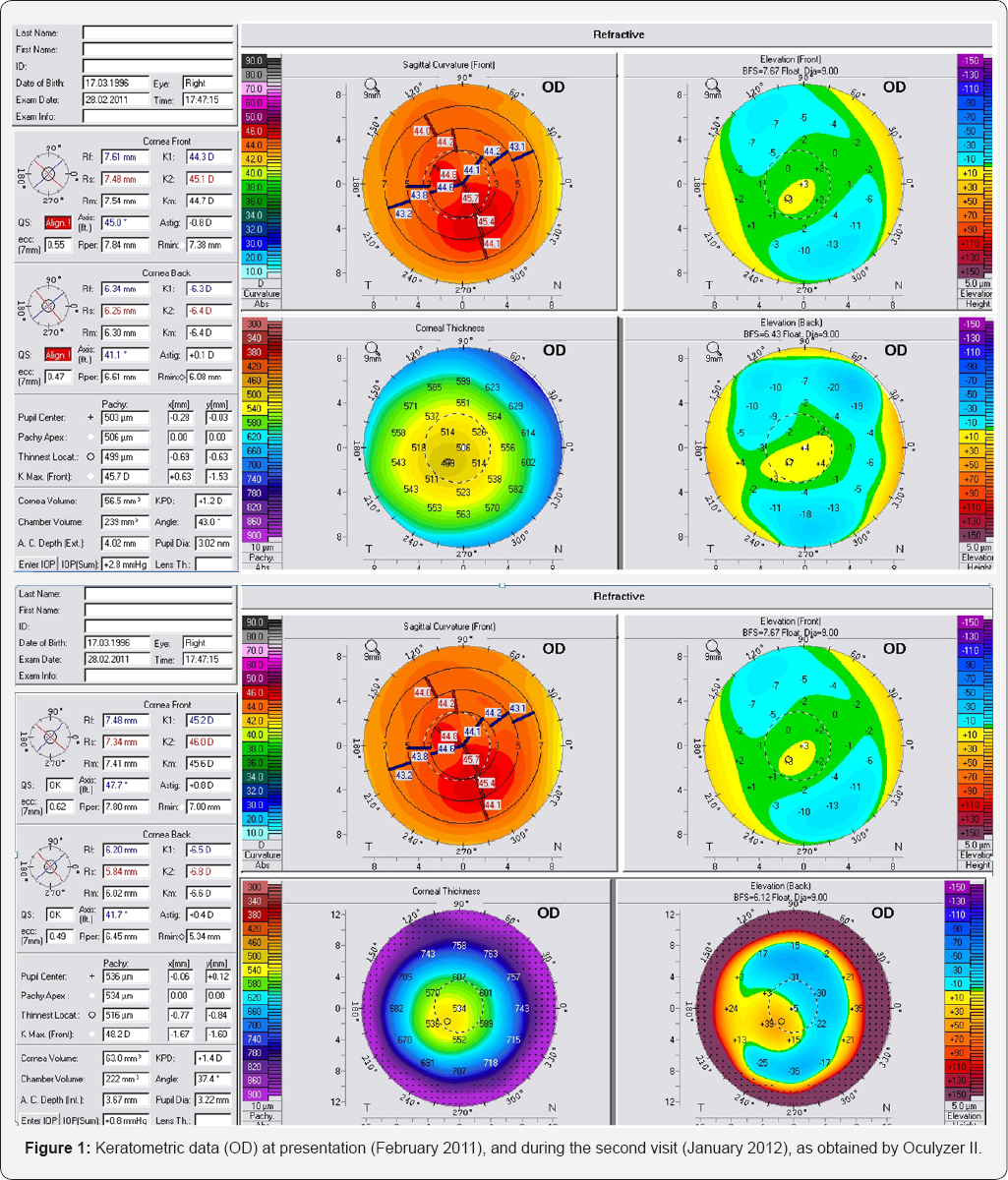
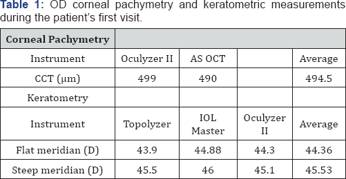
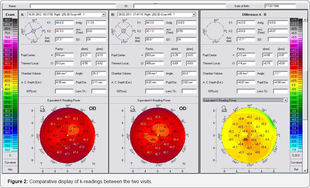
The initial anterior segment parameters recorded during the first visit (late February, 2011) are summarized in (Figure 1 &Table 1). Based on these findings, astigmatism was determined -1.25 D, with the flat axis was determined at 21°. We saw the patient several months later (late January, 2012). The data (Figure 2 & Table 2), indicated keratoconic progression, despite that overall corneal thickness was found increased by an average of 13μim. For example, the difference with the Pentacam (Oculyzer II) preimposed on the old and newer topometric measurement showed a change in the central steepening. For example, sagittal curvature increased, K1 (flat axis) by +0.2 D and K2 (steep axis) by +0.8 D. Likewise, anterior surface elevation was found increased by approximately + 13μim, also there was change between lowest elevation to pupil center (20711, -13 -> +3μiM, 2012 -18 -> +8μim). Based on these findings, astigmatism was determined at 40°

We decided to further evaluate this finding because on topometric terms this finding suggested progression of the keratoconus. We obtained Artemis II+ HF UBM corneal epithelium mapping. Careful evaluation of the UBM findings revealed that the epithelium (Figure 3) was overall thicker compared to a population of 33 patients (50 eyes) [4]. The patient’s epithelium featured mean thickness 56μm (normal population 50.8μim), central thickness 60μm (normal population 52.1μm), and average peripheral thickness 50μm (normal population 49.54μm), while over the cone (located superiorly - temporal) the epithelium was thicker, at 53μm.

Discussion

We feel that this is a good example where the -at first sight- progression of keratoconus is accompanied by epithelium whose thickness is overall thicker than the control population, particularly at the pupil center by a significant amount (+8μm). This would be impossible to establish with Scheimpflug imaging maps alone. It appears the HF Ultrasound is able, by giving the specific epithelium maps to point to that direction. Obviously, for the definite diagnosis of this theory would be re-evaluation of the cornea with Oculyzer and UBM. In case where the epithelium returned to a thinner thickness we would expect that the Pentacam maps would flatten and that would establish that there is no actual progression of the keratoconus. It is interesting to compare the posterior curvature maps of past and current and see in these maps that there is a change in posterior curvature. One has to take into account that the increase of irregularity by the epithelium thickening may be the factor that creates this bias.

Acknowledgement

Dr. Kanellopoulos is a Consultant to Alcon Wave light.

For more Open Access Journals in Juniper Publishers please click on: https://juniperpublishers.com

For more articles in  JOJ Ophthalmology (JOJO) please click on: https://juniperpublishers.com/jojo/index.php

For more about juniper publishers  please click on: https://www.juniperpublishersgroup.com/

Wednesday, 12 February 2020

Preemptive Inferior Fornixstabilizing Procedure to Reduce Chemosis During Lower Eyelid Ectropion Repair: Surgical Technique and Outcomes- Juniper Publishers


Juniper Publishers- JOJ Ophthalmology


Results

A total of 19 patients underwent the inferior fornix suture stabilization procedure at the time of ectropion repair surgery. The average age was 82.5 years old (Range: 63-95), 13 were male (68.4%) and 6 were female (31.6%). Of the 19 patients included in the study, a total of 24 eyes underwent the inferior fornix stabilization procedure (5 OD, 9 OS, 5OU). The average number of postoperative follow-up visits was 2.2 (Range: 1-4), and the average total follow-up length was 8.5 weeks (Range: 1-16). In 79.2 percent (19 of 24) of the cases, no postoperative chemosis was noted following the ectropion repair in combination with the inferior fornix stabilization procedure described above. The incidence of chemosis noted postoperatively was 20.8 percent (5 of 24). The following chemosis classification system described by Weinfeld et al. [4] was used for classifying postoperative chemosis:
  1. Type 1 (acute mild) involves mild edema and inflammation, yellow and/or pink conjunctiva color, absent lagophthalmos, and less than three weeks duration.
  2. Type 2 (acute severe) involves severe edema and inflammation, yellow and/or pink conjunctiva color, lagophthalmos present laterally, and less than three weeks duration.
  3. Type 3 (subchronic) involves mild to severe edema, chronic inflammation, pink color, absent lagophthalmos, and duration of chemosis between three weeks and six months.
  4. Type 4 (subchronic because of lower lid malposition) involves severe edema, moderate to severe inflammation, pink color, lower lid malposition and/or ectropion, and duration lasting until lid malposition is corrected [4].
All five cases of postoperative chemosis were classified as Type 1, acute mild. In all cases, chemosis ultimately resolved without any permanent sequela. No secondary entropion was observed in any case. All patients maintained good function, motility, and cosmesis of lower eyelids, and physician and patient satisfaction was achieved in all cases.


Discussion

This surgical technique may be used for preventing postoperative chemosis, thus limiting the deleterious effects that conjunctival edema may have on wound healing. Indications for this chemosis limiting procedure may be for severe ectropion repair, patients with high likelihood of ectropion repair failure, or those at high risk for postoperative chemosis. Chemosis or severe lid instability on preoperative physical exam, as well as a history of previous chronic chemosis following ocular surgery may provide indication for inferior fornix stabilization at the time of the initial surgery. Additionally, this chemosis limiting procedure may be used for postoperative patients who have chronic chemosis resistant to medical management requiring a return to the operating room for surgical treatment of the conjunctival edema.
Sutures have been used for correcting eyelid malrotation for centuries [11]. Snellen described the Snellen Suture Technique in 1869 for correcting ectropion by passing a suture through the conjunctiva in the inferior fornix and out the skin inferiorly, therefore rotating the eyelid margin. Snellen used two horizontal mattress sutures about 3 mm apart just inferior to the tarsus, nearest the margin of the eyelid. The first was placed at the junction of the outer and middle third of the conjunctiva, and the second at the junction of the inner and middle third [1,12]. A modified Snellen suture technique was described by Laval and Schneider and later by Barrett aimed at correcting inferior prolapsed conjunctiva, differing from Snellen by incorporating the arcus marginalis and inferior orbital rim [13,14]. This incorporation, however, resulted in an increased risk of secondary entropion [10,13]. Malone and Tse described a inferior fornix suture technique for treating postoperative prolapsed conjunctiva using three double-armed 4-0 chromic gut sutures inserted in a horizontal mattress fashion to invaginate prolapsed inferior forniceal conjunctiva. The suture needle was passed through the dome of the prolapsed conjunctiva, into the inferior cul-de-sac, and brought out through the skin 8-9mm below the lash margin, and tied without a bolster. By not incorporating the arcus marginalis and inferior orbital rim, and by passing the suture full thickness through the eyelid, the likelihood of causing eyelid malrotation was likely reduced [10]. Unlike the technique described by Malone and Tse, the inferior fornix suture technique in this study was done at the time of the primary surgery in a preemptive effort aimed at prevention of postoperative chemosis and inferior fornix conjunctival prolapse in high risk patients. Additionally, foam bolsters were used when tying the sutures and the procedure did not include a temporary tarsorraphy.
This simple surgical procedure offers several advantages over medical management alone. It can be used as a corrective treatment for persistent chemosis, and also as a preventative measure for those with a high likelihood of postoperative chemosis. Also, preventing the inferior fornix conjunctiva from prolapsing or repositioning intra operative prolapsed conjunctiva will minimize exposure, inflammation, and epidermalization of the conjunctiva [10].
This study, however, has some limitations. The lack of control arm comparing chemosis incidence following severe ectropion repair without the proposed inferior fornix stabilization procedure restricts us from comparing the natural history of chemosis independent of the inferior fornix stabilization. Future studies of this technique could be performed prospectively. We do, however, feel that this technique significantly reduced the likelihood of postoperative chemosis given that only 20.8 percent of severe ectropion repairs in patients who were at high risk of postoperative chemosis actually manifested chemosis, all of which were characterized as Type 1, acute mild. Additionally, all cases of chemosis ultimately resolved without any permanent sequela. We believe this surgical technique can be used as a successful preventative measure for postoperative chemosis as well as for a surgical treatment option of postoperative chemosis resistant to conservative management.


Acknowledgment/Disclosure


The authors have no proprietary or commercial interest in any materials discussed in this article.


Tuesday, 11 February 2020

Visual Impairment and Smart Cities: Perspectives on Mobility- Juniper Publishers


Juniper Publishers- JOJ Ophthalmology


Abstract

Introduction: Visually impaired people face many problems when it comes to urban mobility, even though law guarantees their rights. Many of them are able to guide well themselves using white canes and tactile devices, but they still needs some help from the others to successfully complete their journey or assignment, which reduces their autonomy or even their safety. When using public transportation, the visually impaired report lack of awareness and sensitivity of people in general. Improvements of labor field are also important in order to evidence the value of the visually challenged manpower to economy, exercising social inclusion and meliorating their self-esteem. Thus, the idea of a smart city is extremely relevant, because it characterizes progress of infrastructure and services using technology, making city administration, education, public security, health service, housing and transportation even more connected and efficient. The purpose of this study is to show that the combination between concepts related to mobility of unsighted people and to smart cities results in benefits for both the visually impaired and the society.
Discussion: Improving visually impaired autonomy in mobility gathering concepts based on smart cities.
Conclusion: The fusion of concepts related to visual impairment and smart cities is extremely beneficial for autonomy, mobility and economy.
Keywords: Visual impairment; Autonomy; Technology; Mobility; lob market; Smart cities, White cane


Introduction

The term "visual impairment" refers to irreversible visual loss, even after medical treatment. World Health Organization (WHO) classifies visual function in 4 levels: normal vision, moderate visual impairment, severe visual impairment and blindness. Moderate visual impairment combined with severe visual impairment under the term "low vision". Individual with this condition, despite the visual loss, is able to plan and/or execute assignments. About 1% of the world population presents some kind of visual deficiency, and more than 90% of those are distributed in third world countries [1].
Visual loss can be either congenital or acquired. A person who had developed blindness during his life has visual memories, thus, such memories are preserved. However, those who were born blind will not have the capacity of forming visual images, but they will develop strategies in order to structure a mental representation of space. Unsighted people normally use sonorous, kinesthetic, tactile, thermal and olfactory information through reminiscent senses [2]. In those cases, moving through places requires sensor-motor-cognitive skills, including perception, codification, learning and space information recall. That assignment can be stressfull, especially for the existence of two factors that directly affects the process of space orientation: environment layout and environment quality information [2].
Even though law guarantees their rights, visually impaired people face many problems when it comes to urban mobility. Many of them are able to guide well themselves using white canes and tactile devices, but they still needs some help from others to successfully complete their journey or assignment, which reduces their autonomy or even their safety. When using public transportation, the visually impaired report lack of awareness and sensitivity of people in general and the need of help to inform bus destination, for example [2].
Absence of great urban mobility results in negative consequences not only for people's life quality, but also for economy, including factors that influence since Brazilian social security till the indexes of productivity and of competitiveness at job market [3]. Is doubtless that mobility technology facilitates the access to visual, tactile, olfactory and sonorous information through systems like Braille, conventional canes, especial boards, etc. It's concession should be considered as an integrant and an indispensable part of the rehabilitation or habilitation process [4].
Nowadays, new mobility technology is emerging such as electronic canes, which integrate the information given by the ultrasound sensor to the traditional cane characteristics, maintaining the touch technique used in independent dislocation [1]. Another example is WAYFINDR® App that uses beacons (proximity device that sends information), Bluetooth, headphones, bone conduction and the smart phone itself, permitting visually impaired people to walk on streets in a more independent way [5].
The concept of a smart city includes also an idea of urban management based on Technology of Information and Communication [3], using hardware and software as sensors, Radio Frequency Identification (RFID), beacons, and other items related to de Internet of Things concept (IoT) [6-8]. Many of the technologies applied resort to Global Positioning System (GPS) for the purpose of collecting location data. However, GPS can suffer interference from obstacles which hind satellite's signal propagation, as indoor places, where signal hardly can be captured with enough quality. Thereby, other forms to localization will be required, as well as inertial sensors or ultrasound pulses [9].
Accordingly, at smart cities will be possible monitoring, localization and geo referencing actions, traffic preventive maintenance, alternative transportation and itinerary and route information. Users also can report urban accessibility features trough their ride, describing street and sidewalk conditions, for an example [3]. The purpose of the present study is to show that the combination between concepts related to mobility of unsighted people and to smart cities results in benefits for both the visually impaired and the society.


Discussion

A person who is blind or has low vision faces many adversities during the day. Those difficulties goes since obstacles in the way till poor infrastructure on educational and on professional fields [5]. Among the most common structural problems are bumpy sidewalks and few or none signaling, making harder and dangerous to cross streets [10]. In general, the visually impaired recur to some instruments that give space orientation on urban mobility and they are directly influenced by the city infrastructure [4].
  1.  White cane pencil tip: ideal for places still unknown. Enables different types of soil identification by vibration and sounds produced [4].
  2.  White cane roller tip: used for space recognition at places without many obstacles [4].
  3.  Walls: are used as a safety reference for the visually challenge [4].
  4.  Lowered sidewalks: offer risks when tactile floor is not present to indicate where the sidewalk starts [4].
  5.  Tactile floor: very helpful, but needs to be perfectly installed in order to preserve its information [4].
  6.  Urban furniture: benches, trashcans, street sings, bus stop, telephone cabins, etc [4].
Another aspect of a poor urban mobility is the negatives consequences for city economy, mentioning the impact on Brazilian social security when it comes to the occurrence of accidents and to the raise of governmental pensions; besides productivity reduction as the employee spends extra time and effort commuting to work [3].
Difficulties are present also on the job market. To professional qualify and to be competitive can be a great challenge for people who suffer from visual loss. Thus, assistant technology is indispensable because it provides more independency for the visually impaired and freedom to do everyday duty in a plainer way. Yet, technology incentive, especially for mobility, is extremely important and even determinant to insert this group in professional, educational and social communication spheres, making them participatory in society [11]. Some projects have already been developed, as WAYFINDR® App. That system recurs location data given by beacons via Bluetooth to define one’s location, then, audio instructions are created to guide the individual trough the space, avoiding dangerous spots, permitting people to reach their destination safer and faster [5].
There are also some projects of the so-called smart canes [1].
  1.  E Touch cane: is known as the "speakerphone cane" .It has Global Positioning System (GPS), voice recognition and headphones, by which the visually impaired, indicates its starting point and destination by voice command [1].
  2.  «Low Cost» electronic cane: it has two sensors that warn the visually impaired when there is some obstacle within a certain distance [1].
  3.  «Smart» electronic cane: created by the Universität Konstanz, this type of electronic cane is able to trace routes and identify signs that have Quick Response codes (QR codes) to help the user cross streets or find establishments [1].
  4.  Smart Cane: developed by students at the University of Michigan, can recognize radio frequency identification tags located along the route [1].
It is important to note that with the advent of generations of communities and services that have the Web conception as a platform, possibilities of information dissemination and data sharing have increased. That fact happened due to popularization of Internet and to increased shared data flow, allowing the user to participate in creation of content. That is a great contribution for many smart cities tools [9]. The collected and processed data make possible to plan some action such as details about the path where the user travels with his smart cane, after processing references about the place. That technology should also be used at environments as school, work or home [3].
A smart city uses information and communication technology resources, providing more interactivity in order to improve its infrastructure and its public services in general, making that administration, education, health service, public security, and housing and transportation sectors even more connected and efficient. Technology is the starting point for a smart city [8].


Conclusion

It is possible to infer that through the fusion between the concepts of Technology of Information and Communication applied in auxiliary devices for people with visual impairment and the idea of smart cities has a positive impact on these people's life quality. Therefore, the greater autonomy of the visually impaired results in greater self-esteem, better security and ensure competitiveness in job market. In addition, infrastructure is benefited with improvements generating more interactivity and development for the city.


Acknowledgement


We are grateful to CNPq for the Technological and Industrial Initiation grant A granted by process no 180180/ 2017-7 for the project “Support magnifying glass with color change and adjustment of light intensity for visual rehabilitation” to student Caio Henrique Marques Texeira.
For more Open Access Journals in Juniper Publishers please click on: https://juniperpublishers.com


Monday, 10 February 2020

A Case of Temporal Arteritis Associated with Atypical Clinical Findings- Juniper Publishers


Juniper Publishers- JOJ Ophthalmology


Introduction

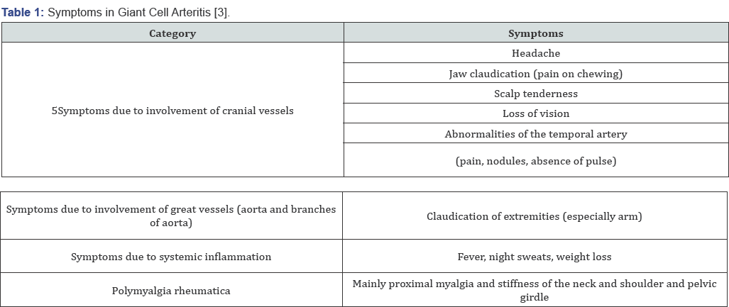
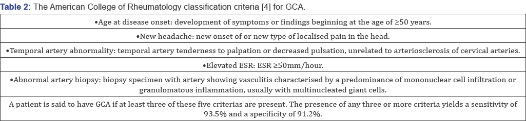
Giant cell arteritis (GCA), is a systemic vasculitis, affects medium and large sized cranial arteries [1], it can cause serious morbidities. Giant cell arteritis (GCA) predominantly affects elderly females [2]. The typical symptoms of new-onset GCA are bitemporal headaches, jaw claudiacation, scalp tenderness, visual disturbances, systemic symptoms such as fever and weight loss, and polymyalgia [3]. The diagnostic assessment comprises laboratory testing (erythrocyte sedimentation rate, C-reactive protein), temporal artery biopsy and imaging studies. The most important complication is permenant and deep vision loss. Diagnosis of TA is difficult if typical symptoms other than visual loss are absent. It can be difficult to diagnose this disease in the absence of typical clinical signs and labaratuary findings. Here we report a silent and atypical temporal arteritis case (Table 1& 2).


Case Report

63 year old hypertensive male patient applied to our hospital with complaint of vision loss in his right eye for two
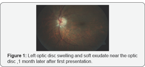
weeks. His visual acquity was perception of hand movements in right eye and 8/10 according to Snellen Chart in left eye. While anterior segment evaluation revealed bilateral nuclear sclerosis, fundoscopic examination revealed papiledema in his right eye. He did not complain of any associated headache, scalp tenderness, jaw claudication or constitutional symptoms such as weight loss, fever, malaise or sweats. We consulted the patient to internal medicine, rheumotology and hematology departments. His clinical and labaratuary findings were unremarkable except thrombocytosis. Following a presumptive diagnosis of non- arteritic ischemic optic neuropathy the patient was hospitalized and treated with intravenous 1000mg methylprednisolone for 3 days. After 3 days, oral prednisolone therapy was started 60mg per day. Oral steroid therapy was tapered 10mg with 3 days interval. However at 10-days' examination visual acquity in his right eye was reduced to loss of light perception. 1 month later, patient presented with sudden vision loss in his left eye, headache and malaise. Fundus examination revealed left optic disc swelling and soft exudates near the optic disc (Figure 1). Floroscein anjiography showed delayed filling at arterial phase and late staining of the optic disc. When labaratuary tests were repeated, erythrocyte sedimentation rate (ESR) was 65mm/ h(normal range: 1-15mm/h) and creactive protein (CRP) level was 15mg/L (normal range: <5 mg/L). According to these findings, with the diagnosis of arteritic ischemic optic neuropathy, intravenous steroid therapy was initiated. Rheumatology department evaluated the patient again and a temporal artery biopsy was performed; focal chronic inflammation and elastic fiber degeneration areas were identified at histologic sections (Figure 2). At the fifth day of intravenous steroid therapy visual acquity in left eye was 7/10 according to the Snellen Chart. The Humphrey Perimetry test revealed a paracentral island in his left eye.Since the visual acquity was at the level of loss of light perception at the right eye we didn't perform the Humphrey Perimetry test for the right eye.


Discussion

Patients with GCA typically present headache, jaw claudication, fever, weight loss, myalgia, arthralgia or malaise. Conversely, patients with occult GCA, first described by Simmons & Cogan [5], present with sudden visual loss without systemic symptoms and signs. Thus, due to the lack of symptoms, diagnosis and treatment of silent GCA may be considerably delayed when compared to typical GCA. Occult GCA , a potential cause of blindness, is defined as ocular involvement of GCA without any systemic symptoms and signs [6]. Ocular symptoms include visual loss, amaurosis fugax, diplopia and eye pain Hayreh et al. [6], described cotton wool spots in up to one third of eyes with visual loss dring the early stages of occult GCA [6]. Scalp abcess, chronic earache, bilateral central retinal artery occlusion, mydriaitic pupil, dry cough, aort anevrism , bilateral submandibular lymphadenopathy, hepatosplenomegaly are some atypic symptoms of GCA. The occult GCA group had higher C-reactive protein levels, a higher platelet count , and lower serum albumin levels [7]. Ophthalmic involvement can occur in up to 50% to 70% of the GCA patients, and this represents an ocular emergency. Arteritic anterior ischemic optic neuropathy (AAION) is the most common type of ophthalmic involvement in GCA and can cause permanent visual loss. Therefore, prompt diagnosis and treatment with a high dose of steroids is essential for these patients. In our patient at the first presentation labaratuary findings were unremarkable and there were no systemic symptoms; therefore at the beginning, our presumptive diagnosis was non-arteritic ischemic optic neuropathy of right eye. 1 month later, sudden vision loss appeared at left eye and he was complaining from headache, scalp tenderness ; ESR and CRP levels were elevated on labaratuary examination at that time. Hayreh et al. [6] found that ESR and CRP levels were relatively lower in patients with occult GCA compared to cranial GCA. On the other hand, Hamidou et al. [7] found higher CRP levels, a higher platelet count, and lower serum albumin levels in occult GCA group. In our patient's first presentation, ESR and CRP levels were within the normal limits. One month later when the other eye was effected ESR and CRP levels were remarkably high (Figure 3].
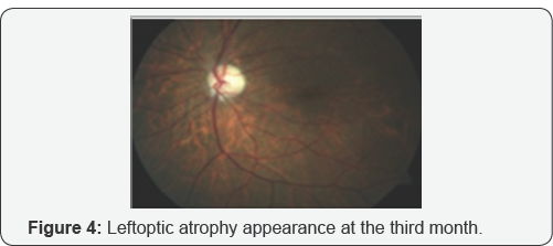
Although Liozon et al. [8] describes occult GCA as a protracted inflammatory response and a relatively benign short term outcome, in our case despite the long term corticosteroid and immunsupresive treatment, visual acquity was loss of light perception level in the right eye. Tan et al. [9] reported a case of GCA involving only the occipital artery which was revealed by magnetic resonance angiography. Chomlak et al. [10] reported a case of GCA with vertebral artery involvement, which was refractory to immunosuppressives. Hocevar et al. [11] claimed that, even early diagnosis and a prompt initiation of steroid did not prevent relapses in GCA. In our case after first attack despite the prompt intravenous steroid therapy one month later the patient consulted with visual loss at his other eye this verified Hocevar’s hypothesis. Cullen et al. [12] reported an occult GCA case in Singapore. Papakostas et al. [13] reported a case of GCA that presented with cotton wool spots and retinal vasculitis affecting small-size retinal arterioles. Shambhu et al. [14] reported an atypical case of GCA presenting as mild upper abdominal pain and generalized weakness in the context of hyponatremia as the presenting manifestation of vasculitis that was subsequently diagnosed by MRI scanning. Cheema et al. [15] described a case had the signs and symptoms consistent with GCA but who had an ESR within the normal limits, 27mm/h. In our case ESR at the first presenting ESR was within the normal limits,too. Labarca et al. [16] reported that patients with hypertension or diabetes at GCA diagnosis have more relapses during follow-up. Kermani et al. [17] reported seven patients with a positive temporal arteritis for GCA had a normal ESR and CRP at diagnosis (Figure 4].
In people older than 55 years, amaurosis fugax or visual loss, development of an acute ocular ischemic lesion with or without elevated erythrocyte sedimentation rate and systemic symptoms, should raise suspicion for giant cell arteritis. The diagnosis of temporal arteritis is easily made when sudden loss is contemporary with other symptoms and raised inflammatory markers. In the absence of known symptoms, in a patient diagnosed as ischemic optic neuropathy, occult temporal arteritis shouldn’t be out of mind. Despite the early diagnosis and treatment final visual acquity may not be satisfactory.


Conflict of Interest

Asfuroglu declares that he has no conflict of interest. Author Koz declares that she has no conflict of interest. Author Ozbalkan declares that she has no conflict of interest. Author Sandikci declares that she has no conflict of interest. Author Ciftci declares that she has no conflict of interest. Author Ozdemir declares that she has no conflict of interest.
This article does not contain any studies with human participant performed by any of the authors.

Informed consent was obtained from all individual participants included in the study.

For more Open Access Journals in Juniper Publishers please click on: https://juniperpublishers.com


Friday, 7 February 2020

Efficacy of Amniotic Membrane Transplantation in Refractory Infective Keratitis Leading to Stromal Thinning, Descematocele and Perforations- Juniper Publishers


Juniper Publishers- JOJ Ophthalmology

Introduction

Diseases affecting the cornea are a major cause of blindness all over the world, second only to cataract in overall importance [1]. One of the commonest corneal causes is Infectious Keratitis. The prevalence of blindness directly resulting from complications of Infective Keratitis is estimated to be 5% [2]. Cases refractory to the medical therapy requires urgent surgical intervention to retrieve the vision and most importantly to salvage eye. Available surgical management in refractory keratitis cases include tissue adhesives, Bandage Contact Lenses (BCL), penetrating or lamellar keratoplasty [3] patch grafts, or conjunctival flaps. Unfortunately, these therapies are associated with a considerable number of complications and address only the tectonic problem, without solving the ongoing infection and inflammation. BCL and conjunctival flaps being a temporary measure does not provide with new collagen to improve corneal thickness and stabilize the cornea. For such situations Penetrating Keratoplasty (PK), Lamellar Keratoplasty (LK) or patch grafts was the only option and is still being used widely. PK and patch grafts performed to seal a corneal perforation may be complicated with synechiae, glaucoma, uveitis, and graft failure in the setting of an inflamed or infected eye [4]. Recurrence of infection in corneal grafts is also challenged. LK being difficult to perform may result in a double chamber between the donor and recipient cornea in some cases. Tissue adhesives may dislodge and are used as a temporary measure, obviating the need for a PK within a few days [5,6].
Preserved human amnion has been successfully used as a biological bandage, promoter of epithelialization, inhibitor of inflammation and angiogenesis, as well as a carrier for ex vivo cultured limbal stem cells [7]. Amniotic Membrane Transplant (AMT) offers the advantage of avoiding potential allograft rejection. Even if corneal transplantation is needed, the success rate is improved if performed on an eye that underwent AMT reducing inflammation [8,9]. Amniotic Membrane (AM) integrates in cornea and thus can be used as a treatment for corneal perforation by restoring corneal stromal thickness so that emergency PK can be avoided, as suitable donor corneal button availability is difficult in every place. Therefore, an alternative management for various stages of infectious keratitis including deep refractory stromal ulcers, descematocele and corneal perforations is reconstruction of the surface with AMT adjuvant with appropriate antimicrobials and supportive medications. In this prospective study AMT in various gravities refractory infective keratitis has been attempted to understand the efficiencies and limitations associated with it.


Methods

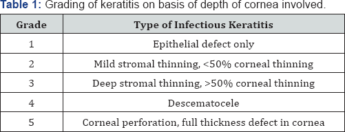
A prospective, interventional study was done on 150 eyes of 150 patients. All patients with refractory (unresponsive to conventional treatments significantly for more than 2 weeks) infective keratitis, advanced infectious keratitis with descematocele and corneal perforation requiring urgent concealment to salvage the eye, were treated with single or multi layered AMT. Patients with non-infective ulcers and perforations were excluded from the study. Corneal ulcer was graded 1-5 according to the depth of corneal involvement on slit lamp biomicroscopy (Table 1). Microbial investigations (staining for bacteria and fungus with culture-sensitivity) were done and antimicrobials started accordingly. B-scan ultra sonography was done in hazy media to rule out involvement of posterior segment. Any systemic (diabetes) or ocular (dacryocystitis) conditions hindering the healing of ulcer or triggering the infection were investigated and managed.
On basis of slit lamp examination at the site of most impact.


Technique

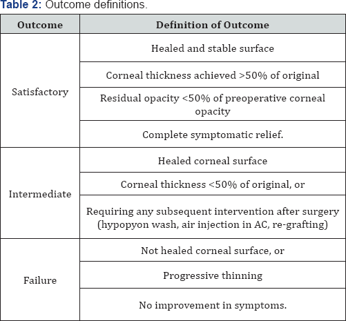
Surgery was performed preferably under sub conjunctival or peribulbar anesthesia. In children or uncooperative patients general anesthesia was used. Debridement of the necrotic tissue was done from and around the ulcer bed. Care was taken to remove the pseudo cornea over the perforation at the end of debridement to prevent leaking of aqueous and thus allowing proper keratectomy. Single layer preserved AM was used in cases of deep stromal ulcer. AM with epithelial side up was spread over the ulcer and trimmed to fit the ulcer. It was secured with continuous or interrupted 10-0 monofilament nylon suture. Descematocele and small corneal perforations up to 4mm were treated with multilayer AMT owing to deep corneal involvement. A sheet of AM, folded over it-self with epithelial side out, filled the ulcer crater and anchored to the healthy ulcer margin with interrupted 10-0 nylon suture. It was covered with a single sheet of AM similarly as in cases of deep stromal ulcers. In large corneal perforations of 4-6mm with extensive surrounding stromal necrosis, margins were not sturdy to hold the suture and there was a risk of cutting-off a corneal bite. In such cases single layer was sutured at limbus to at least provide tectonic support to the eyeball and delaying the need for PK. Side port or paracentasis was made in cases hypopyon and corneal perforation to reform the anterior chamber with air and reposit the prolapsed iris with help of spatula. Anterior synechiae if present were broken to prevent formation of adherent leucoma and thereby secondary glaucoma. Hypopyon if present was washed through the side port and intracameral antibiotic or antifungal was also injected according to sensitivity. At the end a BCL was placed over the cornea to prevent irritation from corneal sutures and maintaining AM in place. Antimicrobial, cycloplegics, ocular hypotensive and lubricating drops were continued along with systemic supportive therapy. Frequent follow-ups were done weekly for 1 month, biweekly till 3 months and monthly till 6 months. Efficacy was monitored on basis of improvement in symptoms and visual acuity, healing of the ulcer by re-epithelization and formation of anterior chamber, achievement of corneal transparency and corneal thickness. Accordingly patient's outcome was described as satisfactory, intermediate and failure (Table 2).


Observation and Results

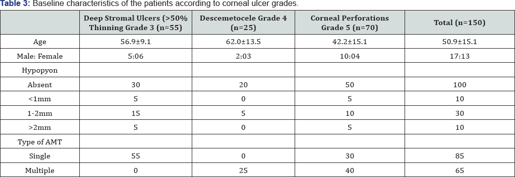
Keratitis was classified (Table 1) according to the depth of the cornea involved into 5 grades. Grades 1 and 2 responded well with medical management, therefore did not require AMT. Grades 3-5 with deeper corneal penetration of infection did not heal merely with medical management, there was an apprehension of corneal thinning and progression of infection, which required AMT. Of the 150 patients who underwent AMT, 55 (36.67%) were deep stromal ulcers, 25 (16.66%) were descematocele and maximum 70 (46.67%) patients were of corneal perforation ranging from 1-6mm. There was no age group or gender preponderance. Symptoms of redness, pain, watering and foreign body sensation (FBS) were collectively present in all the cases. Lid oedema and photophobia were also present in majority of the cases (70.6% and 90% respectively).Presence of discharge was seen in moderately less cases (30%). ranging between 1-2mm and 10 cases (20%) had hypopyon of Hypopyon was present in total 50 (33.3 %) cases where 10 cases >2mm (Table 3).
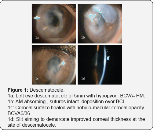
Single layer AMT was done in total 85 cases, all 55 cases of deep stromal ulcer and 30 cases of corneal perforation >4mm with extensive necrosis to provide tectonic support to maintain integrity to eyeball. Roofing with multilayer technique was done in 65 cases, all 25 cases of descematocele and 40 cases of corneal perforation >4mm in largest dimension where neighboring corneal tissue was healthy to hold the corneal sutures (Table 3). Patients were observed in repeated postoperative days. Rapid descent of symptoms was observed after the AMT. There was drastic improvement in pain, lid oedema, FBS and discharge in the first week. Symptoms were barely present in few cases by 1 month, which totally recovered by 3 months in all the cases (Figure 1).
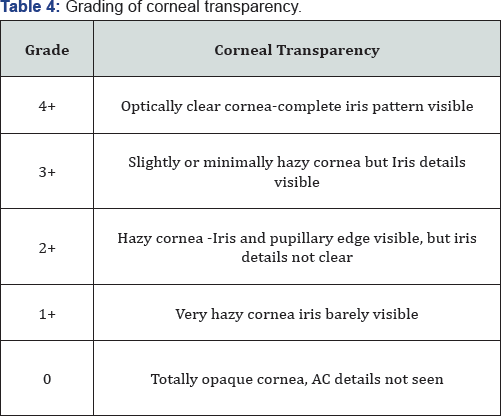
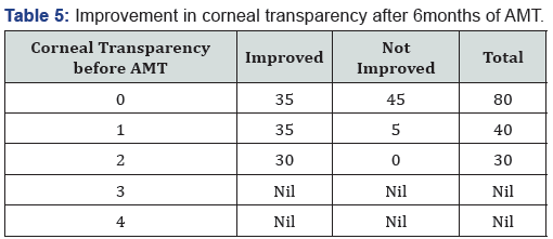
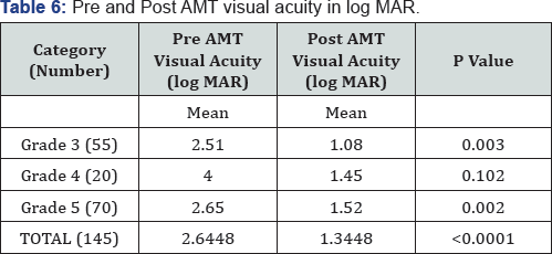
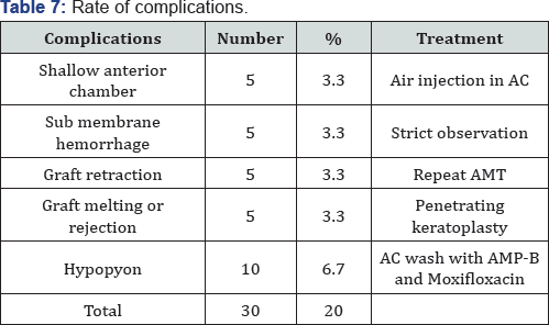
Corneal transparency graded from 0 (leucomatous opacity) to +4 (clear cornea, with no haze) was measured objectively at the site of most impact on slit lamp (Table 4). Improvement was seen in 105 of 150 cases and was statistically significant (p=0.016). However none of the cases improved to +4 transparency that is totally clear cornea (Table 5). Visual acuity was recorded before and after 6 months of treatment in 145 of 150 cases as 5 cases of fungal ulcer failed to heal with AMT (Table 6). Improvement in BCVA when taken collaborate, was extremely significant (p >0.0001). Mild to moderate complications were faced during the entire course of treatment. They were shallow anterior chamber in 5 cases in perforation which was tackled with air injection in anterior chamber and breaking anterior synechiae. Hemorrhage beneath AM in five cases which resolved spontaneously. Graft retraction was seen in five cases for which repeat AMT was done. Hypopyon developed in 10 cases and did not resolve with topical therapy was managed with anterior chamber wash and intracameral moxifloxacin and amphotericin-B respectively (Table 7). Hypopyon did not redevelop in these cases. All the complications were successfully managed with appropriate treatment with no recurrence and good results. Also no re-infection was noted. Graft melting and corneal perforation was seen in 5 cases of fulminant fungal ulcer and required urgent therapeutic PK.
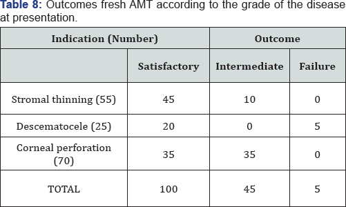
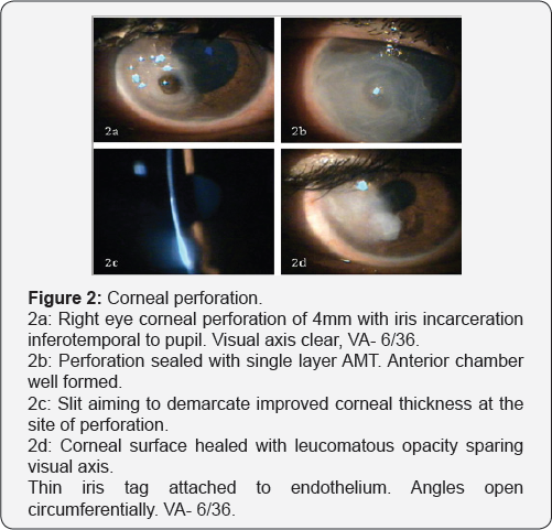
Satisfactory results were seen in 100 of 150 eyes (66.67%), intermediate results seen in 45 cases (23.33%). Failure was noted in 5 cases (3.33%) of fulminant fungal ulcers that showed subsequent corneal perforation requiring Therapeutic PK (Table 8). All the cases in intermediate category which also required subsequent intervention, healed with stable cornea. Thus, successful results were seen in 145 of 150 cases (96.67%) of which in 30 cases subsequent penetrating keratoplasty was done for leucomatous corneal opacity obscuring the visual axis left after healed ulcer (Figure 2).


Discussion

Approximately one-third of cases of infective keratitis require surgical interventions at the acute stage to prevent perforation or spreading of infection [10-14]. Keratoplasty being majorly followed in such situation faces a limitation of availability of good quality donor corneas, mainly in developing countries, recurrence of infection, difficulty in technique and graft rejection. Moreover, for fungal keratitis PK is technique dependent and may also carry a risk of recurrent infection [15].
Thus AMT is sought as an alternative, which has been extensively reported in ophthalmology literature [16-19]. AMT offers the advantage of stimulating re-epithelization, preventing neovascularization and scar formation and avoiding potential allograft rejection. Even if corneal transplantation is needed, the success rate is improved if performed on an eye with reduce infection and inflammation, this can be achieved with AMT [8,9]. In present study complete epithelization was noted in 145 of 150 cases, that is 96.67% success rate. Similar to our study, Chen et al. [20], showed 82.61% success rate, 4 of 23 cases in there study faced AM melting and graft failure requiring therapeutic PK in 3 and delayed healing with vascularization in the other. Kim et al. [21] used multilayer AM in cases of descematocele and corneal perforation. Corneal surface was healed successfully in all cases, and no recurrence of infection or rejection was experienced. Hanada et al. [22] used multiple layers of AM for deeper stromal ulcers down to descemetocele, to restore the normal corneal thickness as well as in corneal perforations from 0.5 to 3mm with or without additional tissue adhesive with high success rates (73-93%). In present study corneal perforations in cases of infectious keratitis up to 6mm have been treated successfully with AMT alone, and 100% corneal epithelization with more than 50% corneal thickness have been achieved in all 70 cases of perforation. In a series by Heiligenhaus et al. [23]. Seven patients with herpes simplex virus or varicella zoster- induced severe ulcerative keratitis, 5 of 7 eyes healed after first AMT [23]. In another study, stromal defect was filled up with multilayer technique proved to be better than monolayer procedure [22,24,25].
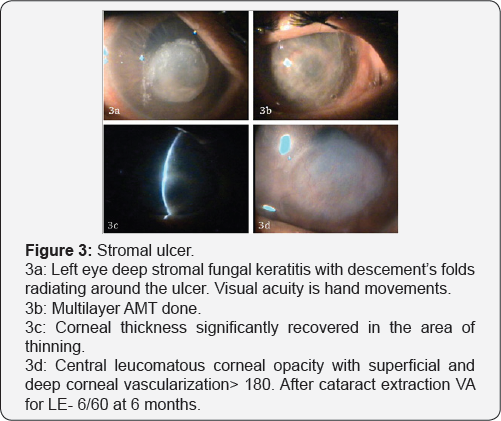
In present study 70% showed significant improvement in corneal transparency and increasing corneal transparency improves the best-corrected visual acuity further emphasizing the healing properties of AMT. Chen et al. [20], preserved useful vision after AMT in cases of fungal keratitis in 52.2% eyes. Kim et al. [21], reported 21 cases of successful AMT in infectious keratitis, in which visual acuity increased except for 5 cases because of irreversible corneal opacity. AMT has come up as a very effective managing technique for refractory ulcers. It aids in permanent healing of the refractory infective keratitis and prepares the cornea for definitive reconstructive procedure if required (Figure 3).


Conclusion


We have found that AMT represents a viable method of treatment to promote healing and prevent progressive melting of refractory infectious keratitis. Besides being cost-effective it’s easy to perform, with a short learning curve. Thus, it might be considered a first-line surgical technique when maximal medical treatment has failed.

For more Open Access Journals in Juniper Publishers please click on: https://juniperpublishers.com


Thursday, 6 February 2020

Medial Rectii Recessions a Surgical Procedure for Bil Alternating Infantile Esptropia in 6 Months Male Twins- Juniper Publishers


Juniper Publishers- JOJ Ophthalmology


Abstract

6 months un identical twins were seen by me in 2002 at qatif central hospital eastern province ks a with parents having noticed bill alternating inward ocular deviation since 2 monthsft normally delivered twins no history of exposure to oxygen birth trauma convulsions jaundice fever or any other cong ocular disorder like 1 sclera cornea 2 keratitis corneal dystophy peters corneal anomaly or limbal corneal dermoid on exam both twins dhad bil alt 15 degrees esotropia no limitation of ocular movements 2 no turning of head towards the direction of paralysed muscle 3 no diplopia 4 no abnormal head posture or false orientation under sedation mydriatic refraction and fundus examination was done ref was equal in both eyes in both eyes so were the normal fundii key words esotropia is inward ocular deviation 2 expotropia is outward ocular deviatin 3 hetrophoria is latenr ocular deviatin 4 infantile is age from birth to 3 years .


Introduction

squint is a common ocular disorder nowadays due to abuse of playing games on mobiles and i pads incidence of ocular deviation has increased recently I saw twins of 6 years of age parents complained that one twin had left eye ocular deviation since 3 months as this twin was constantly playing video games on exam this twin had plus 2d cylinder 130 axis other twin as he was not playing games was normal squints can be 1 esotopia 2 esotropia 3 heterotopias 4 paralytic 5 non paralytic 6 accommodative 7 non accommodative 8 pseudo squint in those who have broad bridge of nose epicanthal folds and wide inter canthal distance results of bill alt infantile esotropia with bill 5mm medial rectii recessions are very satisfactory both 1 from correction of angle of squint and reaction of eye which is minimal [1].


Discussion

Bill alt esotropia presents as 1 crossed fixation 2 uncrossed fixation 3 over action of inf oblique 4 a v patern 5 broad angle 6 covering the dominant eye will make p child to cry so after all investigations under g a both twins were operated under g a exposure of eye done with eye speculmm rotation of eye ensyred with 6 zero silk sutures 5mm recessions of medial rectii one and position of muscle secured on sclera using 6 zero vicoryl suture closure of conjuctival wound done with 8 zero silk sures next day children had very satisfactory correction of angle of deviation and most important thing was that eye reaction was minimal

Conclusion


Bill m rectii recessions is a very satisfactory procedure for cases of bill alt infantile esotropia as we do not cut muscle that is resection so the eye reaction is minimal and secondly correction of eye deviation is good I did this procedure in 2002 even today same procedures is done even after 17 years due to satisfactory modality of this surgical procedure however some complications r seen like 1 under correction 2 over action of inf oblique 3 amblyopia 4 d v d dissociated vertical deviation 5 accommodative element in my cases more than 1 buyers follow up of twins was normal.

For more Open Access Journals in Juniper Publishers please click on: https://juniperpublishers.com