Friday 31 January 2020

Schwartz Jampel Syndrome- A Case Report- Juniper Publishers


Juniper Publishers- JOJ Ophthalmology

Itroduction

Blepharophimosis is a general diminution of palpebral fissure in all its dimensions. The lids usually show ptosis, dystopia canthorum, lateral displacement of the lateral puncti, or abnormalities of the lashes such as ditichiasis or misdirected and stiff lashes. The other ocular defects associated with congenital blepharophimosis include strabismus, nystagmus, amblyopia, microphthalmus, anophthalmus, epicanthus inversus, microcornea and hypermetropia [1,2]. Schwartz-Jampel syndrome, an autosomal recessively transmitted disease, is a rare presentation of blepharophimosis.

Case History

A 2 year old male child, having dysmorphic features was referred from the department of Pediatrics for Ophthalmic assessment. The child was the first born of healthy non- consanguineous parents after an uneventful pregnancy. His mental and motor development was normal and he acquired independent walking at 16 months. Fine pincer grasp developed by 9 months of age. At the age of 2 years the child could talk only two words with meaning. Social development of the child was poor because of his abnormal appearance and poor language development. The parents noted the abnormal facial expression at the age of 18 months.
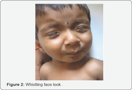
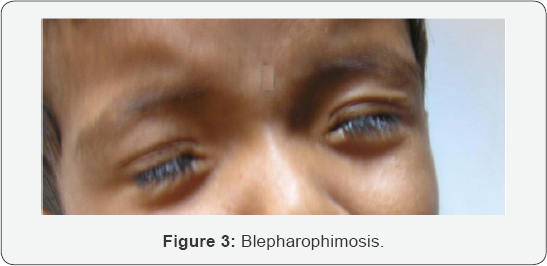
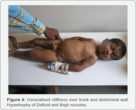
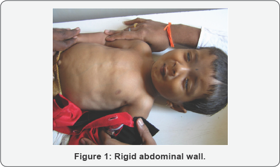
On examination the child had a short stature. The head posture was normal. Forehead did not show excessive wrinkling. The child had blepharophimosis (Figure 1). Lid crease was present. The child also had hypertrichosis. The globe examination was normal. The extraocular movements were normal. There was no refractive error. Fundus was normal. The child demonstrated pursing of lips giving him a 'whistling face' appearance and restricting his mouth opening (Figure 2). The shape of the chest was abnormal with sternal protrusion and sub-costal retraction (Figure 3). There was stiffness of his abdominal wall. The upper and lower limbs demonstrated hypertonia. The deep tendon reflexes were exaggerated. He had a waddling gait. The child had a high pitched voice (Figure 4).


Discussion

Schwartz-Jampel syndrome is a rare autosomal recessively transmitted disease, characterized by generalized myotonic myopathy, typical facial features, skeletal dysplasia, contracture of joints, growth retardation and bone maturation delay [3]. However a few cases showing dominant inheritance have also been reported. It is classified into 3 types based on age and severity
  1. Type 1A
  2. Type 1B
  3. Type 2

Type 1A

The type 1A disease is diagnosed in mid-childhood with recognition of myotonic facies with convex profile, short palpebral fissure, telecanthus, dimpling or quivering of the chin, prominent eyebrows, low hairline, low-set ears, flat base of the nose, micrognathia, microstomia, sometimes high-arched palate. The child exhibits progressive myotonia, muscle wasting and orthopaedic problems with decreased linear growth myotonia plateus by mid childhood. Additional findings reported in a few cases are myopia, hypertrichosis, and strabismus. The continuous myotonia is probably responsible for both muscular hypertrophy and peculiar facial appearance.

Type 1B

Type 1B is more severe than 1A, Bone dysplasia is present at birth. Long bones are shortened, femurs are dumbbell shaped. Bone epiphyses are large and vertebral bodies are flat.

Type 2

Type2 disease is more severe. Onset is neonatal, there is short limb dysplasia and long bones are bowed. Early death is frequent [4].
The diagnosis is predominantly on the basis of the typical dysmorphic facies [5]. EMG showing continuous discharges further supports the diagnosis. The gene defect in SJS type 1 is located in the 1p34-p36 of chromosome 1, whereas it is different in type 2 [6,7]. Perlecan the major proteoglycan of basement membranes is altered in patients with Schwartz- jampel syndrome disease [8]. However, a significant amount of molecular heterogeneity exists, genomically and proteomically, within SJS type 1. Currently no known correlation exists between the specific mutations found and the specific features of a given case However, the new mutations found by Stum et al. In 2006 have been discovered so recently that not enough time has elapsed to explore such possibilities. The new findings should be important tools to help find correlations among genetic variants, perlecan forms and levels, and clinical subtypes. Other facts yet unknown also may influence the severity and the specific characteristics of the disease [5]. The genetic tests for perlecan gene are not easily available in the commercial laboratories.
The child was diagnosed as having type 1A type of Schwartz- Jampel syndrome since the typical facial features became manifest at the age of 18 months. The old pictures of the child taken on his first birthday showed normal facial features. Medications that have been found useful in myotonic disorders such as phenytoin and carbamazipene may help to reduce the abnormal muscle activity. Warm baths are helpful in reducing stiffness. Botox injections are reportedly found useful to relieve blepharospsm.
Patients are generally treated with Carbamizipene 2030mg/kg body weight and most of them show improvement. Carbamazipene probably works by inhibiting neuronal sodium channels and may have direct effects on neurotransmitter systems. Orbicularis oculi myectomy, levator aponeurosis resection and lateral canthopexy are some surgical procedures which may be tried if the response to carbamazipene or botox is not adequate. The parents of the child were educated regarding the genetic nature of the disease and were referred to the geneticist. This particular child has not reported for follow-up as he belongs to a remote village far from our hospital and is probably reporting for follow-up at a nearby city.


Conclusion


Schwartz-Jampelsyndromeisararecauseofblepharophimosis. The condition can be managed with medications in most of the? cases. Surgery may be required if the condition does not improve with drugs.

For more Open Access Journals in Juniper Publishers please click on: https://juniperpublishers.com


Thursday 30 January 2020

Eye Loss Due to Disseminated Molluscum Contagiosum Skin Infection Involving the Eyelids in an Immune Competent Child- Juniper Publishers


Juniper Publishers- JOJ Ophthalmology

Introduction

Molluscum contagiosum is a viral infection of skin and mucous membranes caused by a double-stranded DNA poxvirus. The virus causes a characteristic skin lesion consisting of a single or multiple round pearly white umblicated papules [1]. Molluscum contagiosum is largely if not exclusively a human disease although there are few reported cases in some animals [2]. Distribution is worldwide, but it is more common in areas with hot climate [3]. The virus is transmitted directly through skin to skin contact with other infected patients or indirectly through contact with contaminated fomites such as bath sponges and towel. The virus can also be transmitted to other areas in the same patient by autoinoculation [4]. Although all age groups can be affected, it commonly occurs in two age peaks: children and adults. Children are usually infected by casual contact and young adults infected by sexual contact [5]. Clinically molluscum contagiosum lesions are usually asymptomatic; however, some lesions may become pruritic or tender due to associated eczema or inflammation. There are no systemic symptoms [6]. In most cases lesions resolve spontaneously without treatment over the course of several months [7]. On examination, the skin lesions are round, dome shaped, pearly, flesh colored, firm papules with central umblication. They are usually 2-5mm in diameter (except for giant molluscum which may reach few centimeters). Beneath the umbilicated center is a white, curd-like core that contains molluscum bodies. Lesions may be single or multiple distributed on the skin of the head- including the eye lids, neck, trunk, the limbs, and around the genital area [8]. Rarely, it may involve the palms, the soles, mucous membranes of the mouth, or conjunctiva [9,10]. Immuno compromised patients - children and adults, such as HIV patients and patients on immunosuppressive therapy, tend to have atypical and more wide spread and persistent lesions [5,11].
The diagnosis of molluscum contagiosum is clinically evident by the characteristic appearance of the skin lesion. In atypical or giant lesions, a biopsy can be done to reach diagnosis. Histopathology reveals characteristic intracytoplasmic inclusion bodies (molluscum or Henderson-Paterson bodies) [12]. Other tests include complement fixation test (CFT) and polymerase chain reaction (PCR) [13]. Treatment in healthy individuals is not always necessary because most cases are self limiting. Indications include: relieving symptoms and discomfort, improving cosmetic appearance, persistent lesions, and reduction of autoinoculation and spread to other contacts [14]. Many modalities exist [15]. Treatments can be divided into three categories: destructive- physical and chemical), immune modulators, and antiviral [16].


Case Report

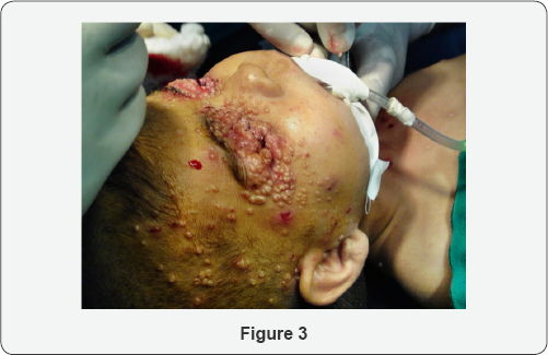
A four years old female child presented to the dermatologist with disseminated skin lesions involving the whole body surface area. The lesions were scattered all over the face, neck, trunk and limbs with larger concentrations around the eyelids- both eyes- and the genital region. The lesions were round pearly white umblicated papules typical of molluscum contagiosum. The patient consulted many dermatologists before she was referred to an ophthalmologist for eye examination. On examination, the lesions were more confluent and concentrated around the eyelids skin and eyelid margins making it difficult to open the balpebral fissure for inspection of the conjunctiva and corneal surface, and the condition was associated with secondary pyogenic infection and discharge around the eyelid margin. Examination under general anesthesia to facilitate eyelids opening and subsequent surgical removal revealed infective keratitis on the right side with profuse pus discharge and extensive corneal stromal ulceration and melting.
The treating dermatologist and ophthalmologist started surgical excision of as many lesions as possible. The eye postoperatively was treated with intensive topical antibiotics eye drops and eye ointment for several days until the infection was resolved and healing of the corneal surface took place. The patient was referred to a pediatrician for the investigation of the possible cause of immune deficiency. The patient did not return subsequently for follow-up (Figure 1-3).


Discussion

Molluscum contagiosum is usually described as a benign and self limiting skin infection that does not always require treatment [17]. However, this may not be the case when the eye is involved [18]. Ocular manifestations may present as a range of complications [1,6,19]. Lesion located on or near the lid margin may give rise to secondary chronic follicular conjunctivitis. Unless the lid margin is examined carefully, the causative molluscum lesion may be overlooked therefore it can be easily misdiagnosed and mistreated. Prolonged follicular conjunctivitis or secondary bacterial infection can result in keratitis usually in the form of fine punctate epithelial erosions or sub epithelial opacities. Corneal vascularisation, scarring and opacification may result in visual acuity loss. Molluscum contagiosum infection commonly involves the face and hands. Itching and scratching facilitate extension of infection to other parts of the same patient; therefore, the disease usually presents as multiple crops and less commonly as a solitary lesion which sometimes becomes a confluent multilobulated giant tumor affecting the eyelid [20,21]. Secondary infection and ulceration can result in permanent scarring.
Molluscum contagiosum is a common pediatric dermatosis in Iraq [22]. Al-Azawi reported a high prevalence of molluscum contagiosum virus (MCV) type I in children age group<10 years [23]. In our clinical practice, molluscum contagiosum infection is wide spread and eye involvement is very common in Iraq. Predisposing factors may include low socioeconomic status, crowding, and low personal hygiene. It affects all age groups especially children in preschool age and primary school age. This highly contagious infection is usually acquired from contact with other infected people. They could be family members or visitors or more commonly other infected children in the neighborhood or schools.
Molluscum contagiosum infections involving the eyelids and periocular area are usually managed by ophthalmologists, and sometimes referred by dermatologists. Although many modalities of therapy are effective in destruction of the virus, the use of substances such as liquid nitrogen or chemicals in the vicinity of the eye may be hazardous [24,25]. Surgical removal by shave excision or curettage is a simple and effective procedure [23]. However, multiplicity of the lesions and young patient age usually necessitate light general anesthesia given by an anesthesiologist in an operation theater and therefore cannot be done as an outpatient office procedure in the minor surgical room. This may result in considerable suffering to the patient and parents and burden on the health care providers [3].


Conclusion

Molluscum contagiosum is not always a self-limiting benign skin infection, but it can cause serious eye complications especially in the third world. Active treatment is indicated to prevent secondary complications and limit the spread of the disease to other people.


Disclosure


The author reports no conflicts of interest in this work.

For more Open Access Journals in Juniper Publishers please click on: https://juniperpublishers.com


Wednesday 29 January 2020

Eye Loss Due to Disseminated Molluscum Contagiosum Skin Infection Involving the Eyelids in an Immune Competent Child- Juniper Publishers


Juniper Publishers- JOJ Ophthalmology


Abstract

Background: Molluscum contagiosum is a viral infection of skin and mucous membranes caused by a DNA poxvirus. It is a common skin infection in children with numerous ocular manifestations.
Case report: Eye loss due to disseminated molluscum contagiosum infection of the skin involving the eyelids in a 4 years old immune competent female child. The clinical history, exanimation, treatment and follow-up are presented.
Conclusion: molluscum contagiosum is not always a self limiting benign skin infection and can cause serious eye complications especially in the third world. Active treatment is indicated to prevent secondary complications and limit the spread of the disease to other people.
Keywords: Viral infection; Molluscum contagiosum; Eye loss


Introduction

Molluscum contagiosum is a viral infection of skin and mucous membranes caused by a double-stranded DNA poxvirus. The virus causes a characteristic skin lesion consisting of a single or multiple round pearly white umblicated papules [1]. Molluscum contagiosum is largely if not exclusively a human disease although there are few reported cases in some animals [2]. Distribution is worldwide, but it is more common in areas with hot climate [3]. The virus is transmitted directly through skin to skin contact with other infected patients or indirectly through contact with contaminated fomites such as bath sponges and towel. The virus can also be transmitted to other areas in the same patient by autoinoculation [4]. Although all age groups can be affected, it commonly occurs in two age peaks: children and adults. Children are usually infected by casual contact and young adults infected by sexual contact [5]. Clinically molluscum contagiosum lesions are usually asymptomatic; however, some lesions may become pruritic or tender due to associated eczema or inflammation. There are no systemic symptoms [6]. In most cases lesions resolve spontaneously without treatment over the course of several months [7]. On examination, the skin lesions are round, dome shaped, pearly, flesh colored, firm papules with central umblication. They are usually 2-5mm in diameter (except for giant molluscum which may reach few centimeters). Beneath the umbilicated center is a white, curd-like core that contains molluscum bodies. Lesions may be single or multiple distributed on the skin of the head- including the eye lids, neck, trunk, the limbs, and around the genital area [8]. Rarely, it may involve the palms, the soles, mucous membranes of the mouth, or conjunctiva [9,10]. Immuno compromised patients - children and adults, such as HIV patients and patients on immunosuppressive therapy, tend to have atypical and more wide spread and persistent lesions [5,11].
The diagnosis of molluscum contagiosum is clinically evident by the characteristic appearance of the skin lesion. In atypical or giant lesions, a biopsy can be done to reach diagnosis. Histopathology reveals characteristic intracytoplasmic inclusion bodies (molluscum or Henderson-Paterson bodies) [12]. Other tests include complement fixation test (CFT) and polymerase chain reaction (PCR) [13]. Treatment in healthy individuals is not always necessary because most cases are self limiting. Indications include: relieving symptoms and discomfort, improving cosmetic appearance, persistent lesions, and reduction of autoinoculation and spread to other contacts [14]. Many modalities exist [15]. Treatments can be divided into three categories: destructive- physical and chemical), immune modulators, and antiviral [16].


Case Report

A four years old female child presented to the dermatologist with disseminated skin lesions involving the whole body surface area. The lesions were scattered all over the face, neck, trunk and limbs with larger concentrations around the eyelids- both eyes- and the genital region. The lesions were round pearly white umblicated papules typical of molluscum contagiosum. The patient consulted many dermatologists before she was referred to an ophthalmologist for eye examination. On examination, the lesions were more confluent and concentrated around the eyelids skin and eyelid margins making it difficult to open the balpebral fissure for inspection of the conjunctiva and corneal surface, and the condition was associated with secondary pyogenic infection and discharge around the eyelid margin. Examination under general anesthesia to facilitate eyelids opening and subsequent surgical removal revealed infective keratitis on the right side with profuse pus discharge and extensive corneal stromal ulceration and melting.
The treating dermatologist and ophthalmologist started surgical excision of as many lesions as possible. The eye postoperatively was treated with intensive topical antibiotics eye drops and eye ointment for several days until the infection was resolved and healing of the corneal surface took place. The patient was referred to a pediatrician for the investigation of the possible cause of immune deficiency. The patient did not return subsequently for follow-up (Figure 1-3).


Discussion

Molluscum contagiosum is usually described as a benign and self limiting skin infection that does not always require treatment [17]. However, this may not be the case when the eye is involved [18]. Ocular manifestations may present as a range of complications [1,6,19]. Lesion located on or near the lid margin may give rise to secondary chronic follicular conjunctivitis. Unless the lid margin is examined carefully, the causative molluscum lesion may be overlooked therefore it can be easily misdiagnosed and mistreated. Prolonged follicular conjunctivitis or secondary bacterial infection can result in keratitis usually in the form of fine punctate epithelial erosions or sub epithelial opacities. Corneal vascularisation, scarring and opacification may result in visual acuity loss. Molluscum contagiosum infection commonly involves the face and hands. Itching and scratching facilitate extension of infection to other parts of the same patient; therefore, the disease usually presents as multiple crops and less commonly as a solitary lesion which sometimes becomes a confluent multilobulated giant tumor affecting the eyelid [20,21]. Secondary infection and ulceration can result in permanent scarring.
Molluscum contagiosum is a common pediatric dermatosis in Iraq [22]. Al-Azawi reported a high prevalence of molluscum contagiosum virus (MCV) type I in children age group<10 years [23]. In our clinical practice, molluscum contagiosum infection is wide spread and eye involvement is very common in Iraq. Predisposing factors may include low socioeconomic status, crowding, and low personal hygiene. It affects all age groups especially children in preschool age and primary school age. This highly contagious infection is usually acquired from contact with other infected people. They could be family members or visitors or more commonly other infected children in the neighborhood or schools.
Molluscum contagiosum infections involving the eyelids and periocular area are usually managed by ophthalmologists, and sometimes referred by dermatologists. Although many modalities of therapy are effective in destruction of the virus, the use of substances such as liquid nitrogen or chemicals in the vicinity of the eye may be hazardous [24,25]. Surgical removal by shave excision or curettage is a simple and effective procedure [23]. However, multiplicity of the lesions and young patient age usually necessitate light general anesthesia given by an anesthesiologist in an operation theater and therefore cannot be done as an outpatient office procedure in the minor surgical room. This may result in considerable suffering to the patient and parents and burden on the health care providers [3].


Conclusion

Molluscum contagiosum is not always a self-limiting benign skin infection, but it can cause serious eye complications especially in the third world. Active treatment is indicated to prevent secondary complications and limit the spread of the disease to other people.


Disclosure


The author reports no conflicts of interest in this work.

For more Open Access Journals in Juniper Publishers please click on: https://juniperpublishers.com

For more Open Access Journals please click on: https://juniperpublishers.com    

Monday 27 January 2020

Hay-Wells Syndrome: Case Report- Juniper Publishers


Juniper Publishers- JOJ Ophthalmology


Abstract

Introduction: Hay-Wells Syndromeis a rare disease with an autosomal dominant transmission. It is a type of ectodermal dysplasia, leading to impaired development of hair, nails, teeth and glands, usually in association with cleft palate and/or lip and ankyloblepharon. This syndrome is present at birth and diagnosis is made based on child phenotypic features. This study aims to describe a case of this syndrome.
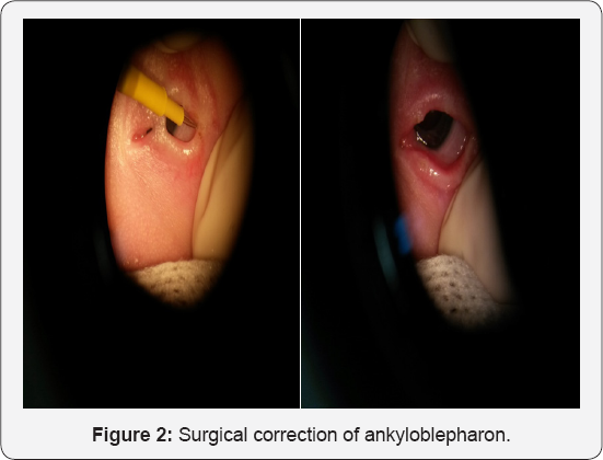
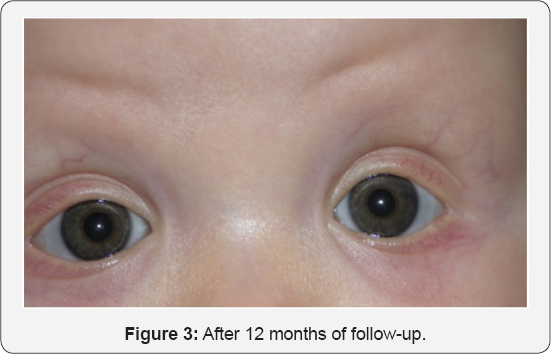
Case report: Authors describe a case of a newborn with no history of parental consanguinity or other similar cases in the family. At birth alopecia, ankyloblepharon, cleftpalate, micrognatia and absent nails were detected. Surgical correction of ankyloblepharon was done. Postoperative period had no complications and after 12 months of follow-up lid edges are completely free.
Discussion: As a genetic disease, treatment is symptom-oriented. Antibiotic ointments can be used in skin lesions. Surgery is reserved for correction of cleft palate/lip and ankyloblepharon. Genetic counseling is recommended.
Keywords: Syndrome; Ectodermal dysplasia; Ankyloblepharon cleft palate and lip


Introduction

Hay-Wells Syndrome, also known as AEC syndrome (ankyloblepharon-ectodermal dysplasia- clefting syndrome), is a rare genetic disease [1], with an autosomal dominant transmission. Sporadic cases have, however, also been described. First described by Hay and Wells in 1976 [2], it was later found that it is caused by a p63 gene mutation, a known regulator of epithelial development/differentiation and homologous of the TP53 oncosupressor gene [3,4]. It is a type of ectodermal dysplasia, leading to impaired development of hair, nails, teeth and glands, usually in association with cleft palate and/or lip and ankyloblepharon. These are considered the cardinal signs of the syndrome by most authors. This syndrome is present at birth and diagnosis is made based on the child's phenotypic features. The authors aim to describe a case of a male newborn with the cardinal signs of this syndrome at birth, as it is a very rare syndrome with just a few cases reported worldwide.


Case Report

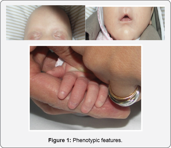
Male white newborn with no history ofparental consanguinity or other similar cases in the family. The baby was born by spontaneous vaginal delivery after an uneventful pregnancy with good prenatal care. At birth alopecia, ankyloblepharon, cleft palate, micrognathia and absent nails were detected (Figure 1a,1b & 1c- Phenotypic features). Anterior segment was normal, with no detected anomalies in the lacrimal apparatus. The rest of the physical examination was normal.
Ophthalmic ultrasound revealed a normal posterior segment. Abdominal and pelvic ultrasound were normal. Head ultrasound showed a small cyst in the pellucid septum and an apparent thickening in optic chiasmal area. Further study with Cranial MRI was normal, thus excluding a septum-optic dysplasia. Blood tests (CBS, electrolytes, hepatic function, kidney function, imunophenotyping and immunoglobulin levels) revealed a decrease in immunoglobulin A (X; normal range X), with no other changes. Hearing tests were normal.
Dermatology consultation recommended topical streoids in scarceeczematous areas and daily mineral ointments. Later hair prosthesis was discussed with the parents. Surgical correction of ankyloblepharon was carried out by ophthalmologists. Tearing of lid bridges was done in the operating room, under general anesthesia, using an electric scalpel in both eyes (Figure 2- Surgical correction of ankyloblepharon). Careful tearing with gentle pulling of the eyelids was carried out in order not to damage the underlying cornea. Post-operative period had no complications and after 12 months of follow-up lid edges are completely free (Figure 3 - After 12 months of follow-up).
Cleft palate was complete, type III. Surgical correction was performed according to Furlow and Von Langenbeck procedures by pediatric surgeons. Blood molecular analysis of TP63 mutations and deletions/duplications was normal; we are still waiting for molecular analysis of mouth mucosa specimen. Unfortunately, it was not possible to have parental authorization to carry DNA analysis in their cells. Speech therapy was also prescribed.


Discussion

Hay-Wells Syndrome, also known as AEC Syndrome (ankyloblepharon-ectodermal dysplasia-clefting), is a rare autosomal dominant disease [1]. Ankyloblepharon, ectodermal dysplasia signs and cleft palate are considered cardinal signs of this syndrome [5]. All of these features were present in our case.
Ankyloblepharon results from fusion, partial or total, of superior and inferior lid edges. In general, eyelids remain closed until the 5th week of gestation, when they open spontaneously. The mechanism underlying this phenomenon is still on debate, but many author point out keratinization as the key [6]. Accordingly, any kind of anomaly occurring between 7th and 15th week of gestation can result in eyelid anatomy changes [6]. Ankyblepharon can also be present in Trisomy 18 and CHAND Syndrome (Curly hair-Ankyblepharon-Nail Dystrophy), being associated with heart malformations, hydrocephaly, imperforated anus and glaucoma. Ankyblepharon should then be a warning sign for the possibility of other important simultaneous diseases [7].
Ectodermal dysplasias are a group of diseases in which there is impaired development of hair, teeth, nails, sweet glands and other structures originating from ectoderm [8,9]. These anomalies, when associated with other malformations, correspond to a group of ectodermal dysplasia syndromes that includes EEC Syndrome (ectodactilia-ectodermal dysplasia- clefting), Rapp-Hodgkin Syndrome and Chand Syndrome, the most important differential diagnosis of Hay-Wells Syndrome [9]. In our case, none of the other malformations were identified, so that Hay-Wells Syndrome diagnosis was straight forward.
Hay-Wells Syndrome patients can have various degrees of alopecia, thin and scarce hair, onicodystrophies, palmoplantar hyperkeratosis, cutaneous pigment changes [10], hypohidrosis, hypodontia, teeth malformations and ear anomalies. Lacrimal canal obstruction is common. Other described features include super numerary nipples, otitis media, hypospadia, middle-facial hypoplasia, hypertelorism, low height, mental retardation, deafness and other ocular deformities.
At birth, a descamative erythrodemia can be seen, with superficial erosion and crusts. Scalp area usually shows an erosive dermatitis, which is a common source of infection, putting these patients at an increased risk of bacterial superinfection and sepsis; this leads to an increase in mortality and morbidity rate in newborns with this syndrome. A lot of Hay-Wells Syndrome cases are erroneously diagnosed as bullous epidermolysis due to the presence of erythrodermia and extensive areas of erosion. It is thought that scalp lesions can disappear with age or lead to alopecia [3].
Although concerns exist about wound healing in patients with this syndrome, there have been no reported instances of wound healing complications. Cleft lip and palate repair can then be performed safely in patients with Hay-Wells syndrome.
This syndrome is caused by a p63 gene mutation, an homologous of p53 oncosupressor gene [3,4], which has a central role in epidermal stratification process, regulating basal keratocyts proliferative capacity. Evidence showing that changes in this gene can be associated with other diseases like EEC and Rapp-Hodgkins syndrome reveal the high pleomorphic effect of p63 gene mutations. Hay-Wells Syndrome results specifically from amino-acid substitution in SAM domain (sterile alpha motif) [3,4].
Timely diagnosis is essential. As a genetic disease with multiple phenotypic features, multidisciplinary action is of main importance and treatment is symptom-oriented. Antibiotic ointments can be used in skin lesions. Surgical treatment is reserved for correction of cleft palate/lip and ankyloblepharon. Genetic counseling is recommended.


Disclosure statement


No sponsorship or funding arrangements relating to research and no conflicts of interest to declare.
For more Open Access Journals in Juniper Publishers please click on: https://juniperpublishers.com


75% of Global Blindness Cases in Southeast Asia- Juniper Publishers


Juniper Publishers- JOJ Ophthalmology

Mini Review

Considerable efforts in many developing countries, through blindness prevention programs, the global number of blind and visually disabled seems to be growing, mainly as an effect of population increase and aging. The population of south East Asia is about 593 million, since the main cause of blindness is cataract and only in this region 283 million people are effected by cataract, 73 million by glaucoma, 52 million by age related macular degeneration, 30 million by corneal opacity and 28 million by diabetic retinopathy. More than 75% of blindness is preventable since the most important factor to understand is that around same number of the blind people could see again if they had eye care facilities in their related areas or regions.
Cataract, childhood blindness, refractive error and low vision, glaucoma, and diabetic retinopathy are the leading causes of blindness. On average throughout south east Asia there is one ophthalmologist for around 200,000 people, and one mid level eye care personnel for around half a million of the population. Most of the eye care personnel are located in large cities and populated towns; on the contrary, around 75% of the population lives in rural areas, thus making access to eye health services is a major challenge.
Road blocks like surgical cost, lack of family support, and failure to understand the need for surgery and other social, infrastructural, and geographic factors, such as availability of ophthalmologists in rural regions particularly and difficulties in accessing eye-care centers with patients having to travel too far. Quality of cataract surgery is also remains a concern, with poor outcomes reaching 40% in some places. In fact, many ophthalmologists do not perform surgery or may be inadequately trained. Cataract, therefore, continues to be a challenge to handle with the need to plan a comprehensive strategy addressing issues related to availability, affordability, accessibility, and acceptability of eye-care services, and improving outcome of cataract surgery in low- and middle-income countries.
Globally, women as compared with men had a larger percentage of blindness caused by cataract. Worldwide, 35.5% of blindness among women was caused by cataract versus 30.1% of blindness among men. It is estimated that blindness and severe impairment from cataract could be reduced by approximately 11% in the low- and middle-income countries if women were to receive cataract surgery at the same rate. Additional focus is needed to bring cataract surgical services to women mainly in low- and middle-income south east Asian countries.
It has been estimated very carefully that half of all the world's blindness is preventable or treatable. Cataract, for example, is well on its way to being eliminated as a major cause of blind-ness in developed nations. Improvements in cataract surgery have resulted in an operation that is 95% successful and is considered one of the safest and most efficacious of all major operations.
Blindness is significantly affects not only the immediate family but by enlarge the whole community. The blindness restricts mobility and requires some assistance for everyday work and task. Consequently, blindness affects the community on a practical level, as children cannot attend school when they become caretakers for blind adults. Thus, countless children are denied the opportunity to receive a basic education, when a sighted adult becomes the caretaker for a blind individual, he or she must stop working partially or complete. This leads to longterm economic and educational repercussions. Consequently the family earning become reduced which put the family in further poverty, frustration, depression and health issues.
To reduce or avoid the blindness in south east Asia an effective ophthalmic services, like cataract surgery, the screening for, and treatment of, diabetic retinopathy. The eye care centres having facilities of high quality, efficient and low-cost cataract surgery is the need of the time. The WHO and world health organizations should have more focus on developing countries or region like south east Asia. The effective eye care services require more coordinated and integrated planning, modules and efforts of services from the primary, secondary and tertiary levels. The need of outreach eye care services for rural areas under able supervision could bring tremendous results.
The eye care organizations are putting substantial efforts to build more hospitals and equip them, but right dedicated volunteers, ophthalmologists team and processes need to place according to the need and demand of area or region to ensure targeted outcome..More over significantly enhancement in resource base, infrastructure, equipment, eye specialists and other ophthalmic workers are needed to emplace along with equal emphasis to build and emplace task management teams to build the right processes and the organizational capability for maximal cost-effective resource utilization. Patient access, affordability and availability of surgical consumables and pharmaceuticals are all part of task management teams.

Focused, clear and transparent strategies are required to reduce global burden of blindness which require a global vision and local implementation. These strategies share a public health focus and community awareness blindness and eye care.

For more Open Access Journals in Juniper Publishers please click on: https://juniperpublishers.com

Thursday 23 January 2020

Prospects of Treating Ocular Hypertension and Glaucoma with Peptidic and Non-Peptide Kinin Mimetic Drugs- Juniper Publishers


Juniper Publishers- JOJ Ophthalmology


Abstract

The endogenously generated small nonapeptide bradykinin (BK) is often associated with inflammation, edema and pain amongst many other functions and pathologies. However, the latter aspects pertain to locally produced BK from circulating plasma precursor polypeptide kinninogen (KNGN). Recent work in ocular tissues and cells have a revealed a novel local synthesis of BK and other kinins from tissue-derived KNGN via action of kallikrien enzymes, quite independent from the blood-derived polypeptide. Furthermore, the whole kininergic system machinery including local KNGN, kallikrein enzymes to generate kinins from KNGN, two sub-types of kinin receptors (Bj-and B2-receptors), and the complete signal transduction pathways coupled to these receptors have been mapped out. Additional work has highlighted a number of downstream signaling and other biological responses that ensue following activation of Ba- and B2 receptors in human ocular cells and tissues. One key aspect to be discussed in detail in this review is the novel finding that BK, peptidergic BK analogs, and especially non-peptide mimetics of kinins (e.g. FR-199097; BKA78), profoundly lower and control intraocular pressure in a number of species, including ocular hypertensive (OHT) monkeys. These novel observations strongly suggest that kinin agonists represent a novel class of ocular hypotensive agents that could be of immense value in treating OHT associated with primary open-angle glaucoma (POAG) and perhaps other forms of glaucoma.


Ocular Hypertension Associated with Glaucoma

At a simplistic level, our knowledge has advanced to a point where it is now clear that elevated intraocular pressure (IOP) results from a fundamental imbalance between the generation of aqueous humor by the ciliary body and its efflux from the anterior chamber of the eye via one of two pathways [1-3]. The most physiologically relevant mechanism of AQH drainage involves the IOP-dependent outflow via the trabecular mesh work (TM) and Schlemm's canal (SC) route [1-3]. The lesser utilized pathways under normal conditions are the uveoscleral [1-3] and ocular lymphatic [4] pathways, but the latter can be engaged by certain drugs such as FP-class prostaglandin analogs (FPGAs) like latanoprost and tafluprost [1-6]. The chronically increased IOP, a condition generally termed as ocular hypertension (OHT), caused by blockage of the AQH drainage TM/SC pathways [7] during the aging process or due to ocular inflammation and deposition of various debris, along with apoptotic death of retinal ganglion cells. can lead to a clinically defined disease called glaucoma [8-10]. This high IOP distressfully distends and traumatizes the whole Whilst, glaucoma is painless and otherwise asymptomatic globe and initiates the death of RGCs and/or breakage of RGC axons at the back of the eye. These elements then cause a retrograde demise of the RGCs leading to severing of nerve fibers connecting the retina to the brain [11-13]. Many deleterious neurotoxic elements (e.g. high levels of extruded glutamate, endothelin, inflammatory cyto-and chemo-kines, noxious gases (e.g. nitric oxide; hydrogen sulfide)) and proteolytic enzymes (e.g. caspases and matrix metalloproteinases) released by activated macrophages [12-19], injured RGCs and interneurons are the culprits responsible for such neurotoxicity/chemically-induced axotomy of the RGCs. Hypoxia and ischemia [20,21] are also involved in the initiation phase of vascular dysfunction-induced death of RGCs since the thinning of the optic nerve at the level of the optic nerve head (ONH) forces the retinal blood vessels attached to the optic nerve to bend thereby restricting blood supply to the retina. While this progressive loss of RGCs occurs over several decades, if OHT is not treated to reduce the IOP the resulting glaucomatous optic neuropathy causes severe visual impairment, reduction of visual acuity and visual field and eventually results in irreversible blindness. Although many forms of glaucoma exist (e.g. open-angle glaucoma; closed-angle glaucoma; exfoliation glaucoma; myopic glaucoma) [8,9,22-26], the most prevalent is primary open-angle glaucoma (POAG) [26-28]. Behind cataracts, POAG is the second leading cause of blindness afflicting several millions of patients. It is estimated that by 2020, the global glaucoma-related blindness will reach ~80 million [26-28]. Elevated IOP and advancing age are the two major risk factors associated with POAG even though genetic factors [26] and race (especially African and Asian heritage) [29], myopia, diabetes and oxidative stress [30-36] and various vascular irregularities and dysfunctions [20,22], and intracranial cerebrospinal pressure [37,38] have been also linked to the development of POAG. The seriousness of POAG is often underestimated since it causes no overt discomfort or pain to the patient and insidiously progresses unnoticed over time. Considerable damage to the retina [33-36] and optic nerve and optic nerve head (ONH) [14,29,39-41] continues unyieldingly leading to scotomatous damage that manifests as loss of peripheral vision followed by a "tunnel vision" syndrome, thereby finally signaling the demise of ~40% of the original million RGCsof the patient and equivalent loss of connections to the brain and visual cortex [8,9,42-45]. Those lost or dying RGCs cannot be resuscitated and their axonal connections revived [46-48], and if left untreated the glaucomatous optic neuropathy due to OHT and oxidative /neurotoxic elements would claim the remaining RGCs causing total blindness [49-53].
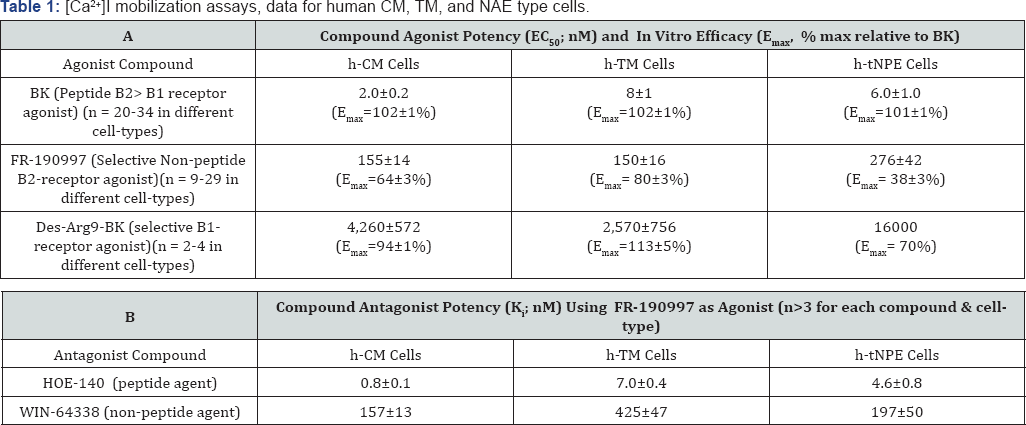
Data are mean±SEM uses a [Ca2+]I mobilization assay in cells derived from several different human donors' eyes. h-tNPE cells are SV40-virus- immortalized human non-pigmented ciliary epithelial cells derived from human ciliary epithelium that respond just like normal primary NPE cells. [145,146,157]
A number of treatment options have been developed to deal with POAG-associated OHT including ocular hypotensive medications [1-3,54-56], laser therapy and surgical interventions [57-64]. Unfortunately, as is the case with most drugs and surgical procedures, these treatment modalities have numerous side-effects (e.g. Burning and stinging, foreign- body sensation, brow-ache, ocular surface dryness, pulmonary hypertension and bradycardia, etc.), and adverse complications associated with them [1-3,54,55]. Additionally, poor compliance [65] and adherence to prescribed topical ocularly administrated medications by the OHT POAG patients (due to forget fulness, poor dexterity,lack of symptomatic pain or other cues due to OHT, poor understanding of the treatment regimen, and perhaps due distrust, etc.) contributes to the progression of the disease process. Similarly, laser therapies, although effective at the beginning, lose their efficacy over time [57-62]. Reports have surfaced that indicate that POAG/ OHT is only controlled in ~50% of patients who received laser treatment, and indeed due to scarring,the procedure often needs repeating within one-five years [57-64]. Indeed, such lasering and filtration procedures also have certain risks of complications and adverse events associated with them. Thus, there remains a continued unmet medical need to discover new and improved eye drop- medications and other novel surgical techniques to help the OHT/POAG patients mitigate and treat their underlying glaucomatous optic neuropathy caused by elevated IOP. To this end, a better understanding of the many complex pathways involved in AQH dynamics [4,7-10] has culminated in the discovery and development of many novel targets and ligands [13,54,55] that can modulate IOP via these targets to accomplish a level of homeostasis of AQH production and drainage. In order to address poor patient compliance, a number of innovations leading to sustained drug-release devices (e.g. implants, punctal plugs or contact lenses) [60-64,66-70] have been developed such that the patient need not remember to self-administer the medication. Likewise, a revolutionary set of novel surgical interventions [57-64] with much reduced surgical time and effort required and minimal adverse events and complications are becoming available [57-64]. These include the following: non-penetrating glaucoma surgery (NPGS), non-invasive glaucoma procedure, minimally invasive micro sclerostomy, blebless ab externo glaucoma surgery, ab externo bleb surgery, and the elegant minimally-invasive glaucoma surgery (MIGS) [60-64] that involve insertions of tiny drainage devices into the anterior chamber of the eye that appear to be highly effective in decreasing IOP [62-64]. Time will tell if indeed these innovations become mainstay treatment options for POAG/OHT patients in the near future. Regardless, however, the ordinary patient who is unable to afford the latter surgical procedures and devices, and those patients who are refractory to or cannot tolerate existing medications, will still require new topically administered drugs to lower and control the IOP in order to preserve their vision.


Present Day Pharmacotherapy for OHT

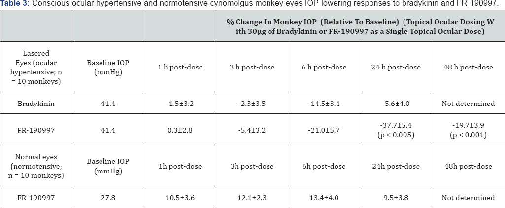
One of the earliest pharmacological agents to be used to lower elevated eye pressure is a muscarinic receptor agonist, pilocarpine [1-3]. It just happened that it promoted egress of AQH through the TM, hence pilocarpine became the first known conventional outflow promoter. Since it strongly constricted the pupil, made accommodation difficult and painful due to brow- ache, and needed to be administered up to 4-times a day, newer drugs with higher efficacy and lesser side-effects were sought and discovered over the next few decades. These included carbonic anhydrase inhibitors such as dorzolamide and brinzolamide, beta-adrenergic antagonists (e.g. timolol; betaxolol) and alpha- adrenoceptor agonists such as brimonidine and para-amino- clonidine [1-3,54,55]. Whilst these drugs lowered IOP well, they required at least twice daily dosing and they primarily inhibited the production of AQH by the ciliary processes of the ciliary body. Eventhough compliance increased and a greater efficacy was achieved, these agents had their own short-comings in terms of side-effects including ocular surface irritation (hyperemia), burning and stinging,allergy and drowsiness (a-agonists), and some pulmonary and cardiac insufficiency (with p-blockers) [13,55] Furthermore, we have learnt that AQH constituents serve important nutritional needs of the tissues inside the anterior chamber of the eye [10], and thus reducing its availability negatively impact the ciliary body, lens, corneal endothelial cells, and may in fact damage the TM and SC endothelial cells. Thus, a major breakthrough in OHT / POAG treatment occurred in the mid-90s when FP-receptor-selective prostaglandin (PG) agonists (PGAs) (e.g. latanoprost; travoprost; bimatoprost; tafluprost; unoprostone isopropyl ester) [3,5,6,71-73] were discovered and introduced into ocular clinical medicine. These PG drugs revolutionized the POAG/OHT treatment paradigm since they required once-daily ocular administration (before bed-time) and were much more potent and efficacious than the existing medications since they created new drainage pathways across the ciliary muscle and sclera (uveoscleral pathway) to help drain the AQH [1-5]. Nevertheless, these novel PGAs had some significant side-effects that included hyperemia, darkening of the iris color and increased pigmentation of the periorbital skin, growth of eye-lashes, deepening of the eye orbit, and in some cases cystoid macular edema [1-5,71-74]. Additionally, some OHT/POAG patients were quite refractory to the PGA drugs such that they required multiple drugs to control their IOPs. Not surprisingly, a multitude of fixed-dose combination products [4,75] containing different dual combinations of various ocular hypotensive drugs (and even a triple combination product) have now become available. However, due to the inherent genetic and biological variation in responses of patients to the different classes of IOP-lowering medications and their relative susceptibility to the side-effects of the drugs, there still remains a great need to hunt for and discover new drugs that are more effective, longer acting, efficacious in majority of OHT patients, and that induce fewer and milder off-target side-effects, thus having a greater therapeutic index than the existing drugs [67,76,77] (Table 3).

Future OHT/POAG Pharmacotherapy

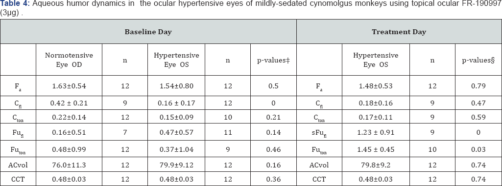
The potential additivity of new pharmacological agents to PGAs in the treatment of OHT and POAG has spurred the recent surge in research for novel agents exhibiting IOP-lowering properties. The realization that POAG is not only caused by elevated IOP since patients with normal IOPs still lose vision [7880], but perhaps is a reflection of enhanced RGC susceptibility to oxidative stress [7-9,12,32-36,42] and apoptotic process [15,47,81-90], has stimulated a renewed interest in finding drugs that have dual or multiplicity of mechanisms of action, including direct potential neuroprotective activity. The latter aspect stems from the finding that agents like betaxolol [91-95] and brimonidine [90,96-99], whilst lowering IOP, also upregulate the release of endogenous neurotrophins [89] in retinal tissues that could heal/rescue some of the RGCs that are compromised from the elevated IOP and oxidative stress. Likewise certain PGAs stimulate blood flow at the ONH in addition to reducing IOP [5,6,100]. Additionally, as the tools to monitor IOP [98,99] have become more accessible at a lower cost and with a greater sensitivity, including round-the-clock monitoring of IOP [99,100], and as new models of OHT/POAG are introduced using various species [48,23-25,101-104], the potential for such innovations to enhance drug discovery have dramatically increased in recent years. The ability to perform AQH dynamic measurements in small laboratory animals like mice [105], and to exploit enucleated and ex-vivo perfused bovine [103,106], porcine [103] and human eye anterior segments, and even whole eye [107], has further accelerated the mechanistic approach to ocular drug discovery and characterization. Accordingly, pharmacological agents that have exhibited ocular hypotensive efficacy in some of these animal/ ex-vivo models include K+- channel openers [108], Na+-K+ -ATPase inhibitor digoxin analogs [109], angiotensin-II receptor antagonists [110,111], renin inhibitors [112], angiotensin converting enzyme (ACE) inhibitors [113-116], ACE-2 activators [117,118], cannabinoids [119], rho- kinase inhibitors [120-122], nitric oxide (NO) donors and their conjugates [123-127], serotonin (5-hydroxy-tryptamine (5-HT)) receptor agonists [103,128,129], hydrogen sulfide donors [130], dopamine receptor agonists [3], melatonin receptor agonists [13], adenosine receptor agonists and antagonists [131], guanylate cyclase activators [123,132], novel EP2 receptor agonists [133,134], dual pharmacophoric PGs encompassing FP and EP3- receptor agonistic properties [135,136], etc. The most recent unexpected discoveries of potential drug candidates for OHT/ POAG treatment pertain to the kallikrein-kinin (KNK) system which will be addressed in detail below (Table 4).
* Values are means±standard deviation. t, comparing hypertensive with contralateral normotensive eyes; �p-values: comparing baseline day with treatment day by Student's two-tailed paired t-test. ACvol, anterior chamber volume, |jl; CCT, central cornea thickness, mm; Cf| fluorophotometric out flow facility, iJl/min/mmHg; Cton, tonographic outflow facility, |j1/min/mmHg; Fa, aqueous flow, |jl/min; FuFI, uveoscleral outflow calculated with Cfl, jl/min; Futon, uveoscleral outflow calculated with Cton jl/min; Times are 30 minutes. FR-190997 (0.01%) was applied as a 30jl drop (total dose of 3jg) to each eye of each monkey Modified from Ref 155.

Kallikrein-Kinin (KNK) System in the Eye

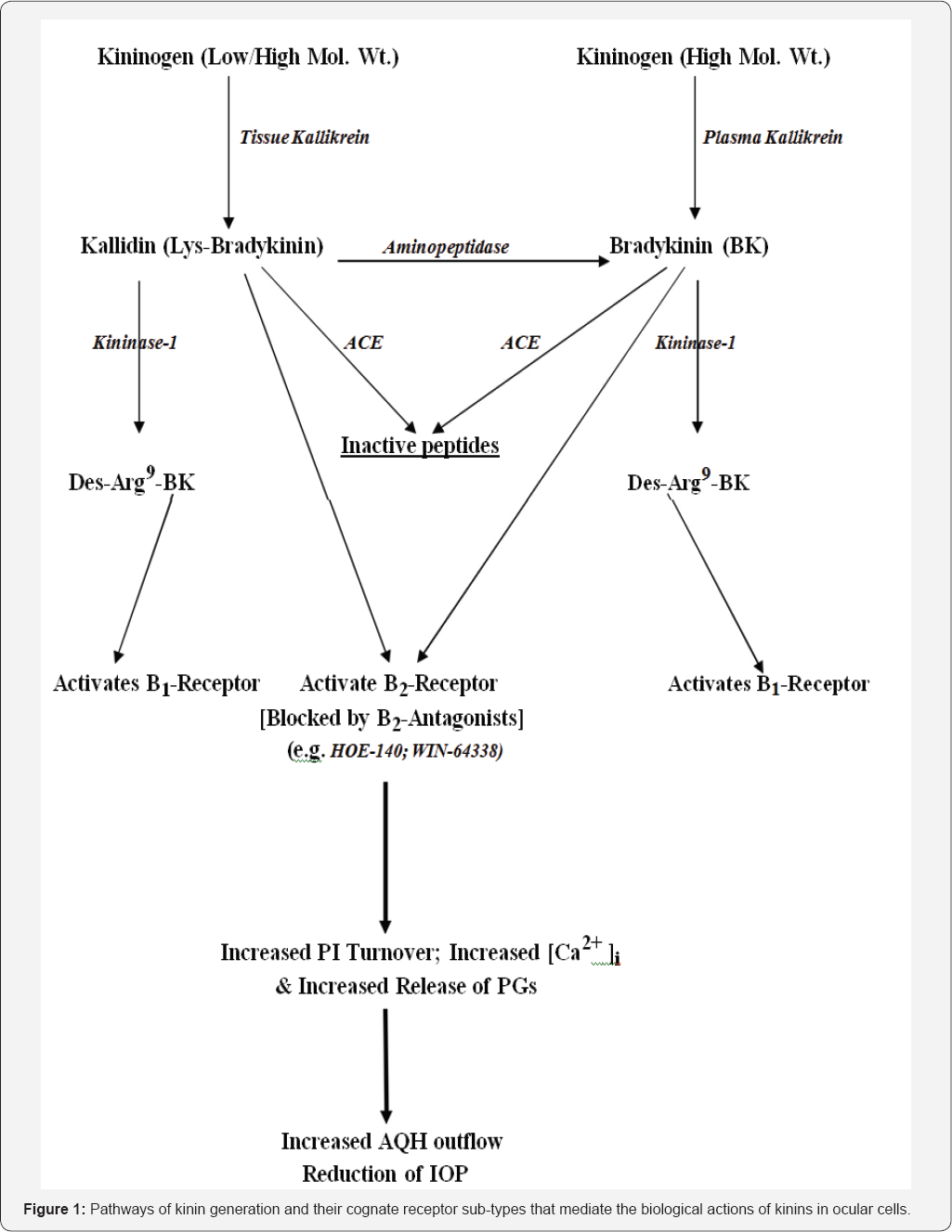
The peptide bradykinin (BK) was discovered many decades ago and the pathway to its generation has since been fully delineated. Today, we know that a precursor polypeptide, kinninogen (KNGN), is cleaved by specific enzymes (kallikreins) to produce a 10-amino acid -and a nine-amino acid containing peptide, Lys-BK and BK respectively [137,138]. Lys-BK is then converted to BK by an aminopeptidase, but both Lys-BK and BK act on the B2-receptor subtype of BK receptors, which is the predominant homeostatic receptor found Under normal physiological situations [137,138]. Kininase-1can convert both Lys-BK and BK to an octapeptide (Des-Arg9-BK) that interacts specifically with B1-receptor subtype of BK receptors which get upregulated during injury, trauma and other deleterious situations [137,138]. ACE inactivates both Lys-BK and BK to small inert peptides (Figures 1, 2). The notoriety surrounding BK and Lys-BK (kallidin) originates from their ability to cause deleterious vasodilation, inflammation, pain and cell proliferation [137,138]. These undesirable effects of kinins have been noted in all parts of the body and either trigger or are manifestations of various underlying diseases ranging from angioedema, diabetes, pulmonary and systemic hypertension, and aneurysms and diabetic retinopathy in the central nervous system and retina [137-139]. It was quite a revelation when various components of the KNK system were found in various compartments of the eye under normal circumstances and associated with cells and tissues of the eye [140,141]. Soon it became clear that BK and perhaps Lys-BK could be formed locally by the actions of tissue-based kallikriens on tissue-derived KNGN thereby creating a paracrine kininergic system within ocular tissues [140,141]. To support this notion further, both B1 and B2-receptor subtypes were found in various ocular cells that were functionally active mediating the actions of BK and related analogs of BK, generating intracellular second messengers [142145] Figure 3 producing further downstream effects such as promoting liberation of PGs [146,147] and causing ocular tissue contraction/ relaxation [148-150], etc. While circulating KNGN and kallikriens in ocular blood vessels do produce BK and Lys- BK to cause the vasodilator and pro-inflammatory effects as in the rest of the body, that system is distinctly different from the tissue-based KNK system in the ocular systems.
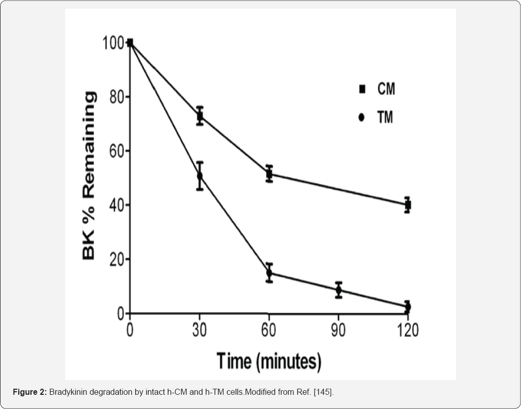
In order to ascribe a role of BK in ocular functions, early researchers administered BK either systemically or topical ocularly (t.o.) or via injections into the anterior chamber of the eye of different species [151-154]. BK either increased or decreased IOP accompanied by local inflammation and/or miosis [148-155].Using ex-vivo isolated bovine [106] and porcine [103,156] eye anterior segments, perfused BK was shown to induce disparate results, either causing decreased outflow or increasing outflow of AQH, thereby adding to the overall confusion. The known metabolic instability of BK in the presence of fluids or exposed tissues/cells [145,157], and possible species differences potentially contributed to these contradictory observations. The need for metabolically stabilized BK analogs or non-peptide BK mimetics was soon realized (see below). The rather paucity of information regarding the ocular BK receptor family in human ocular cells and tissues was slowly overcome by research in the early 80s-90s, including the work oflgic [140], Ma et al. [141], Sharif et al. [142], Wiernas et al. [143,144], and by Webb et al. [106,145,157].
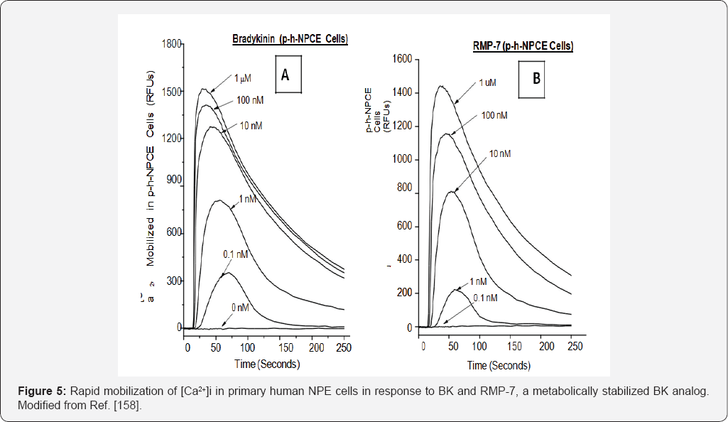
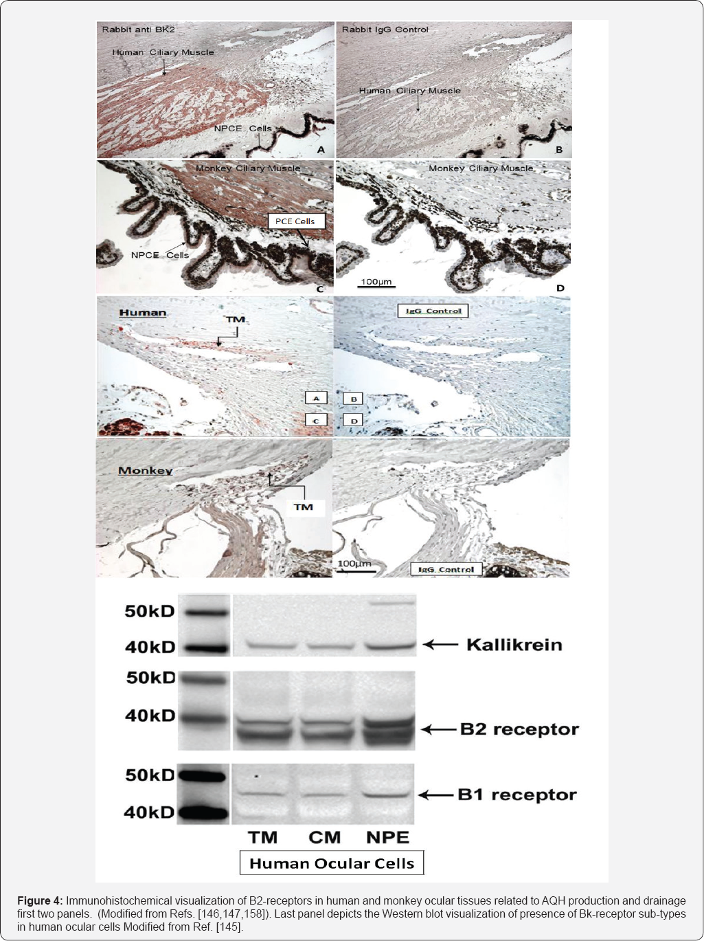
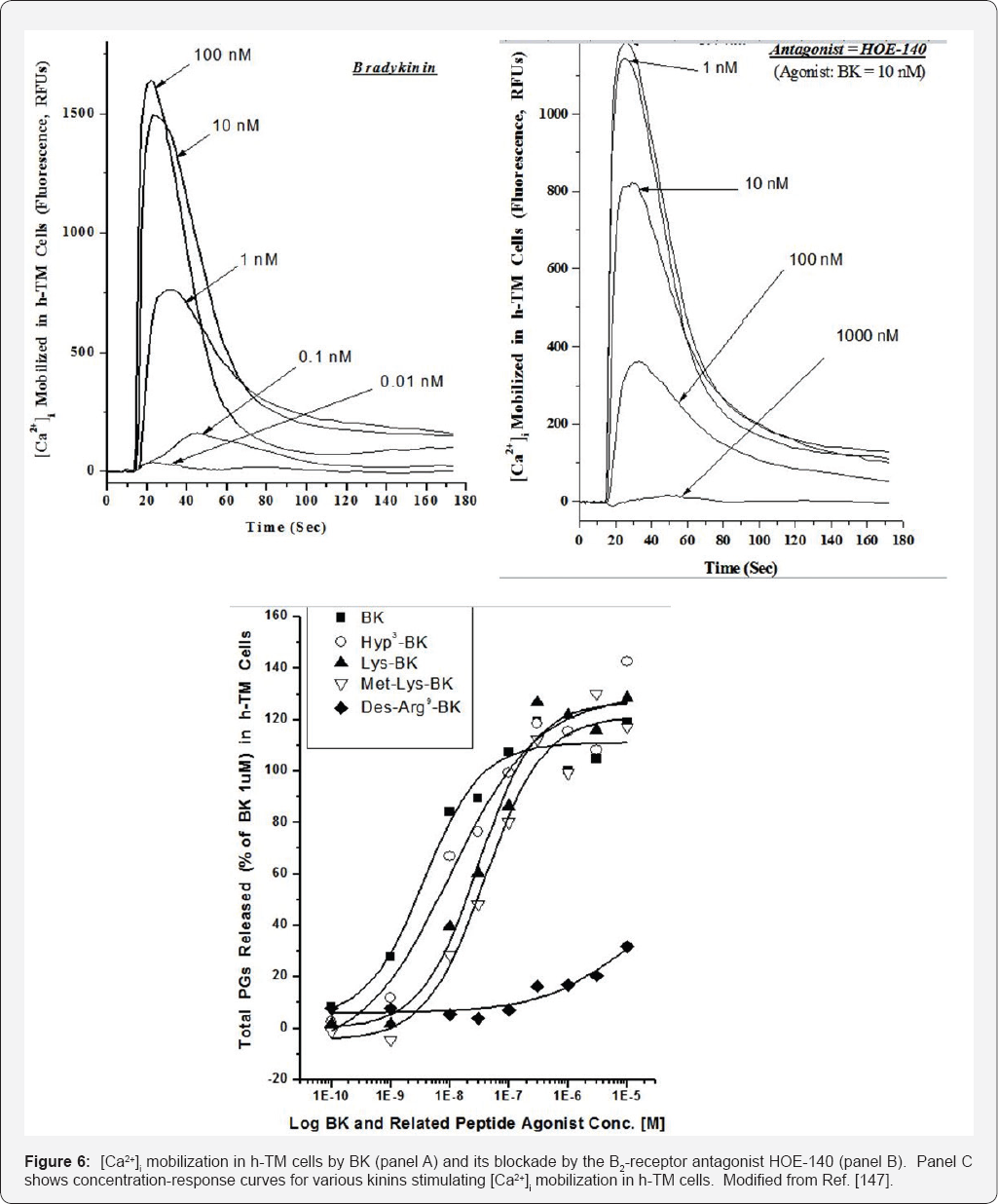
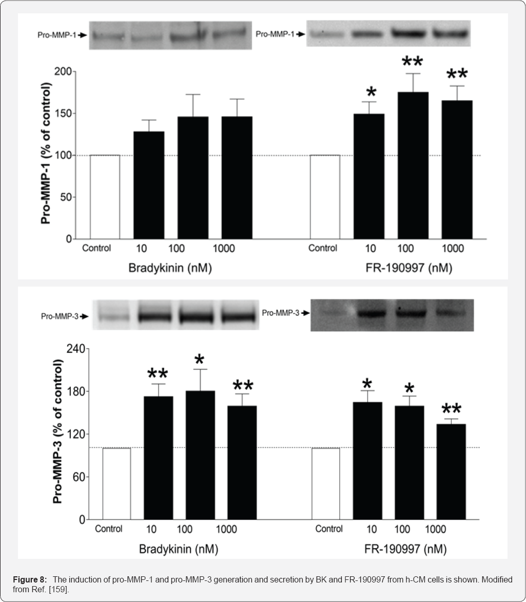
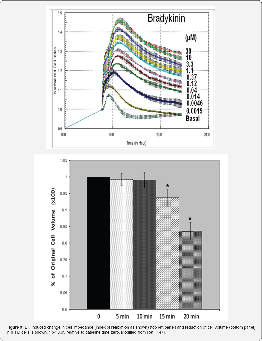
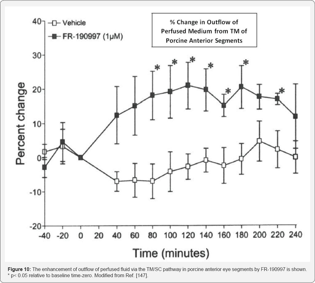
The work of Ma et al. [141] using reverse transcription- polymerase chain reaction (RT-PCR) and Southern blot analyses, and in situ hybridization to localize components of the KNK system in human ocular tissues was affirmed using immunohistochemistry (IHC) [145-147]. For the current subject matter of this review article, it was important to demonstrate the specific distribution and localization of the B2-BK receptor proteins. Accordingly, the presence of B2-BK receptors in human TM [147], ciliary muscle (CM) [146] and non-pigmented ciliary epithelial (NPE) [158] cells was demonstrated by IHC techniques. These are all key tissues involved in the drainage (CM and TM) Figure 4 and production (NPE) of AQH respectively. Importantly, these IHC observations were extended to the cynomolgus monkey anterior chamber tissues [146-148] in order to help correlate functional in vivo data (IOPlowerig; AQH dynamics) with these in vitro observations (see ahead). Next, it was important to determine whether the IHC of B2-receptors bore any linkage to functionality of these proteins in the respective human ocular cells mentioned above. To this end, primary h-NPE, h-CM and h-TM cells were isolated and challenged them with BK, its many peptide analogs and two non-peptide BK-mimetics, FR-190997 and BK2A78. Many of these kininergic compounds stimulated the production of intracellular inositol phosphates [142,156], and all peptide and non-peptide agents liberated endogenous intracellular Ca2+ ((Ca2+)i) from the endoplasmic reticulum in normal h-NPE, h-CM and h-TM cells [146,147,158] Figures 5,7 (and in bovine TM cells [155]) to varying degrees and with different relative potencies, a feature also observed in cells transfected with a cloned human B2-receptor. Additionally, these BK agonists also activated extracellular-regulated kinases-1/2 in h-TM [145] and h-CM [159] cells, and promoted the synthesis and secretion of PGE2 and PGF2a in the latter cells Figure 7. Since Des-Arg9-BK (a selective B1-agonist) was always a very weak agonist in all these biochemical assays, and since two B2-receptor antagonists (H0E-140 and WIN-64338) potently blocked the responses induced by BK, RMP-7 (a stabilized peptide analog of BK), FR-1909997 and BK2A78 [146,147,156,158,160,161], the functionality and pharmacological identification of B2-receptors in these cells were completely confirmed. Additional studies in h-CM and h-TM cells indicated that BK and FR-190997 could also activate intracellular signal transduction pathways to cause the release of various matrix metalloproteinases [106,159] Figure 8 that are known to digest extracellular matrix components such as collagen and thus aid in the efflux of AQH from the anterior chamber to lower IOP, a mechanism previously associated with ocular hypotensive FP-class PGAs [1-3,8,9,53]. Interestingly, BK enhanced the production of cAMP induced by PGE2 in h-TM cells [162], indicating that additional control of TM function by kinins was possible through activation of adenylyl cyclase. The next exciting phase of investigations tried to link these diverse biological actions of kinins in isolated cells of an almost intact organ, in this case enucleated porcine and bovine anterior segments of the eye. While one study initially showed a somewhat decrease in perfused fluid outflow in response to BK in the bovine eye [155], other studies by Webb et al. [106] showed that BK actually robustly stimulated outflow in bovine eyes as did the BK mimetic FR-190997 in other independent experiments using porcine eyes [156]. Mechanistically Webb et al. [106] also showed that this increased outflow by BK was mediated by B2-receptor-induced secretion of MMP-9 since a B2-antagonist and an MMP inhibitor abolished the effects of BK [106].
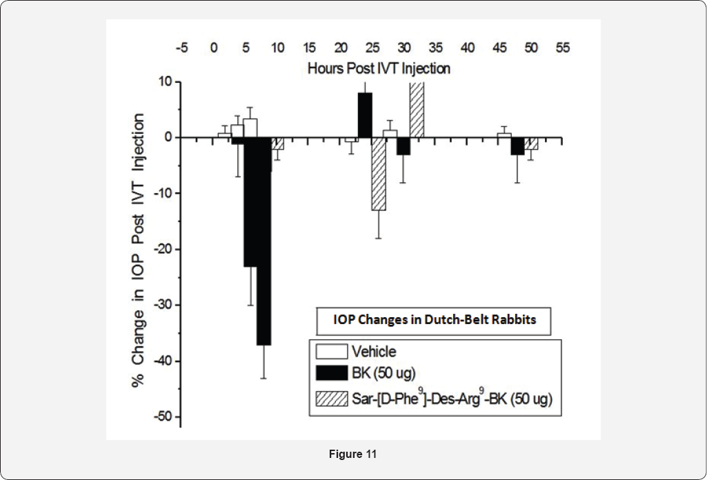
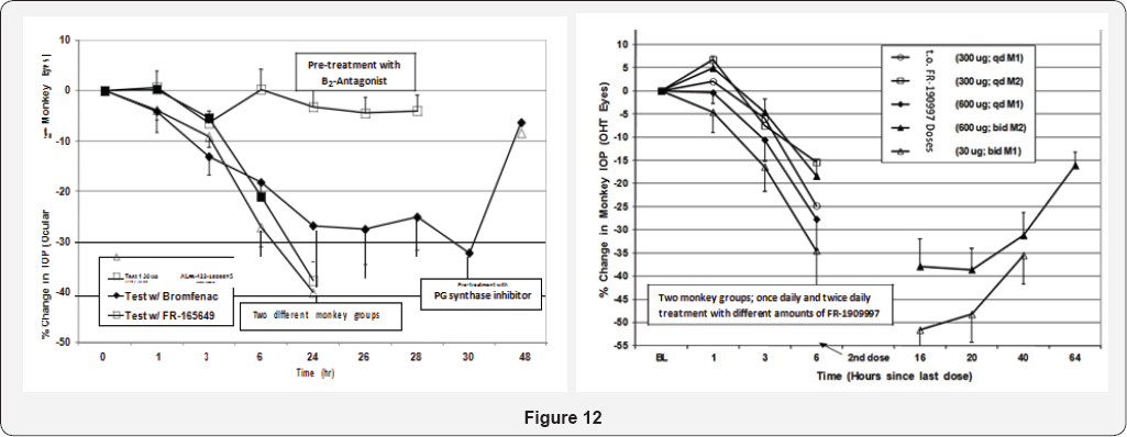
The role of BK in modulating I0P was investigated in a number of species.Topical ocular (t.o.) instillation of BK (50- 100|ig in a 30|il drop) to Dutch-belted rabbits, mixed breed cats, mice, rats, guinea pigs and cynomolgus monkeys (ocularly normotensive or hypertensive) failed to consistently influence IOP to any significant extent. However, intravitreal injection (ivt) of BK (50|ig) in eyes of Dutch-Belted rabbits induced a robustdecrease in I0P up to 8 hrs post-injection [146]. Interestingly, both B1-receptor-agonists, Des-Arg9-BK and Sar(D-Phe9)-Des-Arg9-BK injected ivt, did not alter I0P at all [146,147]. These data further substantiated the fact that only B2- receptors are involved in lowering and controlling I0P without any contribution from B1-receptors, at least in the rabbit. Since ethically and economically, we could not repeat it injection studies using BK in higher animals, and since topical ocular BK was without effect, a different approach was necessitated. Also, even though a metabolically stabilized peptide mimetic of BK (RMP-7) is available, it is still too polar a molecule to be used t.o. For such I0P modulation studies. However, a non-peptidic hydrophobic BK-mimetic, FR-190997 formulated in a standard vehicle was bioavailable when administered topically, and it potently and efficaciously reduced IOP in the conscious ocular hypertensive monkey eyes [156,160]. As little as 1|ig total t.o. Dose induced a 25% IOP reduction for up to 24-hrs after dosing, and dose-dependent reductions out to 48hrs post-t.o. Dosing were possible with a 10|ig dose [156,160]. The ocular hypotensive effects of FR-190997 and BK2A78 were in the realm of what the t.o. PG FDA-approved drugs like TRAVATAN® and Xalatan® produce in the monkey-model and in humans, but only to 24hrs post-dose. The fact that a non-peptide B2- receptor antagonist (FR-165649) completely prevented the I0P- lowering actions of FR-190997 in the 0HT monkey eyes strongly suggested that the B2-receptor was mediating the I0P-lowering actions of FR-190997 [156]. Furthermore, since FR-190997's ocular hypotensive effects were significantly attenuated by prior treatment with a PG-synthesis inhibitor (bromfenac) PGs were involved in mediating at least some of the I0P-lowering activities of this BK-mimetic, this being akin to the in vitro observations with FR-190997 [156,160] and BK [145-147,162]. In ascribing possible mechanisms activated by FR-190997 in its ability to reduce I0P in mildly-sedated 0HT monkeys, it was discovered that a predominant enhancement of uveoscleral outflow of AQH was triggered by FR-190997 [106,156]. However, it would appear that in the porcine isolated anterior chamber model, this compound (and BK in bovine) promoted fluid egress via the TM/SC conventional outflow pathway [106,156]. It remains to be seen whether such observations of robust ocular hypotensive activity of FR-190997 and BK2A78, along with other non-peptide BK-mimetics, can be reproduced in OHT human patients. Since FR-190997 [160] and BK2A78 [161] caused minimal ocular discomfort in conscious Dutch-belt rabbits, mixed breed cats, rats and monkeys after t.o. dosing, and produced no observable systemic or local side-effects, such compounds represent ideal new drug candidates worthy of pursuit in appropriate human clinical trials for determining ocular hypotensive activity. Two physiological observations noted that may limit the future utility of such BK-mimetics to treat OHT/POAG are the apparent tachyphylactic effects of FR-190997 in terms of IOP-lowering at relatively high doses, and a mild anesthetic activity observed on cat corneal surface [160]. However, whether these elements translate to the human ocular system requires further study. As long as low pharmacologically-relevant t.o. doses of FR-190997 and BK2A78 (and their analogs and derivatives [162-164]) are used t.o. in other animal models and in human subjects, it is possible to avoid triggering the above-mentioned "adverse" effects. Further studies on the ocular roles of BK and its analogs and mimetics are eagerly awaited.


Conclusion

Clearly there are now several drugs approved for the treatment of OHT/POAG and a number of new AQH drainage devices either approved or on the horizon for the same purpose of lowering IOP It is the issues of compliance, and the number and relative seriousness of the side-effects, or ineffectiveness and complications of the procedures, that continue to warrant hunt for newer more efficacious and more tolerable medications. The latter quest has resulted in the recent discovery of some new ocular hypotensive agents, including the first generation Kenyan non-peptide mimetics such as FR-190997, BK2A78 and their analogs [162-164].
The studies described in this review have clearly shown the presence of various components of the kininergic system in human and monkey ocular cells and tissues using a variety of techniques. Furthermore, functionally active sub-types of BKreceptor (B1- and B2) also are present in the ocular cells involved in AQH dynamics, Hence, BK and its analogs and mimetics are able to generate a variety of second messengers such as inositol phosphates and intracellular Ca2+ in h-NPE, h-CM and h-TM cells. Activation of this signal transduction pathway, then stimulates the production and secretion of PGs from these cells. These PGs are pivotal in promoting the generation and release of MMPs from CM and TM cells that digest extracellular matrix to create new pathways for AQH to drain from the anterior chamber of the eye leading to lowering of the IOP. Such duality of action of MMPs in response to BK receptor activation probably explains the elevated TM/SC outflow and increase of uveoslceral outflow observed after treatment with FR-190997and the profound IOP-lowering that this compound produces [156,160]. These new non-peptidic kinin mimetic drugs [156,162-164] will hopefully inspire other researchers to use these as templates for synthesizing next generation of ocular hypotensive agents, perhaps with some secondary neuroprotective activity on top of the ocular hypotensive properties. We all await the results of such new discoveries.


Conflict of Interest Statement

Author is an inventor or co-inventor of some granted patents related to the use of BK agonists (peptide and non-peptide) for treatment of glaucoma and the associated OHT, and these are cited in this article. The author, is an adjunct professor at Texas Southern University (Houston, TX) and at University of North Texas Health Science Center (Fort Worth, TX), and has no other conflicts of interest to declare. The intent of this review article is simply to share and expand the knowledge of the ocular roles of Kinins and thus inspire further research in this arena for the discovery of novel drugs and treatments to help combat blinding diseases of the eye, especially OHT/POAG.


For more Open Access Journals in Juniper Publishers please click on: https://juniperpublishers.com